
NEUROPATHIC BLADDER DISORDERS
ANATOMY & PHYSIOLOGY
The Bladder Unit
The functional features of the bladder include
(1) a normal capacity of 400–500 mL,
(2) a sensation of fullness,
(3) the ability to accommodate various volumes without a change in intraluminal
pressure,
(4) the ability to initiate and sustain a contraction until the bladder is empty.
(5) voluntary initiation or inhibition of voiding
despite the involuntary nature of the organ.
The Sphincteric Unit
In both males and females, there are 2 sphincteric elements:
(1) an internal involuntary smooth-muscle sphincter at the
bladder neck.
(2) an external voluntary striated-muscle

The uretrovesical junction
The function of the ureterovesical junction is to prevent backflow of urine
from the bladder to the upper urinary tract.
Nerve supply
The lower urinary tract receives efferent and afferent innervation from both
the autonomic and somatic nervous systems.
•The parasympathetic innervations originates in the second to fourth sacral
segments, The cholinergic fibers supply the bladder .
• The sympathetic nerves originate at T10–L2, The noradrenergic fibers
innervate the smooth muscles of the bladder base, internal sphincter, and
proximal urethra.
• Somatic motor innervation originates in S2–3 and travels to the striated
urethral sphincter .
•There are both somatic and visceral afferents from the bladder and
urethra.

The Micturition Reflex
Intact reflex pathways via the spinal cord and the pons are
required for normal micturition. Afferents from the bladder are
essential for the activation of the sacral center, which then
causes detrusor contraction, bladder neck opening, and
sphincteric relaxation.
The pontine center, through its connection with the sacral
center, may send either excitatory or inhibitory impulses to
regulate the micturition reflex.

URODYNAMIC STUDIES
Urodynamic studies are techniques used to obtain graphic recordings of
activity in the urinary bladder ,Urethral sphincter, and pelvic musculature.
1. Uroflowmetry
Uroflowmetry is the study of the flow of urine from the urethra.
BECAUSE URINARY FLOW RATE IS THE PRODUCT OF DETROSAL ACTION AGAINST
OUTLET RESISTANCE variation from the normal flow rate might reflect dysfunction .
The normal peak flow rate for males is 20–25 mL/s and for females 20–30 mL/s.
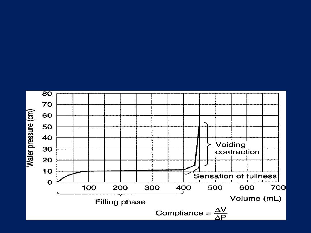
2. Cystometry
Evaluate
The basic factors of normal bladder function
bladder capacity, intravesical pressure against the volume , accommodation, sensation,
contractility, voluntary control.
Cystometry can be done by :
by filling the bladder with water and recording the intravesical pressure against the volume of
water introduced into the bladder.
Urinary sphincteric function can be evaluated by recording the electromyographic(EMG) and
profilometry
3.EMG (electromyographic)
recording the activity of the voluntary component of the sphincteric mechanism
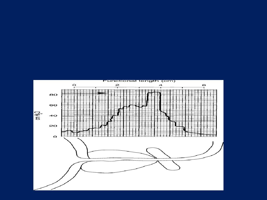
4.Urethral Pressure Profile measurement (profilometry).
recording the activity of both smooth and voluntary components by measuring the intraurethral
pressure of the sphincteric unit.

CLASSIFICATION OF NEUROPATHIC BLADDER
1.Spastic Neuropathic Bladder Neuropathic Bladder
Due to Lesions Above the Sacral Micturition Center
Most lesions above the level of the cord where the micturition
center is located will cause bladder spasticity. Sacral
reflex arcs remain intact, but loss of inhibition from
higher centers results in spastic bladder .

Etiology
A . The lesion is above the pontine micturition center.
Eg: dementia, vascular accidents, multiple sclerosis, tumors, and inflammatory
disorders such as encephalitis or meningitis.
B. Supra sacral Spinal cord injury .
which can be the result of Eg : trauma, herniated intervertebral disk, vascular
lesions, multiple sclerosis, tumor, syringomyelia, or myelitis.
NB Spinal cord injuries at the cervical level are often associated with a condition
known as autonomic dysreflexia (because the lesion occur above the sympathetic
out flow ) .

Features :
(1) reduced capacity,
(2) involuntary detrusor contractions,
(3) high intravesical voiding pressures,
(4) Marked hypertrophy of the bladder wall,
(5) spasticity of the pelvic striated muscle.
(6) autonomic dysreflexia (dyssenergic voiding) in cervical cord
lesions(because the lesion occur above the sympathetic out
flow ).

Clinical Findings
A. SYMPTOMS
The severity of symptoms depends on the site and extent
of the lesion as well as the length of time from injury.
Symptoms include involuntary urination,
frequency, urgency, and also urge incontinence .
B. SIGNS
A complete neurologic examination is most important. The level of the injury needs
to be established,
• assessment of the digital rectal exam, anal tone ,perianal sensation S2S3
• assessment of the sensation Penis S2,outside of the foot S2,big toeS3,sole of foot
S2S3
• assessment of the reflexes
A. Cutaneus bulbocavernous S2-4, cremastric reflexesL1 L2
B. Deep tendon Biceps C5 C6, Triceps C7, KneeL3L4 , Achilis tendon L5S1S2 .
.

C. LABORATORY FINDINGS
Urinalysis patients experience one or more urinary tract
Infections due to catheter , immobilization.
D. X-RAY FINDINGS
Excretory urograms .
The kidneys may show evidence of pyelonephritic scarring,
hydronephrosis, or stone disease.
A trabeculated bladder of small capacity .
E.Cystoscopy
Will show
Small bladder capacity, stones, changes secondary to chronic
infection or indwelling catheters, and the integrity of the
bladder neck and external urethral sphincter can be assessed.

F. URODYNAMIC STUDIES
Combined recording of bladder and urethral sphincter
activity will reveal :
Cystometry:
a low-volume bladder with high filling intravesical pressure ,
involuntary contractions ,high voiding pressures in the
bladder
urethral pressures profile :
normal , high .
EMG :
may show normal, dyssynergy of the external sphincter

2. Flaccid (Atonic) Bladder
Direct injury to the sacral cord segments S2–4 or peripheral
innervation of the bladder results in flaccid paralysis of
the urinary bladder.
Common causes of this type of bladder behavior are trauma,
tumors, tabes dorsalis, and congenital anomalies (eg, spina
bifida,meningomyelocele).
Characteristically:
the capacity is large, intravesical pressure low, and involuntary
contractions absent.

Clinical Findings
A. SYMPTOMS
The patient experiences flaccid paralysis and loss of sensation affecting the muscles and
dermatomes below the level of injury.
The principal urinary symptom is retention with overflow incontinence.
Male patients lose their erections.
B. SIGNS
Neurologic changes are typically lower motor neuron. Extremity reflexes are hypoactive or
absent. Sensation is diminished or absent .
C. LABORATORY FINDINGS
•
Urinalysis patients experience one or more urinary tract Infections due to bladder
catheterization.
•
D. X-RAY FINDINGS
plain film of the abdomen may reveal fracture of the lumbar spine
Excretory urograms should be performed initially to check for calculus, hydronephrosis,
pyelonephritic scarring
Cystogram usually large and smooth walled bladder

E. Cystoscopy
bladder should be large and smooth walled.
F. URODYNAMIC STUDIES
The urethral pressure profile and EMG: reflects normal ,
high or low smooth and striated sphincter .
Cystometry : large bladder capacity with low intra vesical
pressure, involuntary detrusor contractions are weak
or absent.
Voiding is accomplished by straining or by the Crede
maneuver, and there is a large volume of residual
urine.
Crede maneuver : applying sustained pressure over the
bladder

High-pressure sphincter
A .With High-pressure bladder :
If the bladder pressure overcomes the sphincter pressure , the
patient leaks urine (incontinence).
If the sphincter pressure is higher than the bladder pressure during
voiding , bladder emptying is inefficient (retention, recurrent UTIs).
B. With Low-pressure bladder
If The bladder simply unable to generate enough pressure to empty
(retention, recurrent UTIs).
Low-pressure sphincter
A. With High-pressure bladder
The bladder will only be able to hold low volumes of urine before
leaking (incontinence).
B. With Low-pressure bladder
The patient may be dry for much of the time. They may, however, leak
urine (incontinence) when abdominal pressure rises (e.g. when
coughing, rising from a seated position).

DIFFERENTIAL DIAGNOSIS OF NEUROPATHIC BLADDER
•
Cystitis
•
Chronic urethritis
•
Vesical irritation secondary to psychic disturbance
•
Myogenic damage
•
Interstitial cystitis
•
Cystocele
•
Infravesical obstruction

Spinal Shock
•
Intermittent catheterization using strict aseptic technique has proved to be the
best form of management of bladder rehabilatation. This avoids urinary tract
infection as well as the complications of an indwelling catheter (eg, urethral
stricture, abscess, erosions, stones).
•
Cystogram is needed to rule out reflux
•
Urodynamic study should be repeated every 3 months as long as spasticity is
improving and then annually to check for complications of the upper urinary tract.
•
Control infection, a fluid intake of at least 2–3 L/ day should be maintained (100–
200 mL/h) if at all possible.
•
Renal and ureteral drainage are enhanced by moving the patient frequently, with
ambulation, these measures improve ureteral transport of urine, reduce stasis,
and lower the risk of infection.
•
prophylaxis for calculus formation (eg, reduction of intake of calcium and oxalate
and elimination of vitamin D in the diet).

TREATMENT OF NEUROPATHIC BLADDER
The goal of treatment of any form of neuropathic bladder to
restore low-pressure activity to the bladder, In doing so, renal
function is preserved, continence restored, and infection more
readily controlled.
We are always afraid from the effect on the upper U. tract, so the spastic
bladder is more serious .
Spastic Neuropathic Bladder
1 Behavioral therapy
A
.
Education
B. Life style and dietary modification
C. BLADDER TAINING
To consider a bladder rehabilitated to a functional state, a patient should be able to go 2–3
hours between voiding and not be incontinent during this interval.
Voiding is initiated using trigger techniques—tapping the abdomen suprapubically , tugging
on the pubic hair, squeezing the penis.
They may be helped by low dose anticholinergic medication (to decrease intravesical
pressure)or by neural stimulation.

If the functional capacity of the bladder is so small, and involuntary voiding can occur
,satisfactory training of the bladder cannot be achieved, and alternative measures
must be taken
2 . Pharmaclogic therapy
A oral
anticholinergic agents eg oxybutinin,tolterodin
tricyclic antidepressant imipramine,duloxitine
B
INTRAVESICAL INSTILLATION OF BOTULINUM-A TOXIN which inhibit neuromuscular
release at N-M junction.
3 . Neurostimulation of sacral nerve roots
to accomplish bladder evacuation
(bladder pacemaker)
4 . Sacral rhizotomy
Conversion of the spastic bladder to a flaccid bladder
5
.
Augmentation cystoplasty
6
.
Urinary diversion
for irreversible, progressive upper urinary tract deterioration.
A variety of procedures are available, including the standard ileal conduit,
cutaneous ureterostomies.
7
.
A permanent indwelling catheter
with or without ant cholinergic medication.
8
.
A condom catheter
and a leg bag in males if residual urine volumes are small
and the patient does not have bladder pressures above 40 cm of water on
urodynamic evaluation.

Flaccid Neuropathic Bladder Treatment
1 Behavioral therapy
A.
Education
B.
Life style
C.
BLADDER TAINING bladder evacuation can be accomplished by
Voiding should be tried every 2 hours by the clock to avoid embarrassing leakage.
Raise intra-abdominal pressures by straining, using the abdominal and diaphragmatic
muscles .
Crede maneuver by manual suprapubic pressure.
2. INTERMITTENT CATHETERIZATION
regular intermittent catheter drainage every 3–6 hours. This technique eliminates residual
urine, helps prevent infection, avoids incontinence, and protects against damage to the
upper urinary tract. It simulates normal voiding and is easily learned and adapted by
patients.
3. PARASYMPATHOMIMETIC DRUGS
The stable derivatives of acetylcholine assist the evacuation of the bladder.
Bethanechol chloride is the drug of choice.

High sphincter pressure
At level of smooth sphincter
Pharmacologic therapy α-Adrenergic antagonists
Transurethral resection or incision
At level of striated sphincter
Pharmacologic therapy Benzodiazepines Baclofen
Botulinum toxin (injection)
Urethral overdilation
Surgical sphincterotomy
Urethral stent
Circumventing the Problem
Intermittent catheter
Continuous catheterization

Low sphincter pressure
A. Behavioral therapy
Education
life style
Bladder training Timed bladder emptying or prompted voiding
Pelvic floor physiotherapy
B. Electrical stimulation
C.
Pharmacologic therapy α-Adrenergic agonists Tricyclic antidepressants
D. Nonsurgical periurethral bulking agent
E.
Sling procedures
F.
TVT,TOT
G. Artificial urinary sphincter
H. Circumventing the Problem
Intermittent catheterization
External collecting devices
Continuous catheterization

COMPLICATIONS OF NEUROPATHIC BLADDER
1.Infection
2.Hydronephrosis
3. Calculus
A number of factors contribute to stone formation in the
bladder and kidneys.
Bed rest .
Inadequate fluid intake .
Catheterization .
Subsequent infection .

4.Renal Amyloidosis
Secondary amyloidosis of the kidney is a common cause of death in patients with neuropathic
bladder.
5.Sexual Dysfunction
6.Autonomic Dysreflexia
Autonomic dysreflexia is sympathetically mediated reflex behavior .
The phenomenon is seen in patients with cord lesions above the sympathetic outflow from
the cord
PROGNOSIS
The greater threat to the patient with a neuropathic bladder is progressive renal damage
(pyelonephritis, calculosis , and hydronephrosis). Advances in the management of the
neuropathic bladder, together with better follow-up of patients at regular intervals, have
substantially improved the outlook for long-term survival.

The end
Thank you