Lecture 6
Thorax
د.رندعبداللطيف
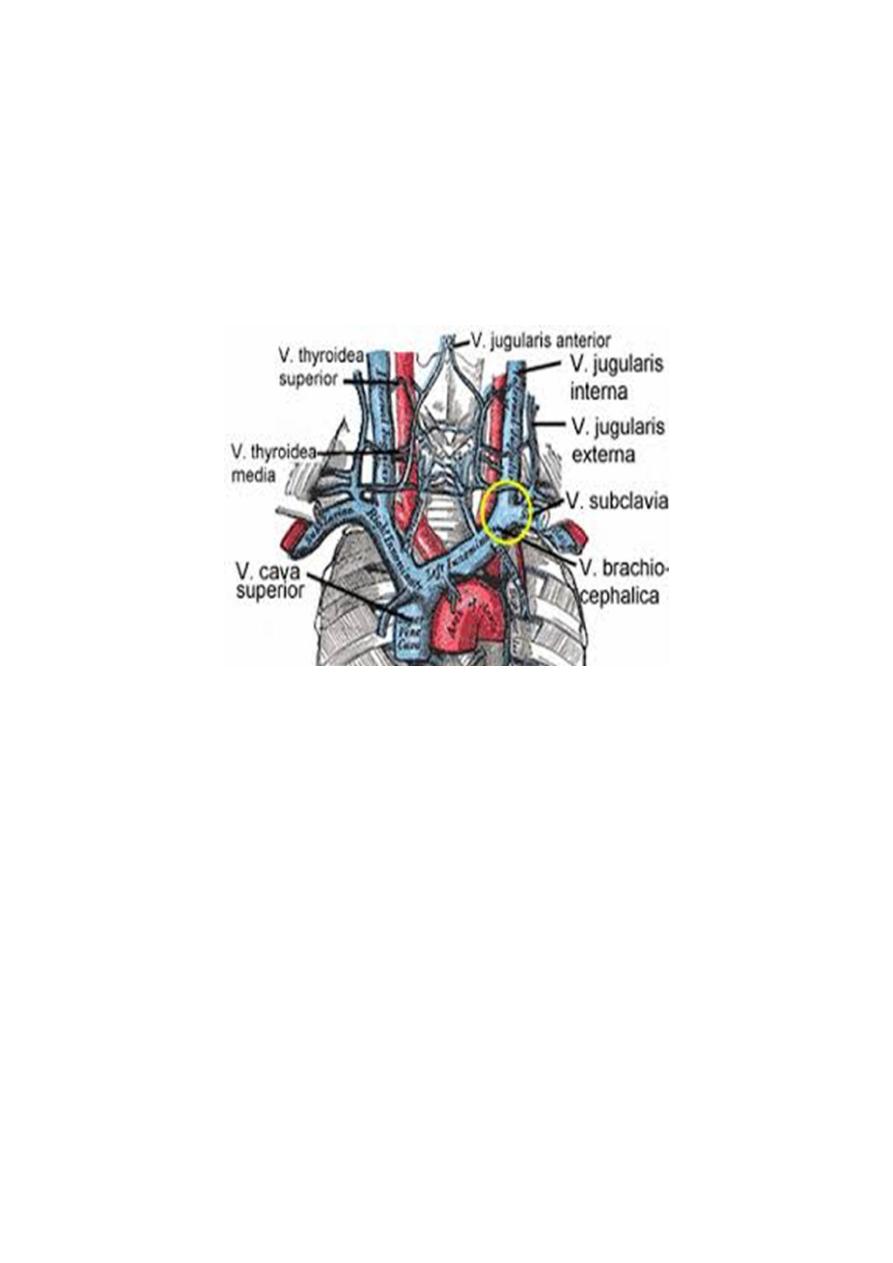
Brachiocephalic Veins
The right brachiocephalic vein is formed at the root of the neck by the union
of the right subclavian and the right internal jugular veins. The left
brachiocephalic vein has a similar origin. It passes obliquely downward and to
the right behind the manubrium sterni and in front of the large branches of the
aortic arch. It joins the right brachiocephalic vein to form the superior vena
cava.
Superior Vena Cava
The superior vena cava contains all the venous blood from the head and neck
and both upper limbs and is formed by the union of the two brachiocephalic
veins. It passes downward to end in the right atrium.
Azygos Vein
The origin of the azygos vein is variable. It is often formed by the union of the
right ascending lumbar vein and the right subcostal vein. It ascends through the
aortic opening in the diaphragm on the right side of the aorta to the level of the
fifth thoracic vertebra. Here it arches forward above the root of the right lung to
empty into the posterior surface of the superior vena cava. The azygos vein has
numerous tributaries:
1- the eight lower right intercostal veins
2- the right superior intercostal vein
3- the superior and inferior hemiazygos veins
4- Numerous mediastinal veins

Lecture 6
Thorax
د.رندعبداللطيف
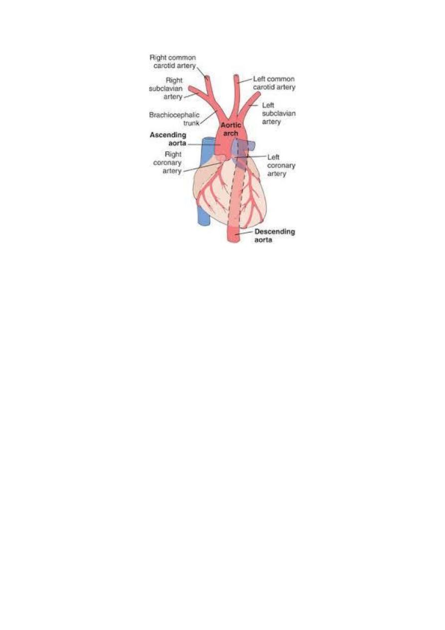
Aorta
The aorta is the main arterial trunk that delivers oxygenated blood from the
left ventricle of the heart to the tissues of the body. It is divided for purposes of
description into the following parts: ascending aorta, arch of the aorta,
descending thoracic aorta, and abdominal aorta.
Ascending Aorta
The ascending aorta begins at the base of the left ventricle and runs upward and
forward to come to lie behind the right half of the sternum at the level of the
sternal angle, where it becomes continuous with the arch of the aorta.
Arch of the Aorta
The arch of the aorta is a continuation of the ascending aorta. It lies behind the
manubrium sterni and arches upward, backward, and to the left in front of the
trachea. It then passes downward to the left of the trachea and, at the level of the
sternal angle, becomes continuous with the descending aorta.
Branches
1- The brachiocephalic artery arises from the aortic arch. It divides into the right
subclavian and right common carotid arteries
2- The left common carotid artery
3-The left subclavian artery
Descending Thoracic Aorta
The descending thoracic aorta lies in the posterior mediastinum and begins as a
continuation of the arch of the aorta on the lower border of the body of the T4
(opposite sternal angle). It runs downward in the posterior mediastinum,
inclining forward and medially to reach the anterior surface of the vertebral
column. At the level of the T12, it passes behind the diaphragm (through the
aortic opening) in the midline and becomes continuous with the abdominal
aorta.
Branches
1-Posterior intercostal arteries are given off to the lower nine intercostal spaces
on each side.
2-Subcostal arteries are given off on each side and run along the lower
border of the 12th rib to enter the abdominal wall.
3-Pericardial, esophageal, and bronchial arteries are small branches that are
distributed to these organs.
Lecture 6
Thorax
د.رندعبداللطيف
Pulmonary Trunk
The pulmonary trunk conveys deoxygenated blood from the right ventricle of
the heart to the lungs. It leaves the upper part of the right ventricle and runs
upward, backward, and to the left. It is about (5 cm) long and terminates in the
concavity of the aortic arch by dividing into right and left pulmonary arteries. it
is enclosed in the fibrous pericardium and a sheath of serous pericardium
together with the ascending aorta.
Branches
1-The right pulmonary artery runs to the right behind the ascending aorta and
superior vena cava to enter the root of the right lung.
2-The left pulmonary artery runs to the left in front of the descending aorta to
enter the root of the left lung
The ligamentum arteriosum
A fibrous band that connects the bifurcation of the pulmonary trunk to the
lower concave surface of the aortic arch. It is the remains of the ductus
arteriosus, which in the fetus conducts blood from the pulmonary trunk to the
aorta, thus bypassing the lungs. The left recurrent laryngeal nerve hooks around
the lower border of this structure. After birth, the ductus closes. If it remains
patent, aortic blood will enter the pulmonary circulation, producing pulmonary
hypertension and hypertrophy of the right ventricle.

Lecture 6
Thorax
د.رندعبداللطيف
Vagus Nerves
The right vagus nerve descends in the thorax, first lying posterolateral to the
brachiocephalic artery. It passes behind the root of the right lung and assists in
the formation of the pulmonary plexus. On leaving the plexus, the vagus
passes to the posterior surface of the esophagus and takes part in the formation
of the esophageal plexus. It then passes through the esophageal opening of the
diaphragm behind the esophagus to reach the posterior surface of the stomach.
The left vagus nerve descends in the thorax between the left common carotid
and the left subclavian arteries. It then crosses the left side of the aortic arch
and is itself crossed by the left phrenic nerve. The vagus then turns backward
behind the root of the left lung and assists in the formation of the pulmonary
plexus. On leaving the plexus, the vagus passes onto the anterior surface of the
esophagus and takes part in the formation of the esophageal plexus. It then
passes through the esophageal opening in the diaphragm in front of the
esophagus to reach the anterior surface of the stomach.
Branches
Both vagi supply the lungs and esophagus. The right vagus gives off cardiac
branches. The right recurrent laryngeal nerve arises from the right vagus in the
neck and hooks around the subclavian artery and ascends between the trachea
and esophagus.
The left vagus gives origin to the left recurrent laryngeal nerve which arises
from the left vagus trunk as the nerve crosses the arch of the aorta. It hooks
around the ligamentum arteriosum and ascends in the groove between the
trachea and the esophagus on the left side (Fig. 3.6). It supplies all the muscles
acting on the left vocal cord (except the cricothyroid muscle, a tensor of the
cord, which is supplied by the external laryngeal branch of the vagus).
Lecture 6
Thorax
د.رندعبداللطيف
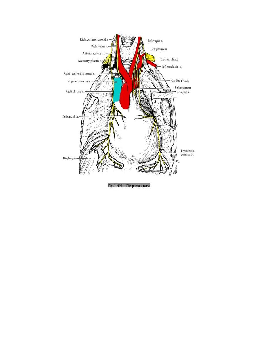
Phrenic Nerves
The phrenic nerves arise from the neck from the anterior rami of the C3, C4,
and C5 cervical nerves.
The right phrenic nerve descends in the thorax along the right side of the right
brachiocephalic vein and the superior vena cava. It passes in front of the root of
the right lung and runs along the right side of the pericardium. It then descends
on the right side of the inferior vena cava to the diaphragm. Its terminal
branches pass through the caval opening in the diaphragm to supply the central
part of the peritoneum on its underaspect.
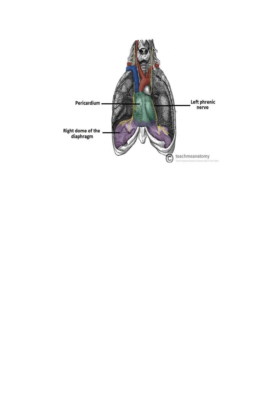
The left phrenic nerve descends in the thorax along the left side of the left
subclavian artery. It crosses the aortic arch and here crosses the left vagus nerve.
It passes in front of the root of the left lung and then descends over the left
surface of the pericardium, which separates the nerve from the left ventricle. On
reaching the diaphragm, the terminal branches pierce the muscle and supply the
central part of the peritoneum on its underaspect.
Lecture 6
Thorax
د.رندعبداللطيف