


1
B
AGHDAD COLLEGE
2015-2016
L
ASER IN OPHTHALMOLOGY
LASER: acronym (or abbreviation) from Light Amplification by Stimulated
Emission of Radiation.
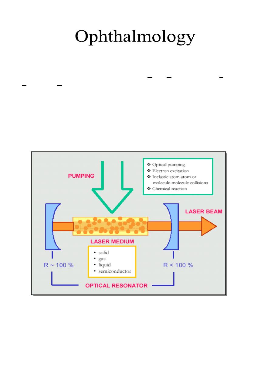
Physics of laser:
The optical resonator or called "Laser chamber" is composed of two reflecting
mirrors at the end of this chamber, and in-between there is a laser medium, which
could be solid, liquid, gas or semiconductor. Around this chamber, there is
pumping source to stimulate laser production. Pumping source is either optical,
electrical or chemical.
Criteria of laser:
1- Monochromatic: all waves have the same wavelength.
2- Coherent: waves have the same phase, so there is constructive interference in-
between.
3- Collimated (Parallel waves).
Lecture: 25
Dr. Najah
2
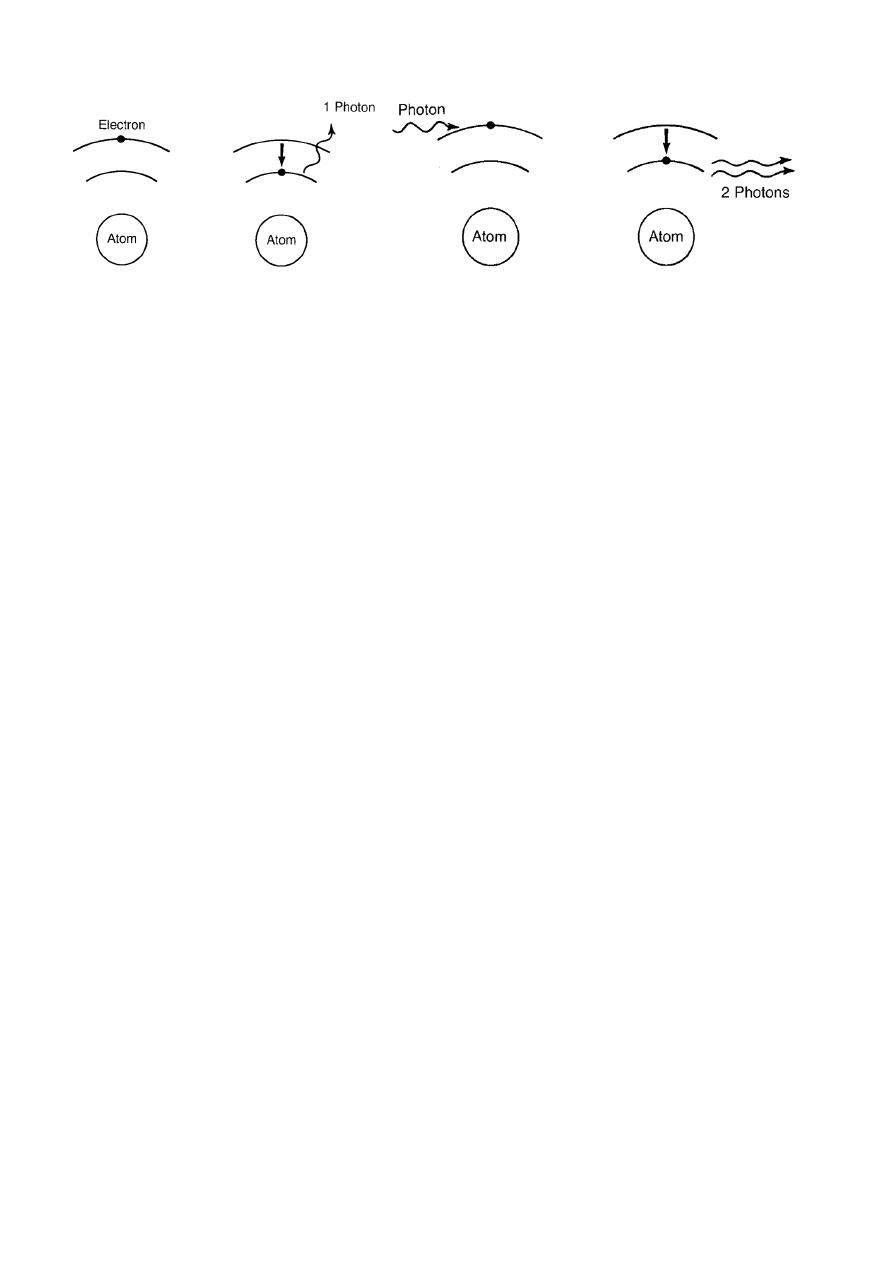
Theoretical basis of laser:
Any electron descend from high energy level to low energy level emits a
photon, but in pumping we create a large number of electrons present at high
energy level at same time that emit high energy producing laser. Then those are
amplified by reflection and re-reflection by the two mirrors (one is totally
reflecting [100%] and the other is partially reflecting [<100%]).
The effect of laser is called "Tissue-laser interaction", and it is in ophthalmology
mainly of three types:
1- Photo-coagulation.
2- Photo-disruption.
3- Photo-ablation.
Laser parameters:
1- Power of laser.
2- Pulse duration.
3- Exposure time.
4- Spot size.
P
HOTOCOAGULATION
:
It is a type of tissue-laser interaction, where there is absorption of energy from
tissue to create a thermal effect. The laser is absorbed by normal photo-sensitizers
(pigments like melanin, xanthophylls and haemoglobin), which absorb energy and
create the thermal effect.
1-Elevation of tissue temperature up to 42° is called hyperthermia.
2- Elevation of tissue temperature up to 60° causes tissue necrosis and
photocoagulation.
3- Elevation of tissue temperature up to 100° causes evaporation.
4- Elevation of tissue temperature up to 150° causes carbonization.
5- Elevation of tissue temperature up to 300° causes melting.
So, we need the 2
nd
effect (photocoagulation)
Types of Laser used in photocoagulation:
1- Argon laser: it is of two types; blue light (488 nm) and green light (514.5 nm),
its medium is gas.
2- Frequency doubled YAG laser: 532nm, its medium is solid.

3
3- Diode laser: red light (760-810 nm) and it is semiconductor medium.
4- Krypton laser: red light, its medium is gas.
5- Dye laser: its medium is fluid.
Uses of photocoagulation:
1- Treatment of diabetic retinopathy: panretinal photocoagulation or focal
photocoagulation depend on diabetic pathology.
2- Prophylaxis of peripheral retinal degeneration as in retinal holes and
breaks.
3- In treatment of POAG. (laser Trabeculoplasty)
4- Cyclodestructive procedure.
P
HOTODISRUPTION
:
It is a tissue-laser interaction induced by plasma formation and dynamic
hydrolysis by hydrolytic shocking waves, which lead to perforation of tissue. Type
of laser used is Nd-YAG laser (1064 nm)
Uses of photodisruption:
1- Peripheral iridotomy using Nd-YAG (1064nm).
2- Posterior capsulotomy: thickening of the posterior capsule developed in 50%
of patients within 3 years after cataract surgery.
3- Cutting of inflammatory membrane in front of intraocular lens.
4- Vitreous bands can be cut by using photodisruption.
5- Cyclo-destructive for treatment of intractable type of glaucoma.
P
HOTOABLATION
:
We have two mechanisms:
Create chemical reaction that leads to break up of bonds between molecules and
increase the volume or the distance between these molecule causing volume
stress and then ejection of tissue outside. There is no thermal effect. The laser
used here is Excimer laser (Argon + Florid) 132 nm.
Uses of photo ablation:
It is used in photorefractive surgery by precise removal of corneal tissue for
treatment of hypermetropia, myopia and astigmatism. Also can be used to create
corneal tract to implant stromal rings in treatment of keratoconus and can be used
for removal of superficial corneal opacification e.g. bands keratopathy (ca
+2
deposits occur in old age, hypercalcemia, and juvenile rheumatoid arthritis
associated with uveitis).