
Nervous Tissue

Nervous tissue :-
is responsible for transport of nerve impulses (motor and
sensory impulses)
it is formed by a network of more than 100 million of nerve
cells (neurons) , nerve fiber and nerve ending.
nerve tissue develops from ectoderm.

•
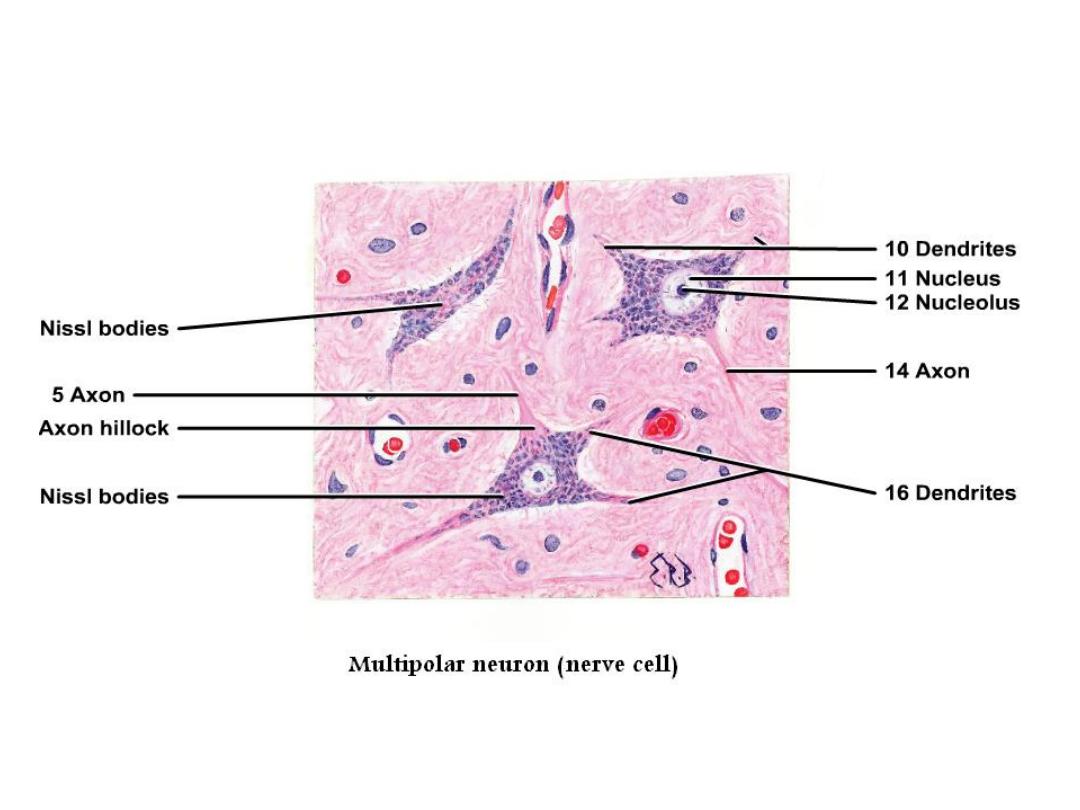
Nerve cells (neurons):- are responsible for reception, transmission
and processing of stimuli and release neurotransmitters and are
consist of:-
•
dendrites:- which are multiple elongated processes specialized for
receiving stimuli from environment
•
cell body:- or (perikaryon) contain nucleus, cytoplasm and Nissl’s
bodies; the large granular bodies consisting of rough endoplasmic
reticulum and ribosomes, that occurs in nerve cell bodies and
dendrites and are the site of protein synthesis.
•
axon:- single process specialized in generating or conducting nerve
impulse to other cells (neuron, muscle, gland)

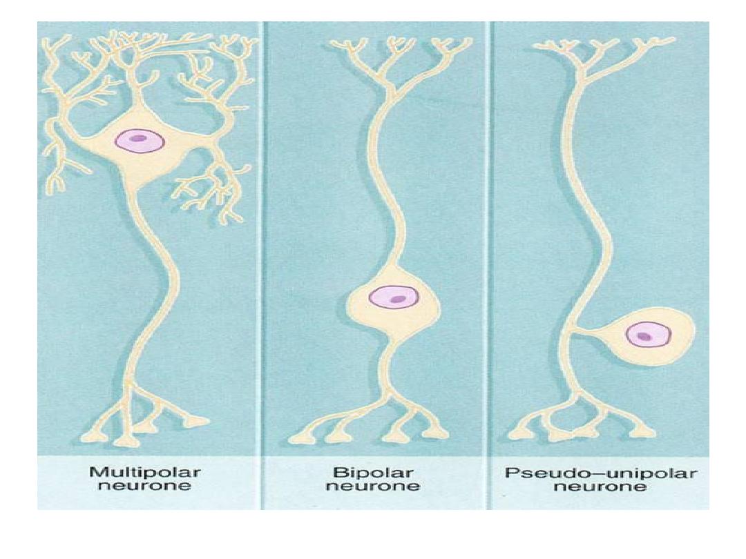
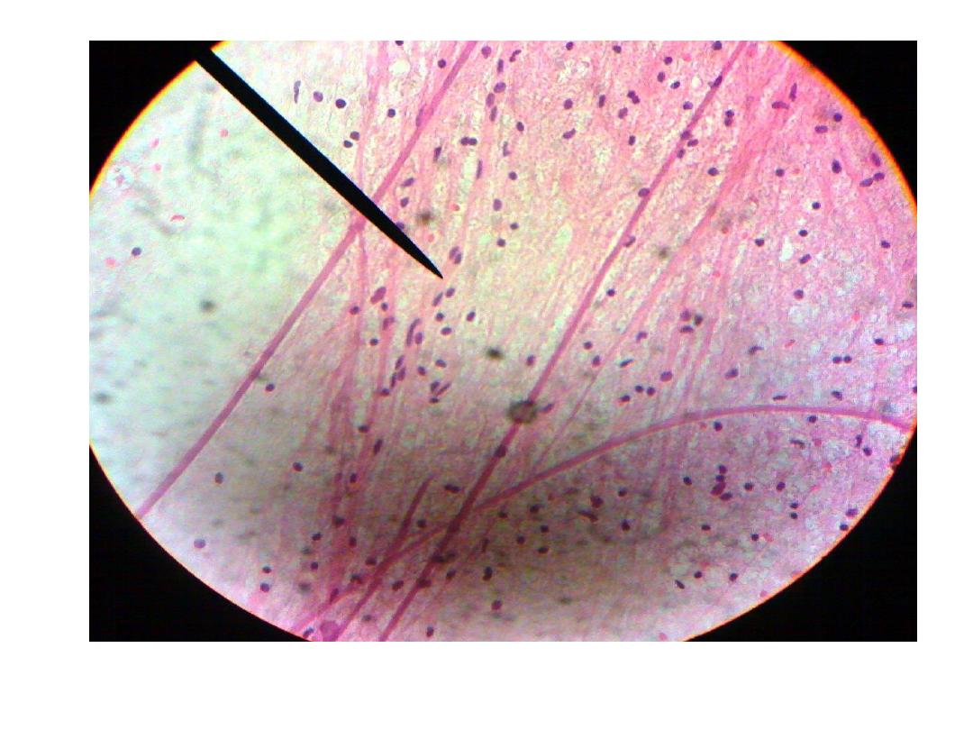
Nerve cell is classified into 3 types according to the
numbers of processes:
• Multipolar which have more than 2 processes. Most
neurons of the body are multipolar.
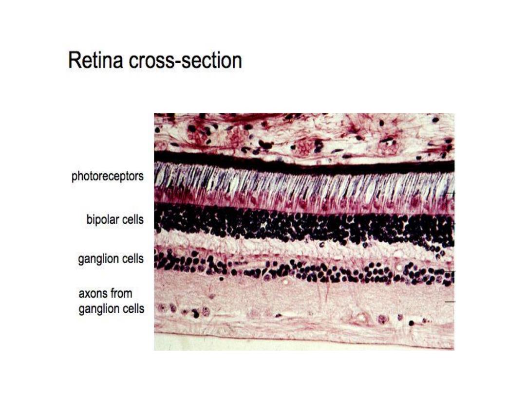
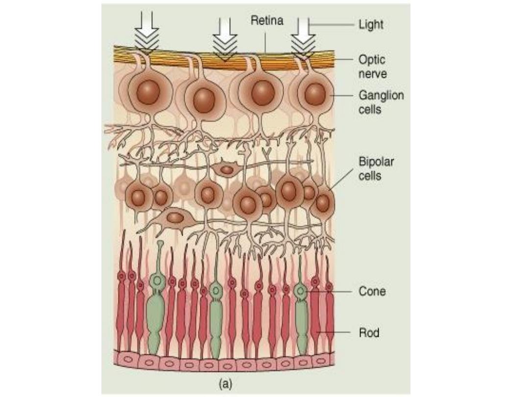
• Bipolar which have 2 processes. Found in retina and
olfactory mucosa.
• Pseudounipolar which has a single process and it is
divided to 2 branches. Found in spinal ganglia and
cranial ganglia.
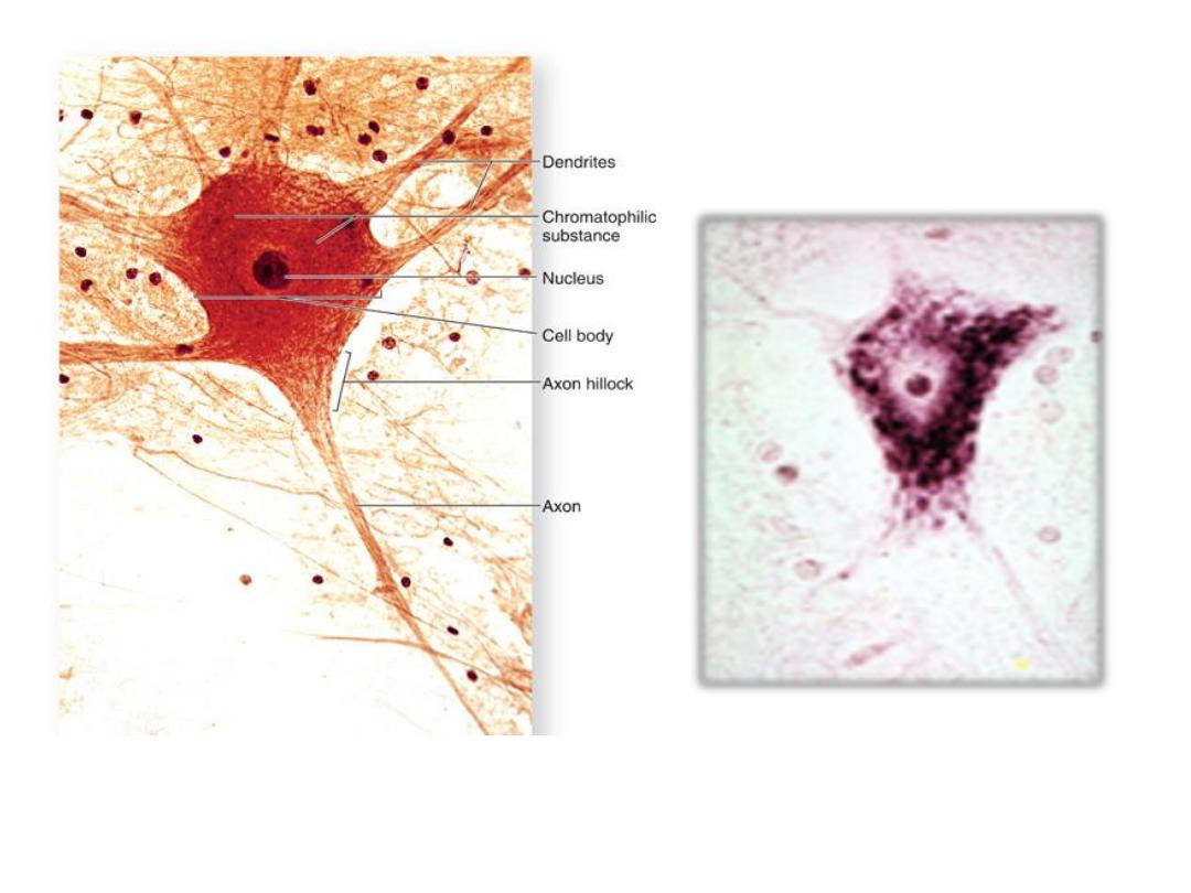
Neuron with Nissl’s
bodies
Multipolar Neuron
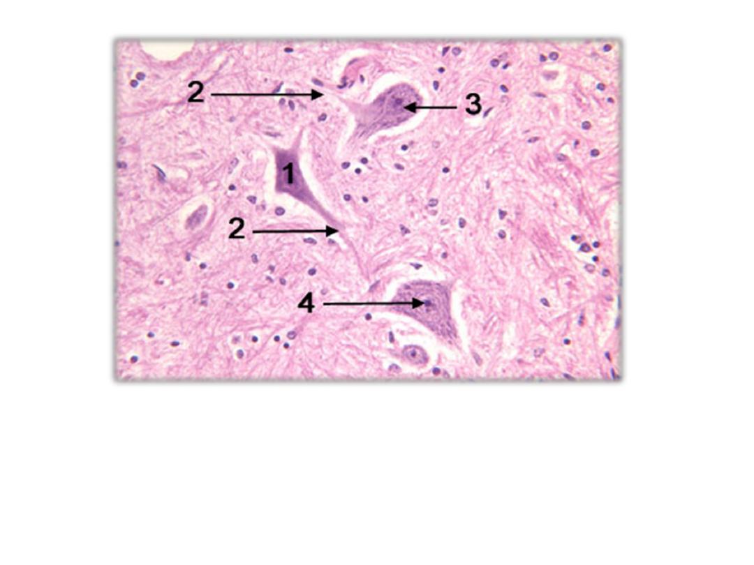
Pseudounipolar neuron

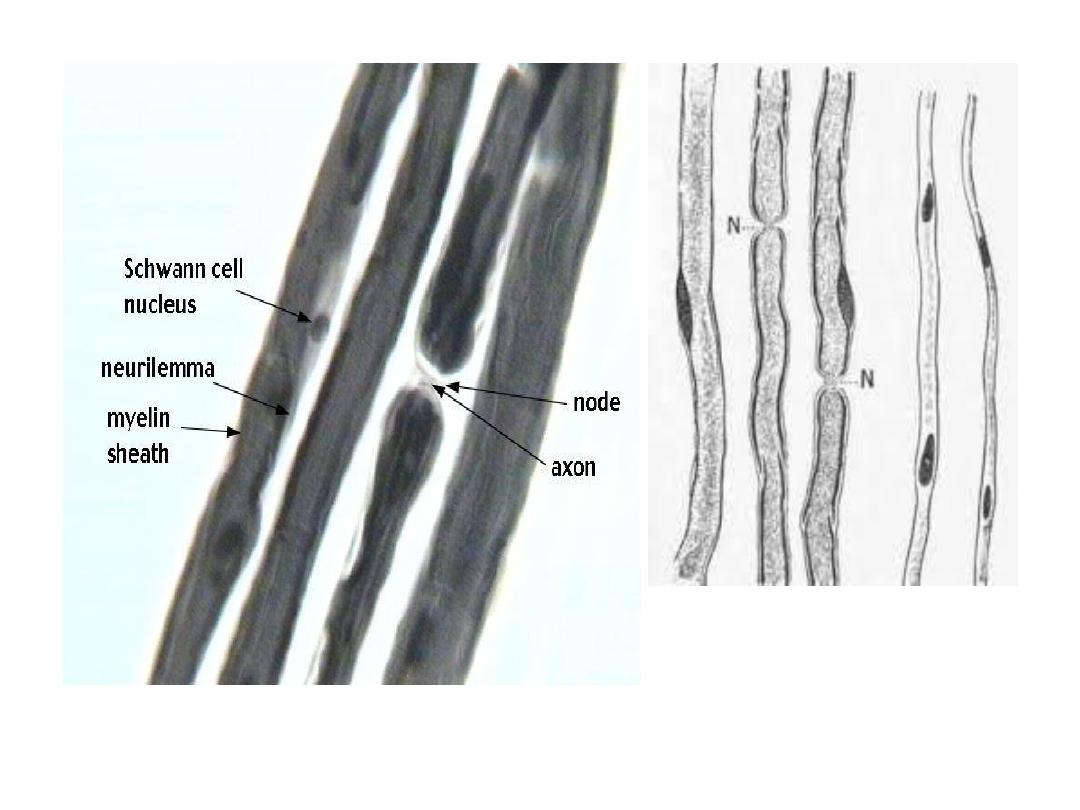
• Fibers: - consist of axons enveloped by special
sheath of Schwann cell. And classified to:-
• Myelinated fibers: - are the fibers which enveloped
with multilayer of Schwann’s plasmalemma and unite
to form myelin sheath and the space between 2
Schwann cells is called the node of Ranvier. Found
mainly in PNS.
• Un myelinated fibers: - the axons are enveloped with
simple cleft of Schwann cells found in CNS.
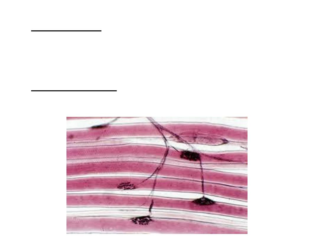
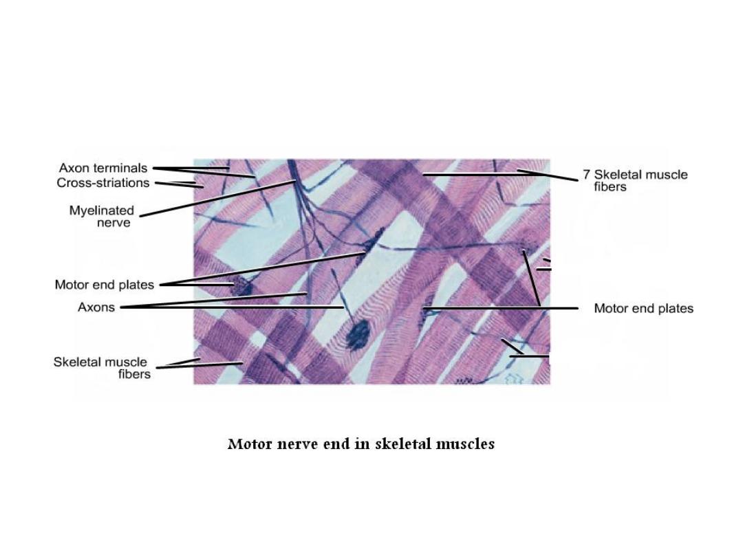
• Nerve ending: - the nerves end either in muscle or connective
tissue or epithelial tissue. Therefore they are of two type
sensory nerve ends or motor nerve ending.
• Motor nerve end: - in which nerve fiber end in striated
muscles and becomes unmyelinated and branch and end with
dents.

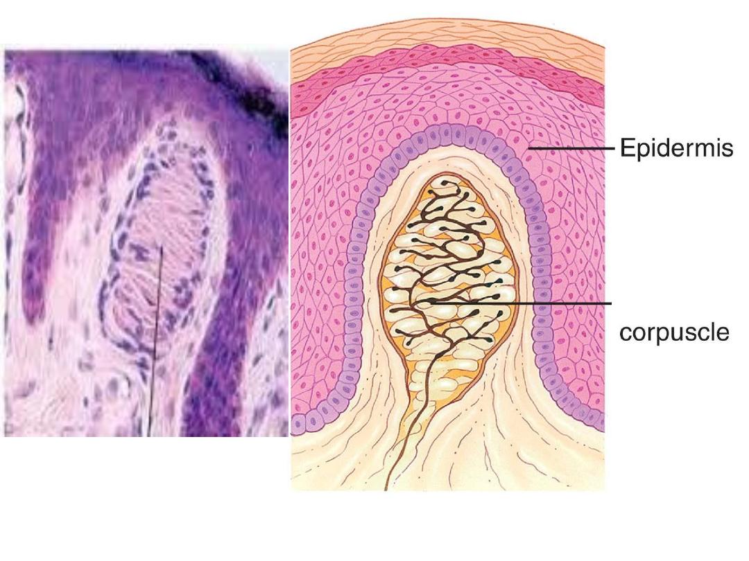
• Meissner’s corpuscles:-
are small encapsulated pressure-sensitive sensory receptors
responsible for detecting a light touch to the skin, found in the
dermis of skin. They are most concentrated in thick hairless skin,
especially at the finger pads. (and foot, eyelid , lips).
•
Meissner’s corpuscles are oval shape, the receptors consist of
flattened supportive cells arranged as horizontal lamellae and
representing specialized Schwann cells, surrounded by a connective
tissue capsule.
• A single un myelinated nerve fiber meanders between the lamellae
and throughout the corpuscle in helical meaner.
Meissner
’s
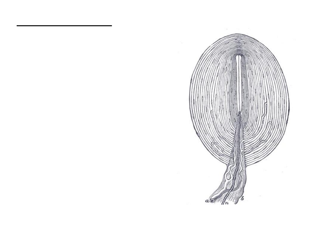
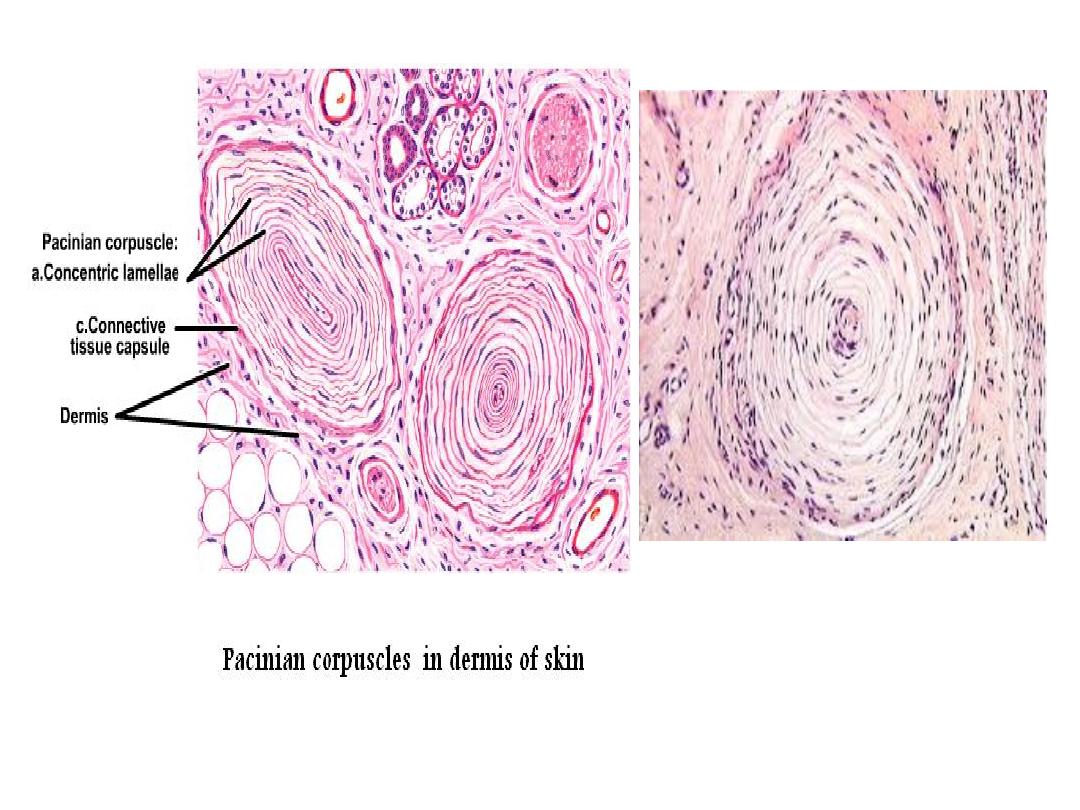
Pacinian corpuscle: -
L
arger and fewer in number than
meissners’.
Large encapsulated sensory receptor
responsive to pressure or coarse touch,
vibration and tension found in deep skin
layer, ligament. These organs consist of
delicate capsule enclosing many
concentric lamellae of flattened cells. The
center of the corpuscle is a neurite of
single and unmyelinated nerve at the
receptive region
.
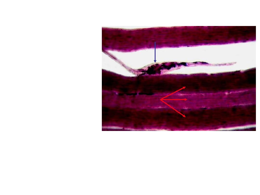
Neuromuscular spindle:
•
is a sensory receptor
within the belly of a
muscle that primarily
detects changes in the
length of this muscle.
• It is a fusiform end organ
in skeletal muscle in
which afferent and a few
efferent nerve fibers
terminate
• it contains from 3 to 10
modified striated muscle
fibers (intrafusal fibers)
that are much smaller
than the ordinary muscle
fibers.

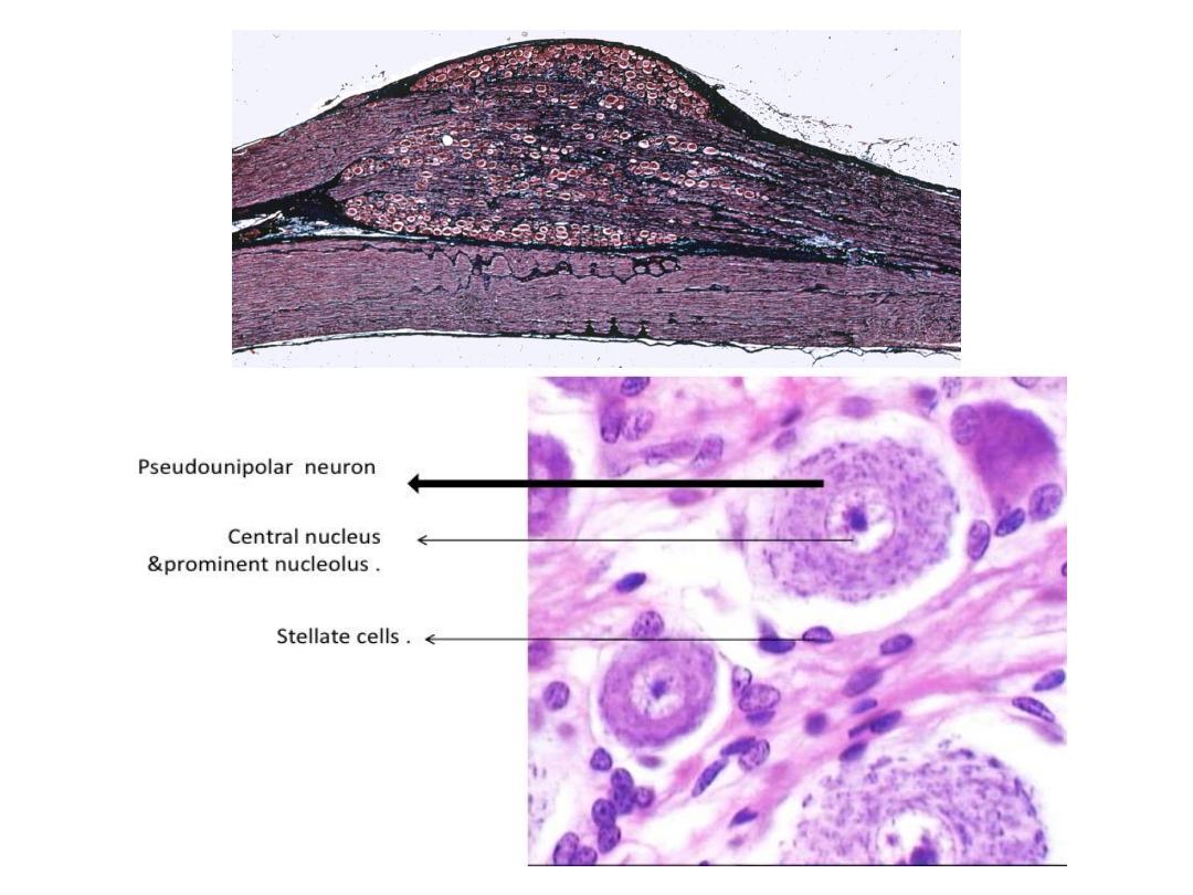
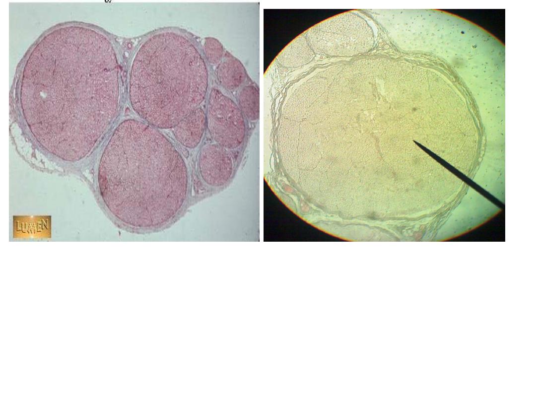
• Spinal ganglion: -
are aggregations of neuron’s cell bodies
located outside CNS and spinal g. lie on the posterior nerve
roots of spinal cord.
• They contain the cell bodies of primary sensory neurons which
are psuendounipolar and they surrounded by satellite cells
(provide structural and metabolic support ) and there is a
fascicle of nerves passing to the centre of ganglion and whole
ganglion is encapsulated by condensed supporting tissue
which is continuous with perineural and epineuria sheaths of
the peripheral nerves.


• Nerve trunk: - nerve fibers grouped in bundles to
form the nerves.
• Nerves have an external fibrous coat of dense
connective tissue called epineurium.Which also fills
the space between the bundles of nerve fibers which
called perineurium.
• The endoneurium consist of a thin layer of reticular
fibers produced by Schwann cells.
• The layers protect the nerve from aggression and act
as barrier to the passage of macromolecules.

Spinal cord
• The spinal cord is the most important structure connect between the
body and the brain.
• The spinal cord is a cylindrical structure of nervous tissue composed
of white and gray matter
• The cord is sheathed with the pia, arachnoid and dura. The dura is the
tough outer sheath, the arachnoid lies beneath it, and the pia closely
adheres to the surface of the cord.
• A transverse section of the adult spinal cord shows white matter in the
periphery, gray matter inside, and a tiny central canal filled with CSF
at its center.

• gray matter is a region containing cell bodies – shaped like the
letter “H” or a “butterfly” and is divided into dorsal or
posterior horn, lateral horn and ventral or anterior horn.
• The two “wings” of the butterfly are connected across the
midline by the dorsal gray commissure and below the white
commissure
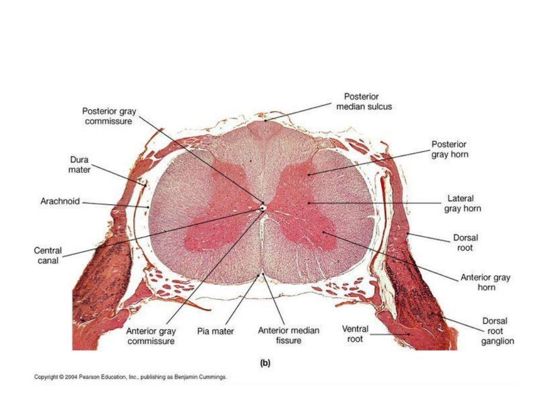
C.S in spinal cord