ANATOMY
HEAD & NECK
Dr. Nawfal K. Al-Hadithi
Lec . 2
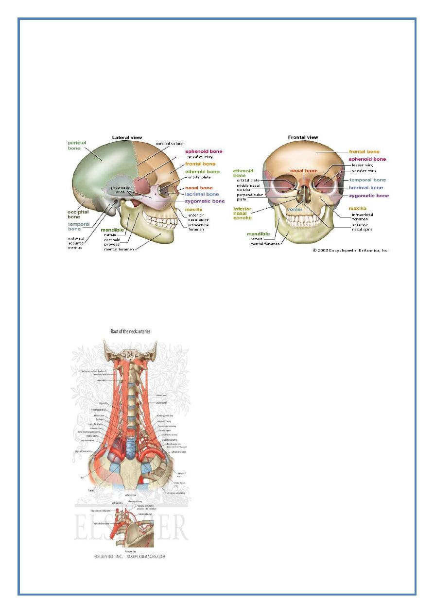
The Cranium
By : Ali Kareem
مكتب اشور لالس تنساخ
2013 – 2014
Head & Neck Dr. Nawfal K. Al-Hadithi
Anatomy
2
Lec. 2 The Cranium
THE CRANIUM
The calvaria :
Is the inside of the vault of the skull, it shows some landmarks:
1- The groove for superior sagittal sinus:
- This long depression grooves the sagittal suture from its inside &
broadens as it goes backward ending on the internal occipital
protuberance.
2- The lateral blood lakes:
- On each side of the above sinus near the vertex lie a variable-size
depression which marks the lateral blood lakes.
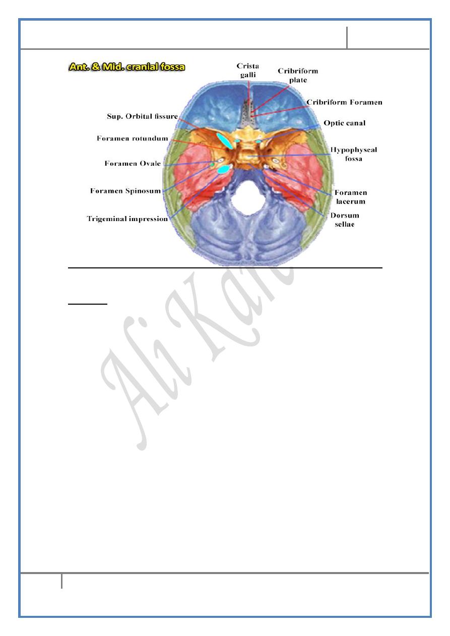
The anterior cranial fossa
:
The floor :
- Formed by the orbital plate of the frontal bone (anterior 2/3)
completed posteriorly by the lesser wing of sphenoid
- A gap in the midline between the two orbital plates is closed by the
cribriform plate of the ethmoid bone & crista galli
- The floor of this fossa forms the roof of the orbit (laterally), the
ethmoidal air cells (intermediate) & the nasal cavity (in the
midline)
Boundaries :
- Anteriorly & laterally : the frontal bone
- Posteriorly : Body & the free edge of the lesser wing of sphenoid
Stigmata :
1- The frontal crest:
- A midline sharp ridge between the cribriform plate & the anterior
wall of the fossa
- It gives attachment to the falx cerebri
2- The crista galli:
- The midline ridge which is centered on the cribriform plate
- It represents the upper part of the vertical plate of the ethmoid
- Gives attachment for the falx cerebri
3- The cribriform plate:
- Is the horizontal plate of the ethmoid which roofs the nasal cavity
Head & Neck Dr. Nawfal K. Al-Hadithi
Anatomy
3
Lec. 2 The Cranium
- It is perforated by many foramina for the passage of the olfactory
rootlets
- The ethmoidal foramina also lie in this plate laterally
4- The anterior clinoid process:
- As the posterior free border of this fossa curves medially, it will
end in a sharp projection faces posteriorly called the ACP
- For the attachment of the tentorium cerebelli
The middle cranial fossa :
The floor :
- Formed anteriorly by the greater wing of sphenoid completed
laterally by the squamous temporal & posteriorly by the petrous
temporal
- The narrow midline part of the floor which connects the two fossae
is formed by the body of sphenoid
- The floor of the fossa roofs the infratemporal fossa laterally & the
sphenoidal air cells in the midline
Boundaries :
- Anteriorly : sphenoidal wings separated from each other by the
superior orbital fissure
- Laterally : the greater wing of sphenoid & squamous temporal
bones curves from the floor upward to form the lateral wall of the
fossa .
Head & Neck Dr. Nawfal K. Al-Hadithi
Anatomy
4
Lec. 2 The Cranium
- Posteriorly : the petrous bone closes it.
Stigmata :
1- The hypophyseal fossa:
- The midline deep notch in the sphenoid body to lodge the pituitary
gland
- The fossa with its anterior wall (tuberculum sellae) & posterior
wall (dorsum sellae) look like the Turkish saddle & therefore this
complex is called the sella turcica
2- The middle clinoid processes: Two shallow projections anterior to
the pituitary fossa & posterior to the optic sulcus
3- The dorsum sellae: is the back of the pituitary fossa.
4- The posterior clinoid processes: are the two projections that project
from each side of the dorsum sellae.
5- The optic canal:
- A rounded foramen medial to the anterior clinoid process leads
from the MCF to the orbit
- Transmits the optic nerve & the ophthalmic artery
6- The optic sulcus (groove): The shallow sulcus which connects the
the two optic canals.
7- The foramen rotundum:
- A rounded foramen in the anterior wall of the fossa
- Transmits the maxillary nerve from the MCF to the spheno-
palatine fossa
Head & Neck Dr. Nawfal K. Al-Hadithi
Anatomy
5
Lec. 2 The Cranium
8- The superior orbital fissure:
- The elongated gap between the two wings of the sphenoid in the
anterior wall of the fossa
- It leads to the orbit & transmits the III, IV, VI nerves & the
ophthalmic division of V nerve with the superior orbital veins.
9- Foramina ovale, spinosum & lacerum: discussed.
10-
The groove for the middle meningeal artery:
- A clear groove starts in the floor of the fossa from foramen
spinosum
- Divides into anterior & posterior grooves according to the branches
of the middle meningeal artery
11-
The arcuate eminence:
- A laterally placed shallow eminence in the anterior wall of the
petrous bone
- It is produced by the underlying superior semicircular canal of the
internal ear
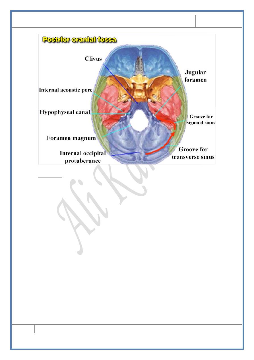
The posterior cranial fossa
:
The floor :
- Formed mainly by the occipital bone completed laterally by the
temporal bone.
Boundaries :
- Anteriorly : the occipital bone as it forms the dorsum sellae in the
midline and petrous temporal bone lateral to it
- Laterally : the mastoid part of the temporal
- Posteriorly : the occipital bone
Head & Neck Dr. Nawfal K. Al-Hadithi
Anatomy
6
Lec. 2 The Cranium
Stigmata :
1- Foramina magnum, jugular & hypoglossal: discussed.
2- The internal acoustic meatus:
- An oval foramen located in the anterior wall of the fossa
- Transmits the facial, vestibulo-cochlear nerves & nervus
intermedius together with the labyrinthine artery
3- The vestibular aqueduct:
- Very small slit lies postero-lateral to the internal acoustic meatus
- Transmits the endolymphatic duct of the membranous labyrinth
4- Internal occipital protuberance: lies opposite to the external one on
the inner aspect of the occipital bone.
5- Grooves for the transverse sinuses: From the internal occipital
protuberance two grooves pass one to each side of the protuberance
representing the transverse sinuses
6- Grooves for the sigmoid sinuses:
- Start at the root of the petrous bone laterally
- Groove the deep surface of the mastoid bone
- Descend down in an S-shape deep groove to end in the jugular
foramen
7- The internal occipital crest:
- A blunt crest from the internal occipital protuberance to the
foramen magnum in the midline
Head & Neck Dr. Nawfal K. Al-Hadithi
Anatomy
7
Lec. 2 The Cranium
- For the attachment of the falx cerebelli
8- The cerebellar fossae:
- Lie on each side of the internal occipital crest
- They lodge cerebellar hemispheres
ANATOMY OF THE VERTEBRA
The vertebra is composed of the following parts :
1- The body :
- Has the shape of a short cylinder with rough upper & lower
surfaces except for the smooth rounded circumference
- Bodies are separated & bound to each other by the intervertebral
discs at the upper & lower surfaces
- They are also connected to each other by the anterior & posterior
longitudinal ligaments at the anterior & posterior surfaces
respectively
2- The pedicles :
- The short processes that project from the posterolateral aspect of
the body
- Vertebral notches indent the upper & lower parts of the pedicle so
when vertebrae articulate with each other these notches will
produce the intervertebral foramina for passage of the spinal nerves
3- The laminae :
- The broad flat bony blades that converge posteomedially from the
pedicles to meet at the root of the spinous process
- They are connected to each other along their upper & lower
surfaces by ligamenta flava
4- The spine :
- Slopes posteroinferiorly from the junction of the two laminae in the
midline
- They are connected with each other along their upper & lower
borders by the interspinous ligaments & at their tips by the
supraspinous ligaments which are deficient in the cervical region
(ligamentum nuchae replaces them).
5- The transverse processes :
Head & Neck Dr. Nawfal K. Al-Hadithi
Anatomy
8
Lec. 2 The Cranium
- Two processes project laterally from the junction of the pedicle &
lamina between the articular processes
- They are connected with each other along their upper & lower
borders by the weak intertransverse ligaments
6- The articular processes :
- Two superior & two inferior projections from the roots of the
transverse processes
- Their shape and direction govern thetype of movement in the
region
THE CERVICAL REGION
Atypical cervical vertebrae are the 1st (atlas), 2nd (axis) & 7th (vertebra
prominence), all the remaining are typical.
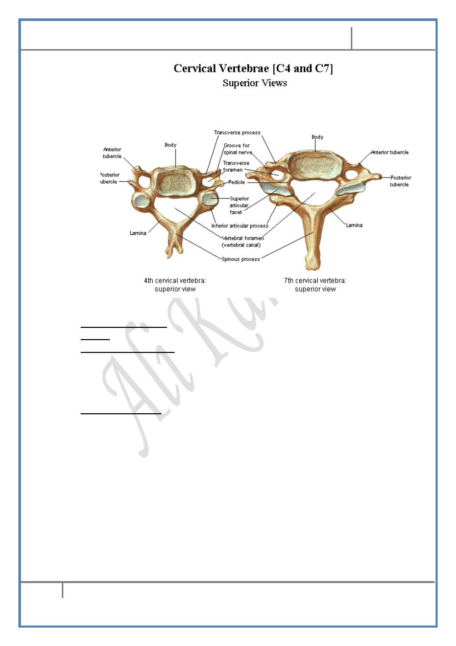
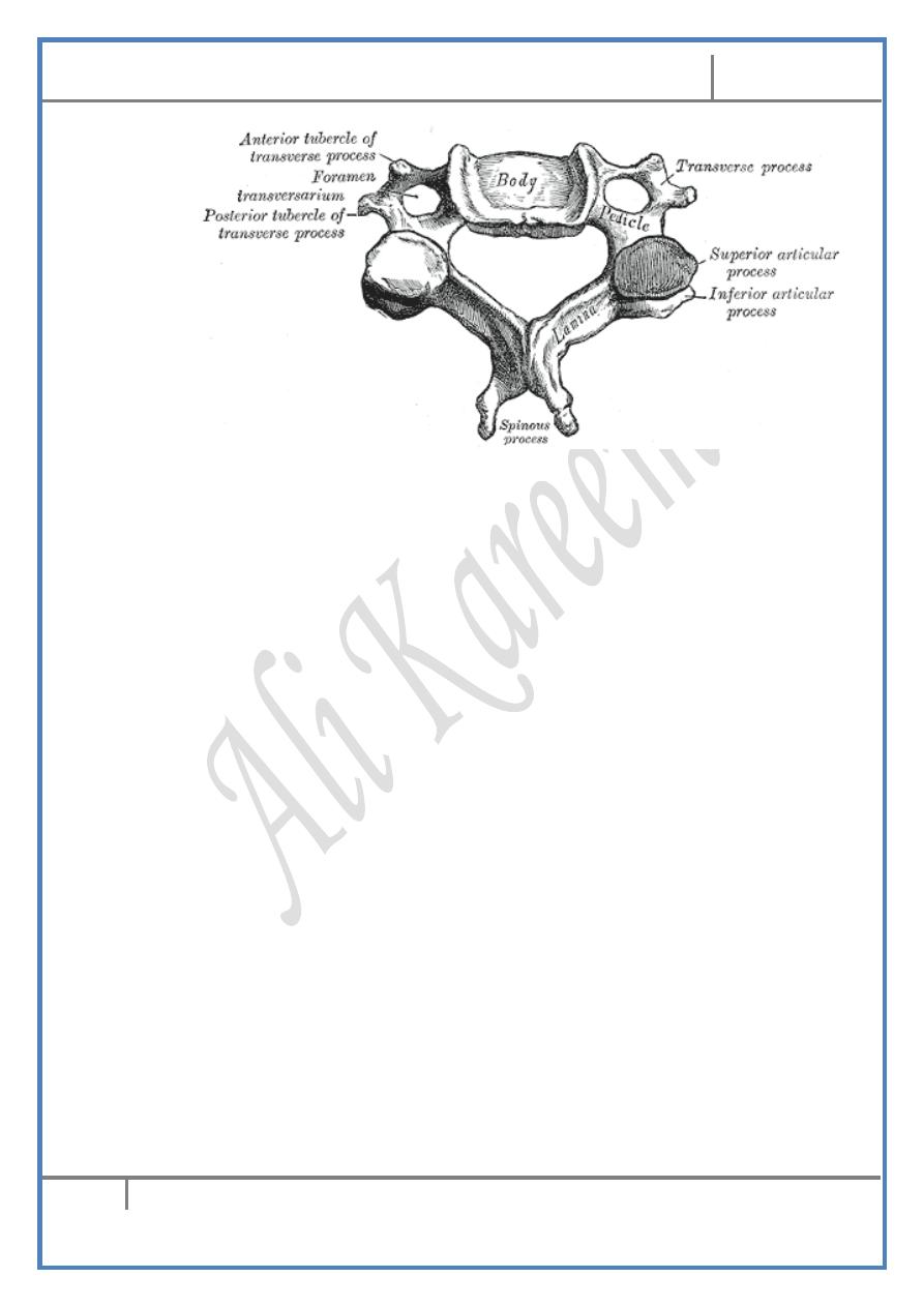
The anatomy of the TYPICAL cervical vertebra :
A typical cervical vertebra has the following characteristics :
1- Bodies :
- Small, rectangular & have superior surfaces which are concave
transversely with projecting lips on either side
- These lips articulate with a corresponding convexity in the inferior
border of the vertebra above forming the joints of Lushka
- There is no vertebral region in the column which has direct
articulation between vertebral bodies but the cervical region.
2- Pedicles :
- Project from the vertebral bodies midway between the superior &
inferior surfaces so that the superior & inferior notches are equal in
size
- In the other regions the pedicle is nearer to the superior than
inferior surface
- This modification is to fit the extra spinal nerve in the region
Head & Neck Dr. Nawfal K. Al-Hadithi
Anatomy
9
Lec. 2 The Cranium
3- Vertebral foramina are large & triangular in shape.
4- Spines are short & bifid.
5- Transverse processes :
- Short with prominent tubercles
- Carry foramina transversaria which transmit vertebral vessels &
vertebral nerve plexus except that of C7 which transmit vertebral
veins only.
6- Articular surfaces :
- Flat & oval permitting free movement at these joints
Atypical cervical vertebrae :
The atlas (C1) :
1- Because it has lost part of its body which fused with the axis as the
odontoid process & has no spine it looks like an oval bony ring.
2- The atlas has a short anterior & a longer posterior arches with two
lateral masses carrying superior & inferior articular processes.
Head & Neck Dr. Nawfal K. Al-Hadithi
Anatomy
10
Lec. 2 The Cranium
3- The anterior arch carries an anterior tubercle in the midline at its
anterior surface for the attachment of the anterior longitudinal
ligament & a facet on its posterior surface for articulation with the
anterior surface of the odontoid process of C2.
4- The posterior arch carries a groove on its superior surface
immediately behind the lateral mass for passage of vertebra artery
& C1.
5- Superior articular facet is large, kidney-shape & concave facing
upward, medially & posteriorly.
6- Inferior articular facet is nearly flat & oval fitting the superior
articular facet of C2.
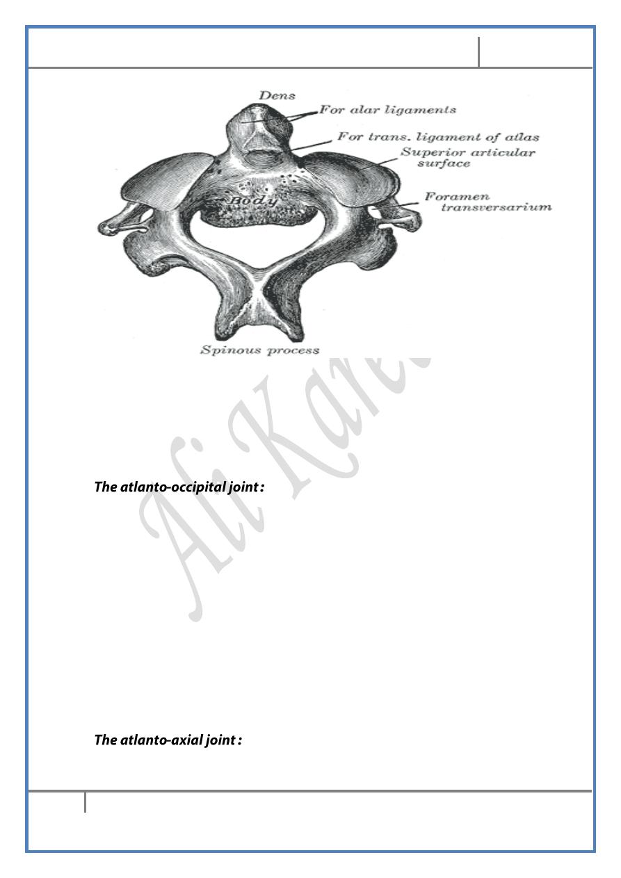
The axis (C2) :
1- *The part of the body of C1 which fused with the axis is represented
by the odontoid process (dens) which rests on the anterior part of the
atlantic body.
*This constitutes the pivot around which the skull & atlas rotate on the
axis.
*For this reason, the anterior surface of the dens carries an oval facet
which articulates with a corresponding facet on the back of the
anterior arch of the body of atlas.
*Structures which hold the dens in position are the cruciate together
with the apical & alar ligaments.
2- Pedicles & laminae are heavy & broad.
3- Spine is very broad, heavy & bifurcated.
4- Superior articular facet is large, flat & faces superolaterally, while the
inferior resembles the inferior facets of other cervical vertebrae.
Head & Neck Dr. Nawfal K. Al-Hadithi
Anatomy
11
Lec. 2 The Cranium
Vertebra prominence (C7) :
1- Spine is long, prominent & not bifid.
2- Transverse processes are larger than other vertebrae.
3- Transverse foramina are smaller than other vertebrae & transmit
vertebral veins only. Occasionally they are absent.
- The two occipital condyles represent the two lateral parts of a
single ellipsoid surface the transverse diameter of which is longer
than the antero-posterior one.
- These articulate with the superior facets of atlas so the general
shape of the joint looks like an egg lies on its side in an egg-saucer,
so the only permitted movement for this egg is to rotate around the
longer axis of the ellipse therefore the only permitted movement in
this joint is head nodding (flexion – extension of the skull)
- The articulation between the superior articular facets of C1 &
occipital condyles are synovial joints with strong lax capsules
- The anterior & posterior atlanto-occipital membranes close the
gaps between the corresponding atlantic arches & the base of skull
around foramen magnum
- This joint is formed of :
Head & Neck Dr. Nawfal K. Al-Hadithi
Anatomy
12
Lec. 2 The Cranium
1- The lateral atlanto-axial joints : TWO joints between the superior
articular facets of axis with the inferior of atlas. These are synovial joints
with lax capsule to permit rotation of the atlas carrying the skull on them.
2- The median atlanto-axial joint :
- TWO joints, the 1st between the anterior surface of the dens & the
facet on the back of the anterior arch of atlas
- The 2nd between the back of the dens & the fibro-cartilagenous
face of the transverse limb of the cruciate ligament
- These are synovial joints with thin capsules.
- The cruciate ligament :
1- The transverse limb :
- Is thick & strong band passes behind the dens
- It is attached between two tubercles located on the medial aspect of
the lateral masses of atlas
- The part in relation to the dens is a fibro-cartilagenous face
forming a joint with the back of the process
2- The longitudinal limb :
- From the back of the body of axis, a smaller band passes upward to
be inserted in the anterior edge of foramen magnum
The apical ligament : is a single slender ligament passes from the apex
of the dens to the anterior margin of foramen magnum.
- Alar ligaments: are two small but strong ligaments diverge
superolaterally from each side of the apical ligament to be attached
to the medial surface of the corresponding occipital condyles.
- Membrana tectoria :
- Is the upward extension of the posterior longitudinal ligament
which broadens as it passes upward from the back of the body of
axis to be attached within the anterior edge of foramen magnum,
where it will be continuous with dura mater. It is the most posterior
structure in this joint & its ligaments
Edited & Published by : Ali Kareem