Lecture 6
Thursday 3/10/2013
Prof. Dr.H.D.El-Yassin 2013
1
Secretion and Absorption in the Large
Intestine
Aim and objectives of lecture 6:
1. to describe the mechanism of absorption, secretion and formation of feces in the
large intestine
2. to state the two processes are attributed to the microbial flora of the large intestine
3. to give examples of some clinical digestve disorderes
The large intestine is the last attraction in digestive tube and the location of the
terminal phases of digestion. It functions in three processes:
• Recovery of water and electrolytes from ingesta: By the time ingesta reaches
the terminal ileum, roughly 90% of its water has been absorbed, but considerable
water and electrolytes like sodium and chloride remain and must be recovered by
absorption in the large gut.
• Formation and storage of feces:
• Microbial fermentation: The large intestine of all species teems with microbial life.
Those microbes produce enzymes capable of digesting many of molecules that to
vertebrates are indigestible, cellulose being a premier example. The extent and
benefit of fermentation also varies greatly among species.
Absorption, Secretion and Formation of Feces in the Large Intestine
1. Absorption: water, sodium ions and chloride ions
2. Secretion: bicarbonate ions and mucus
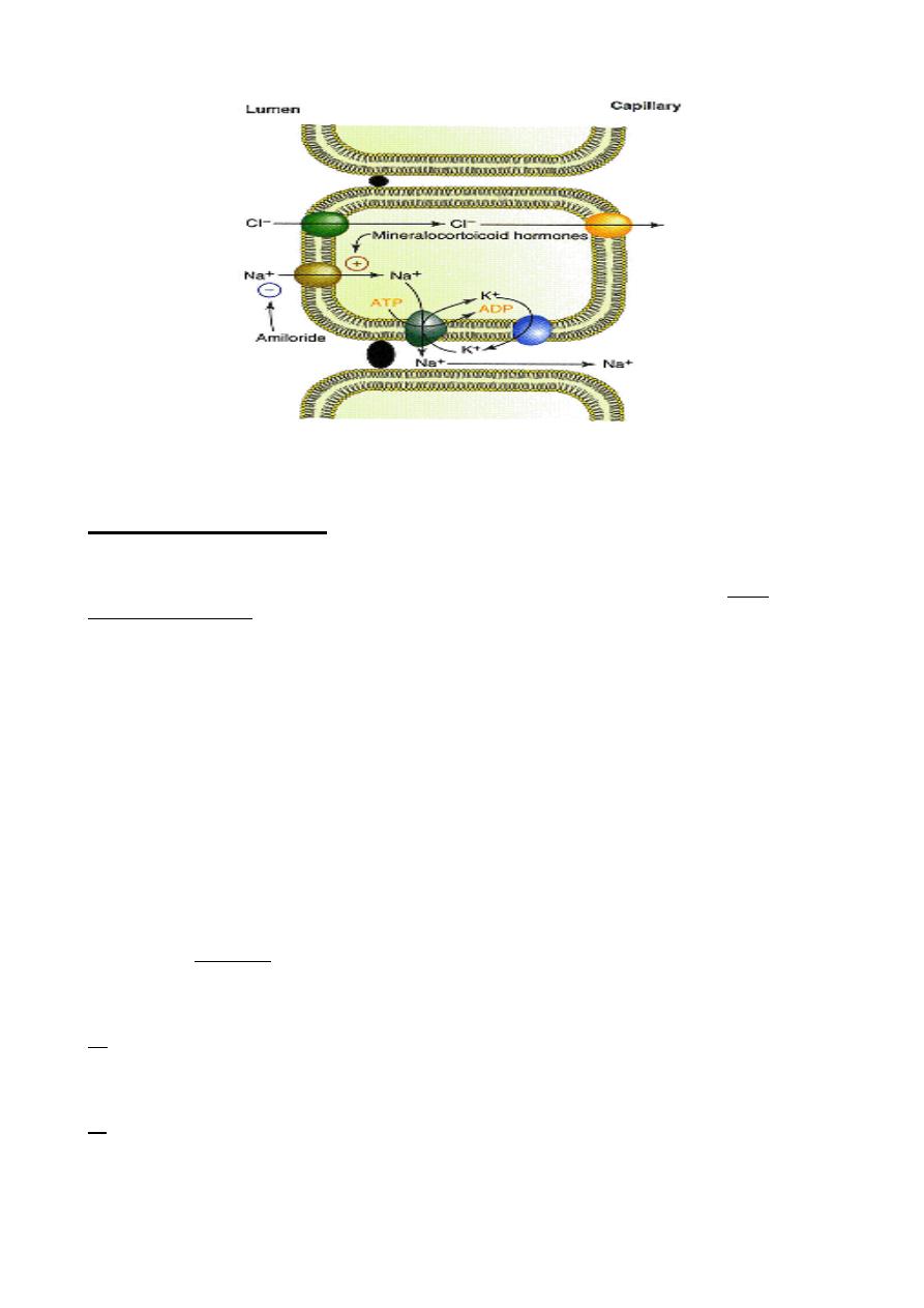
Water, as always, is absorbed in response to an osmotic gradient. The mechanism
responsible for generating this osmotic pressure is essentially identical to what was seen
in the small intestine - sodium ions are transported from the lumen across the epithelium
by virtue of the epithelial cells having very active sodium pumps on their basolateral
membranes and a means of absorbing sodium through their lumenal membranes. The
colonic epithelium is actually more efficient at absorbing water than the small intestine and
sodium absorption in the colon is enhanced by the hormone aldosterone.
Chloride is absorbed by exchange with bicarbonate. The resulting secretion of bicarbonate
ions into the lumen aids in neutralization of the acids generated by microbial fermentation
in the large gut.

Lecture 6
Thursday 3/10/2013
Prof. Dr.H.D.El-Yassin 2013
2
Model for electrogenic NaCl absorption in the large intestine
This Na
+
flux is electrogenic; that is, it is associated with an electrical current, and it can be
inhibited by the diuretic drug amiloride at micromolar concentrations
Microbial Fermentation
Fermentation is the enzymatic decomposition and utililization of foodstuffs, particularly
carbohydrates, by microbes
The large intestine does not produce its own digestive enzymes, but contains huge
numbers of bacteria which have the enzymes to digest and utilize many substrates. In all
animals, two processes are attributed to the microbial flora of the large intestine:
1. Digestion of carbohydrates not digested in the small intestine
Cellulose is common constituent in the diet of many animals, including man, but no
mammalian cell is known to produce a cellulase. Several species of bacteria in the large
intestine synthesize cellulases and digest cellulose.
Colonic bacteria rapidly metabolize the saccharides, forming gases, short-chain fatty
acids, and lactate. The major short-chain fatty acids formed are acetic acid (two carbon),
propionic acid (three carbon), and butyric acid (four carbon). The short-chain fatty acids
are absorbed by the colonic mucosal cells and can provide a substantial source of energy
for these cells. The major gases formed are hydrogen gas (H
2
), carbon dioxide (CO
2
), and
methane (CH
4
). These gases are released through the colon, resulting in flatulence, or in
the breath. Incomplete products of digestion in the intestines increase the retention of
water in the colon, resulting in diarrhea.
2. Synthesis of vitamin K and certain B vitamins
Synthesis of vitamin K by colonic bacteria provides a valuable supplement to dietary
sources and makes clinical vitamin K deficiency rare. Similarly, formation of B vitamins by
the microbial flora in the large intestine is useful to many animals. They are not absorbed
in the large intestine, but are present in feces.
Q Beans, peas, soybeans, and other leguminous plants contain oligosaccharides with
(1,6)-linked galactose residues that cannot be hydrolyzed for absorption, including sucrose
with 1, 2, or 3 galactose residues attached . What is the fate of these polysaccharides in
the intestine?
A These sugars are not digested well by the human intestine but form good sources of
energy for the bacteria of the gut. These bacteria convert the sugars to H
2
, lactic acid and
short-chain fatty acids. The amount of gas released after a meal containing beans is
especially notorious.

Lecture 6
Thursday 3/10/2013
Prof. Dr.H.D.El-Yassin 2013
3
Digestive Disorders
1. Stomach and Intestine
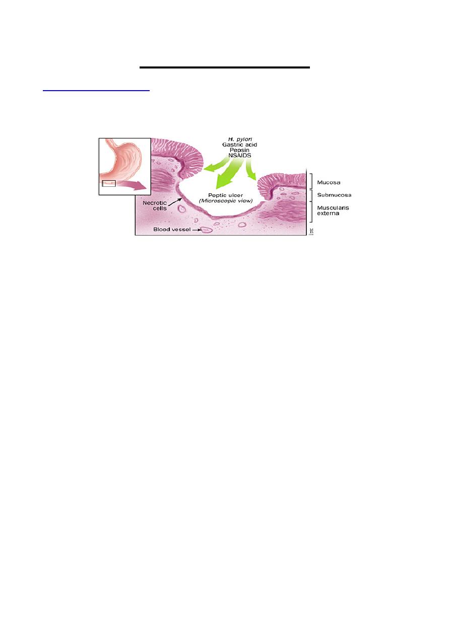
Peptic ulcers
Peptic ulcer is a general term that refers to ulcers occurring in the lower esophagus, the
stomach, or the duodenum (upper part of the small intestine).
What is the difference between a duodenal ulcer and a gastric ulcer?
A duodenal ulcer is a break in the lining of the upper part of the small intestine (the
duodenum); a gastric ulcer is a break in the lining of the stomach.
What causes duodenal and gastric ulcers?
Duodenal ulcers are much more common than gastric ulcers. The primary cause of
duodenal ulcers is increased production of acid by the stomach.
Gastric ulcers, on the other hand, are thought to be caused by changes in the stomach
lining that make it more susceptible to damage by the acid normally produced by the
stomach.
Factors in the development of peptic ulcers include:
• Helicobacter pylori
Research shows that most ulcers develop as a result of infection with bacterium called
Helicobacter pylori (H. pylori).
• Smoking
Studies show smoking increases the chances of getting an ulcer, slows the healing
process of existing ulcers, and contributes to ulcer recurrence.
• Caffeine
Caffeine seems to stimulate acid secretion in the stomach, which can aggravate the pain
of an existing ulcer. However, the stimulation of stomach acid cannot be attributed solely
to caffeine.
• Alcohol
Although no proven link has been found between alcohol consumption and peptic ulcers,
ulcers are more common in people who have cirrhosis of the liver, a disease often linked
to heavy alcohol consumption.
• Stress
Mucos HCO
3
content creates a "micro-environment" around surface cells to prevent acid
damage, but its secretion is inhibited by adrenergic input (prominent in stress!)
• Acid and pepsin
It is believed that the stomach's inability to defend itself against the powerful digestive
fluids, hydrochloric acid and pepsin, contributes to ulcer formation.
• nonsteroidal anti-inflammatory drugs (NSAIDs)
These drugs (such as aspirin, ibuprofen, and naproxen sodium) make the stomach
vulnerable to the harmful effects of acid and pepsin.

Lecture 6
Thursday 3/10/2013
Prof. Dr.H.D.El-Yassin 2013
4
2. Bile and the Biliary System
Gallstones (Cholelithiasis)
There are two major types of gallstones, which form due to distinctly different pathogenetic
mechanisms.
1. Cholesterol Stones
About 90% of gallstones are of this type. These stones can be almost pure cholesterol or
mixtures of cholesterol and substances such as mucin.
The key event leading to formation and progression of cholesterol stones is precipitation of cholesterol
in bile. There are clearly important genetic determinants for cholesterol stone formation. There is
also an important gender bias in development of stones - the prevalence in adult females is two
to three times that seen in males and use of contraceptive steroids is a risk factor for
development of gallstones.
2. Pigment Stones
Roughly 10% of human gallstones are pigment stones composed of large quantities of bile
pigments, along with lesser amounts of cholesterol and calcium salts. The most important risk
factor for development of these stones is chronic hemolysis from almost any cause - bilirubin
is a major constituent of these stones. Additionally, some forms of pigment stones are associated
with bacterial infections. Apparently, some bacteria release deconjugate bilirubin, leading to
precipitation as calcium salts.
b. Jaundice
Jaundice, is yellowing of the skin, sclera (the white of the eyes) and mucous membranes
caused by increased levels of bilirubin in the human body. Usually the concentration of
bilirubin in the blood must exceed 2-3mg/dL for the coloration to be easily visible. Jaundice
comes from the French word jaune, meaning yellow.
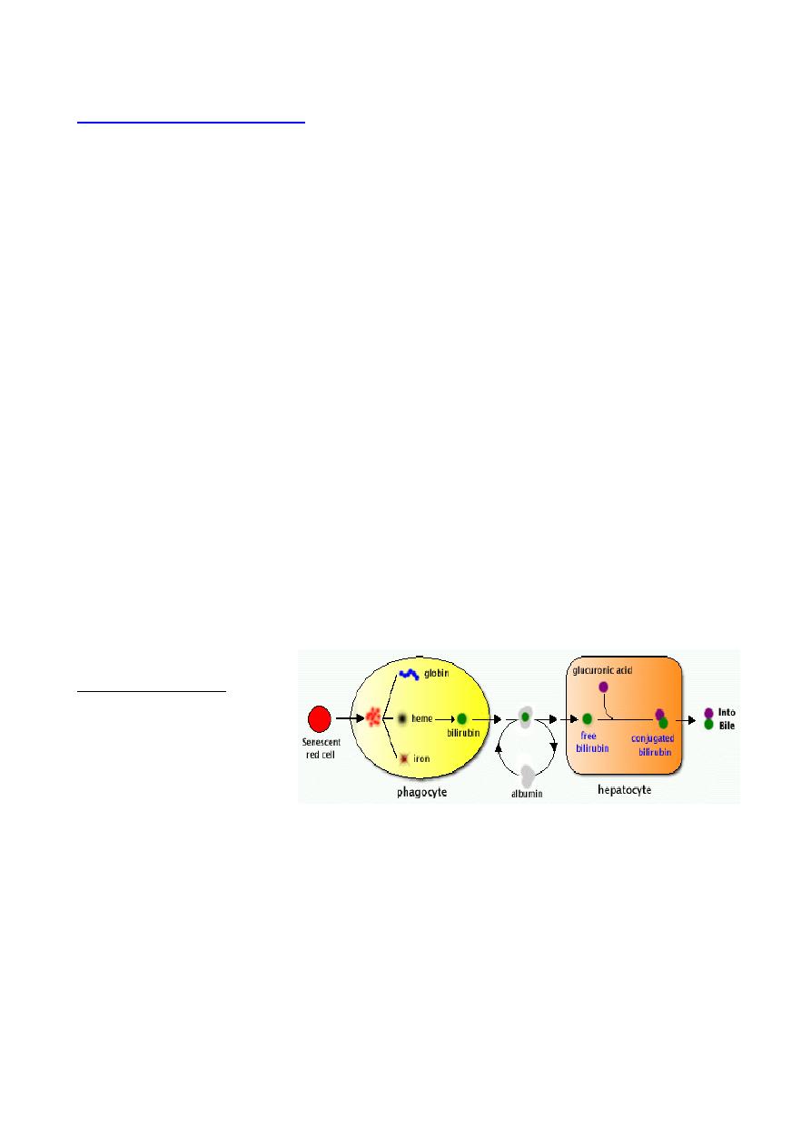
Causes of jaundice
When red blood cells die,
the heme in their
hemoglobin is converted to
bilirubin in the spleen. The
bilirubin is processed by the
liver, enters bile and is
eventually excreted through faeces.
Consequently, there are three different classes of causes for jaundice. Pre-hepatic or
hemolytic causes, where too many red blood cells are broken down, hepatic causes
where the processing of bilirubin in the liver does not function correctly, and post-hepatic
or extrahepatic causes, where the removal of bile is disturbed.
1. Pre-hepatic
Pre-hepatic (or hemolytic) jaundice is caused by anything which causes an increased rate
of hemolysis (breakdown of red blood cells). Malaria can cause jaundice. Certain genetic
diseases, such as glucose 6-phosphate dehydrogenase deficiency can lead to increase
red cell lysis and therefore hemolytic jaundice. Defects in bilirubin metabolism also present
as jaundice.
2. Hepatic
Hepatic causes include acute hepatitis, hepatotoxicity and alcoholic liver disease.
Jaundice commonly seen in the newborn baby is another example of hepatic jaundice.

Lecture 6
Thursday 3/10/2013
Prof. Dr.H.D.El-Yassin 2013
5
• Neonatal jaundice
Neonatal jaundice is usually harmless: this condition is often seen in infants around the
second day after birth, lasting till day 8 in normal births, or to around day 14 in premature
births. Serum bilirubin normally drops to a low level without any intervention required: the
jaundice is presumably a consequence of metabolic and physiological adjustments after
birth. Infants with neonatal jaundice are typically treated by exposing them to high levels of
colored light to break down the bilirubin. This works due to a photo oxidation process
occurring on the bilirubin in the subcutaneous tissues of the neonate. Light energy creates
isomerization of the bilirubin and consequently transformation into compounds that the
new born can excrete via urine and stools.
3. Post-hepatic
Post-hepatic (or obstructive) jaundice, also called cholestasis, is caused by an interruption
to the drainage of bile in the biliary system. The most common causes are gallstones in
the common bile duct and pancreatic cancer in the head of the pancreas.
The van den Bergh test:
When a mixture of sulphanic acid, hydrochloric acid and sodium nitrite (diazo reagent) is added to serum containing an
excess of biliriubin glucuronide a reddish-violet color results, the maximum color intensity being reached within 30
seconds (direct reaction) (for hepatic and post hepatic jaundice)
When the same above reagents are mixed with serum containing an excess of billirubin itself or bilirubi-protien complex
no color develops until alcohol is added, then the reddish-violet color appears.(indirect reaction) (for pre hepatic jaundice)
Note: the addition of alcohol solvent provides the means of solution for the water insoluble bilirubin which is thus enabled
to react with the diazo reagent
3. Intestine
Diarrhea is an increase in the volume of stool or frequency of defecation. It is one of the
most common clinical signs of gastrointestinal disease, but also can reflect primary
disorders outside of the digestive system
There are numerous causes of diarrhea, but in almost all cases, this disorder is a
manifestation of one of the four basic mechanisms described below.
1. Osmotic Diarrhea
Absorption of water in the intestines is dependent on adequate absorption of solutes. If
excessive amounts of solutes are retained in the intestinal lumen, water will not be
absorbed and diarrhea will result. Osmotic diarrhea typically results from one of two
situations:
• Ingestion of a poorly absorbed substrate: The offending molecule is usually a
carbohydrate or divalent ion. Common examples include mannitol or sorbitol, epson
salt (MgSO
4
) and some antacids (MgOH
2
).
• Malabsorption: Inability to absorb certain carbohydrates is the most common deficit in
this category of diarrhea, but it can result virtually any type of malabsorption. A
common example is lactose intolerance resulting from a deficiency in the brush border
enzyme lactase. In such cases, a moderate quantity of lactose is consumed (usually as
milk), but the intestinal epithelium is deficient in lactase, and lactose cannot be
effectively hydrolyzed into glucose and galactose for absorption. The osmotically-active
lactose is retained in the intestinal lumen, where it "holds" water.
• A distinguishing feature of osmotic diarrhea is that it stops after the patient is fasted
or stops consuming the poorly absorbed solute.
2. Secretory Diarrhea
Large volumes of water are normally secreted into the small intestinal lumen, but a large
majority of this water is efficiently absorbed before reaching the large intestine. Diarrhea
occurs when secretion of water into the intestinal lumen exceeds absorption.
Many millions of people have died of the secretory diarrhea associated with cholera. The
responsible organism, Vibrio cholerae, produces cholera toxin, which strongly activates

Lecture 6
Thursday 3/10/2013
Prof. Dr.H.D.El-Yassin 2013
6
adenylyl cyclase, causing a prolonged increase in intracellular concentration of cyclic AMP
within crypt enterocytes. This change results in prolonged opening of the chloride channels
that are instrumental in secretion of water from the crypts, allowing uncontrolled secretion
of water.
Exposure to toxins from several other types of bacteria (e.g. E. coli heat-labile toxin)
induce the same series of steps and massive secretory diarrhea that is often lethal unless
the person or animal is aggressively treated to maintain hydration. In addition to bacterial
toxins, a large number of other agents can induce secretory diarrhea by turning on the
intestinal secretory machinery, including:
• some laxatives
• hormones secreted by certain types of tumors (e.g. vasoactive intestinal peptide)
• a broad range of drugs (e.g. some types of asthma medications, antidepressants,
cardiac drugs)
• certain metals, organic toxins, and plant products (e.g. arsenic, insecticides,
mushroom toxins, caffeine)
In most cases, secretory diarrheas will not resolve during a 2-3 day fast.
3. Inflammatory and Infectious Diarrhea
The epithelium of the digestive tube is protected from insult by a number of mechanisms
constituting the gastrointestinal barrier, but like many barriers, it can be breached and
often associated with widespread destruction of absorptive epithelium. In such cases,
absorption of water occurs very inefficiently and diarrhea results. Examples of pathogens
frequently associated with infectious diarrhea include:
Bacteria: Salmonella, E. coli, Campylobacter
Viruses: rotaviruses
Protozoa:
4. Diarrhea Associated with Deranged Motility
In order for nutrients and water to be efficiently absorbed, the intestinal contents must be
adequately exposed to the mucosal epithelium and retained long enough to allow
absorption. Disorders in motility than accelerate transit time could decrease absorption,
resulting in diarrhea even if the absorptive process per se was proceeding properly.
Conclusions:
1. Regarding the the mechanism of absorption, secretion and formation of feces
a. Water, as always, is absorbed in response to an osmotic gradient
b. Sodium absorption in the colon is enhanced by the hormone aldosterone
c. Chloride is absorbed by exchange with bicarbonate
d. The resulting secretion of bicarbonate ions into the lumen aids in
neutralization of the acids generated by microbial fermentation in the large
gut
2. Two processes are attributed to the microbial flora of the large intestine:
a. Digestion of carbohydrates not digested in the small intestine
b. Synthesis of vitamin K and certain B vitamins
