Lec.2
د.معن الخالصي
2013-
2014
Dr.alkhalisy Embryology
Page 1-7
Trilaminar Germ Disc (3
rd
week of development)
The characteristic events in the 3
rd
week of gestation are:
1- Gastrulation: is the process where the three germ layers of the embryo
(ectoderm, mesoderm & endoderm) will be formed.
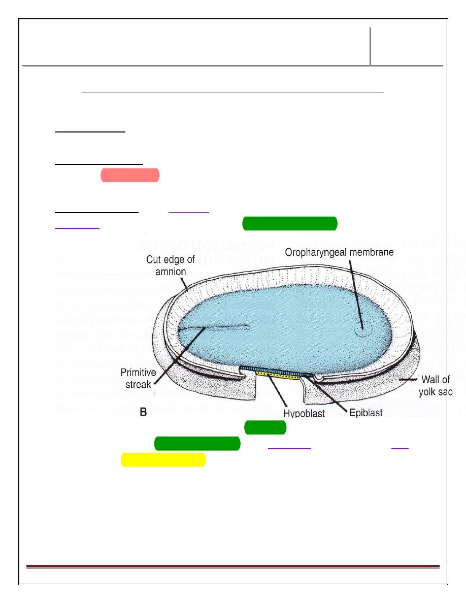
2- Primitive streak: which is ill defined line appeared on the surface of epiblast,
but at the 15-16 days embryo, it become clear visible groove with slightly
bulging region on both sides
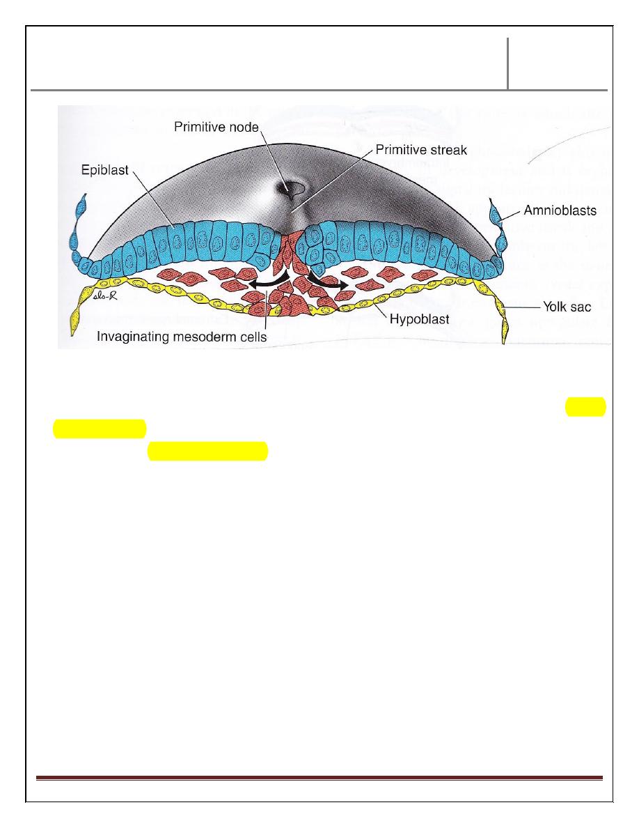
3- Primitive node: at the cephalic end of primitive streak, there’s slight
elevation over an area surrounding the small primitive pit.
o In this week (3
rd
), cells of epiblast will be migrate toward primitive streak, before
arrival to it, it becomes flask shaped cell, then detached from epiblast &slip
inside the streak passing between epiblast & hypoblast.this inward movement
called invagination.
Lec.2
د.معن الخالصي
2013-
2014
Dr.alkhalisy Embryology
Page 2-7
o
o Epiblast cell migration is under control of fibroblast growth factor 8 (FGF8)
which is a growth factor, protein in nature, synthesized by streak cells control
cells migration of epiblast by down regulating E-cadherin, which is protein bind
epiblast cells together.
o FGF8 control cell specification into mesoderm by regulating bachyury (T)
expression.
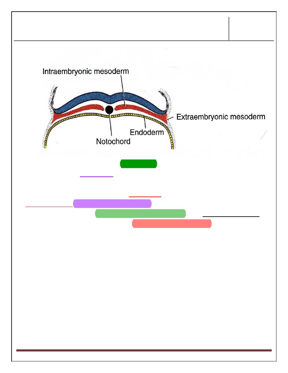
o Once the epiblast invaginate through the streak, it will form three creat, three
types of cells, then layer:-
1- Some displace hypoblast forming endoderm.
2- Other remains in epiblast forming ectoderm.
3- Other remains between endoderm & ectoderm forming what is called
mesoderm.
o Therefore, epiblast through gastrulation process is the source of all germ layers,
from which all tissue of the body will be form, these are ectoderm, mesoderm &
ectoderm.
Lec.2
د.معن الخالصي
2013-
2014
Dr.alkhalisy Embryology
Page 3-7
As process of migration continue & cell move between epiblast & hypoblast they
begin to spread laterally & cranially. Gradually they migrate beyond the disc &
lie in contact with extra embryonic mesoderm covering yolk sac & amnion.
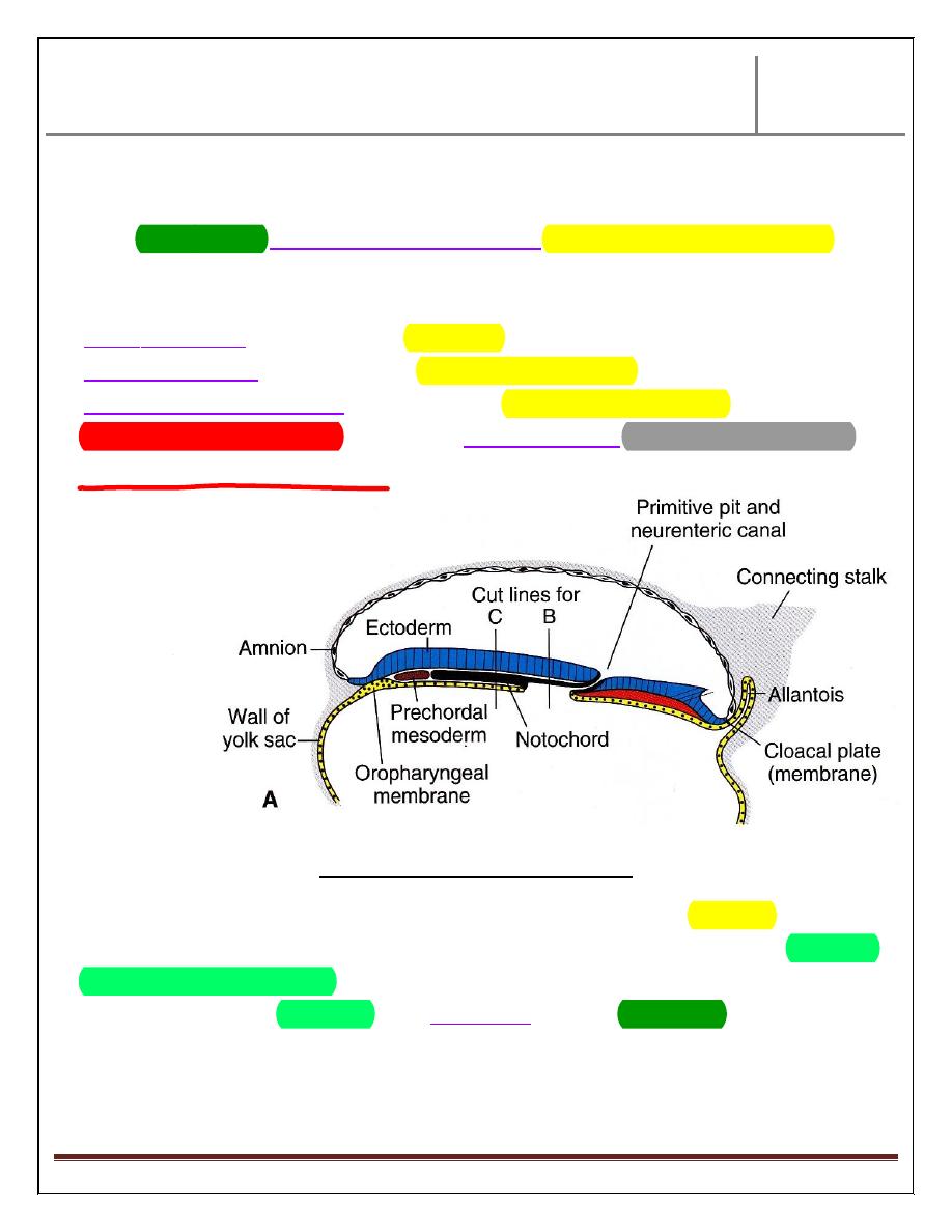
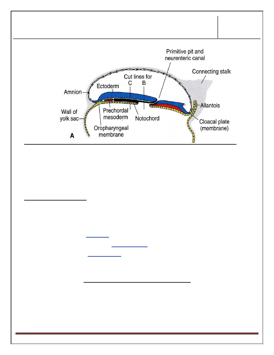
In cephalic direction there is what is called prechordal plate. Which form
between notochord & oropharyngeal membrane.
Prechordal plate later on will be forebrain.
Notochordal cord later on will be central axial skeleton.
Oropharyngeal membrane later on will be opening of oral cavity. The
oropharyngeal membrane forms from tightly adherent endoderm & ectoderm
with no intervening mesoderm.
Formation of notochord
Pre-notochordal cells, invaginating in the primitive pit move forward till
reaching the prechordal plate. These invaginated pre-notochordal cells become
intercalated in endoderm forming the notochordal plate.
With time the plate detached from endoderm forming solid cord called
notochord. This form midline axis & serve as basis of the axial skeleton.
Lec.2
د.معن الخالصي
2013-
2014
Dr.alkhalisy Embryology
Page 4-7
At the point of primitive pit, an indentation in the epiblast will form leaving a
canal temporarily connecting amniotic cavity with yolk cavity. This canal called
neurenteric canal.
Clocal membrane: formed at the caudal end of embryonic disc. It consists of
tightly adherent endoderm with ectoderm with no intervening mesoderm. When
this membrane appears the posterior wall of yolk sac form small diverticulum
extend into connecting stalk, called allantois or allantoenteric diverticulum. This
(allantois) appears around 16
th
day of development & in lower vertebrates, the
allantois serves as a reservoir for excretion product of renal system.
Lec.2
د.معن الخالصي
2013-
2014
Dr.alkhalisy Embryology
Page 5-7
Fate of gastrulation:
Cells migrate through primitive pit will be arranged as follows:
1- Cranially: become prechordal plate & notochord.
2- Laterally: become paraxial mesoderm.
3- Mid-streak region: become intermediate mesoderm.
4- Caudally: become lateral plate mesoderm.
Growth of Embryonic Disc
The embryonic disc changes from flat round structure into broad cephalically ,
narrows caudally & round elongated structure.
The primitive streaks continue to supply new cell till end of 4
th
wk.
In the cephalic part, germ layer begins differentiation in the mid third wk.
Lec.2
د.معن الخالصي
2013-
2014
Dr.alkhalisy Embryology
Page 6-7
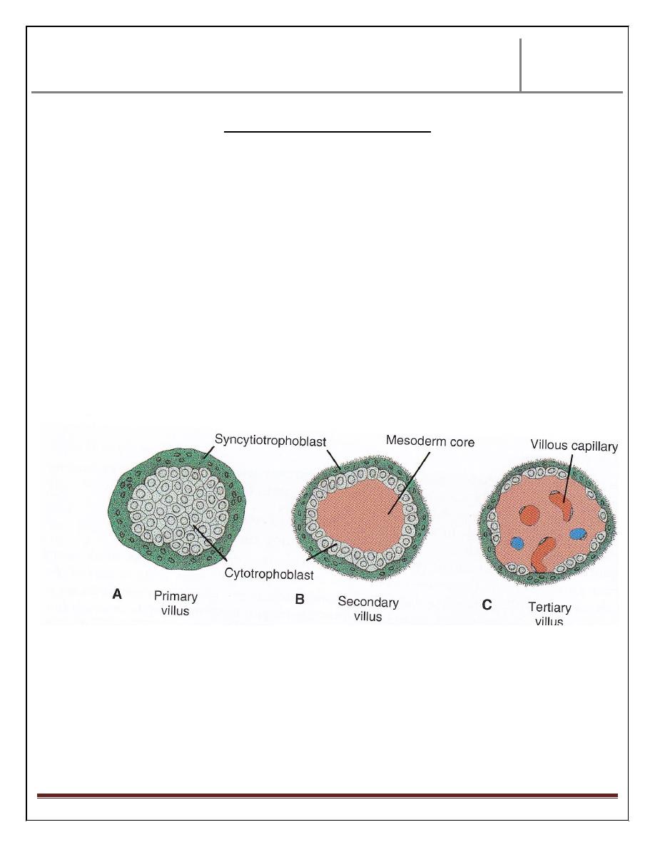
Development of Villi
By beginning of the 3
rd
wk, primary villi appear consist of core of
cytotrophoblast covered by syncytial layer .
Then secondary villi appear when mesodermal core covered by single layer of
cytotrophoblast ,which in turn covered by syncytial layer.
At the end of the 3
rd
week in the core of mesoderm of secondary villi, small
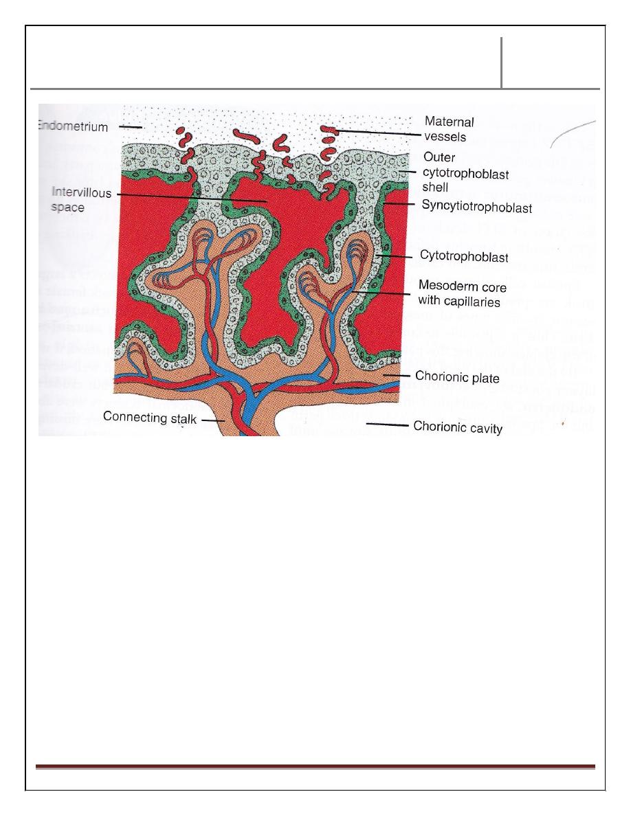
blood vessels will appears forming tertiary villi or definitive placental villus.
Here capillary will develop in the mesoderm of chorionic plate & in the
connecting stalk.
At the end of 4
th
wk when the heart beats, the villus system is ready to supply
embryo by O2& nutrients.
Lec.2
د.معن الخالصي
2013-
2014
Dr.alkhalisy Embryology
Page 7-7
THE END