
Muscular System
Prof. Dr. Malak A. Al-yawer
Department of Anatomy/Embryology Section

At the end of this lecture, the medical
student will be able to
State the embryonic origin of skeletal muscle
Distinguish between somitomeres and somites
Define mesodermal domains of the embryo
Define lateral somitic frontier
State the embryonic significance of the lateral somitic frontier for the
development of dermis and ribs
Describe the embryonic origin of innervation of myotomes
State the new and old concepts of muscle development
Define myogenesis & list their steps
Define patterning of muscles
State the embryonic origins of head musculature
State the embryonic origin of limb musculature
Describe the embryonic origin of cardiac muscles
Describe the embryonic origin of smooth muscles
State some clinical correlates
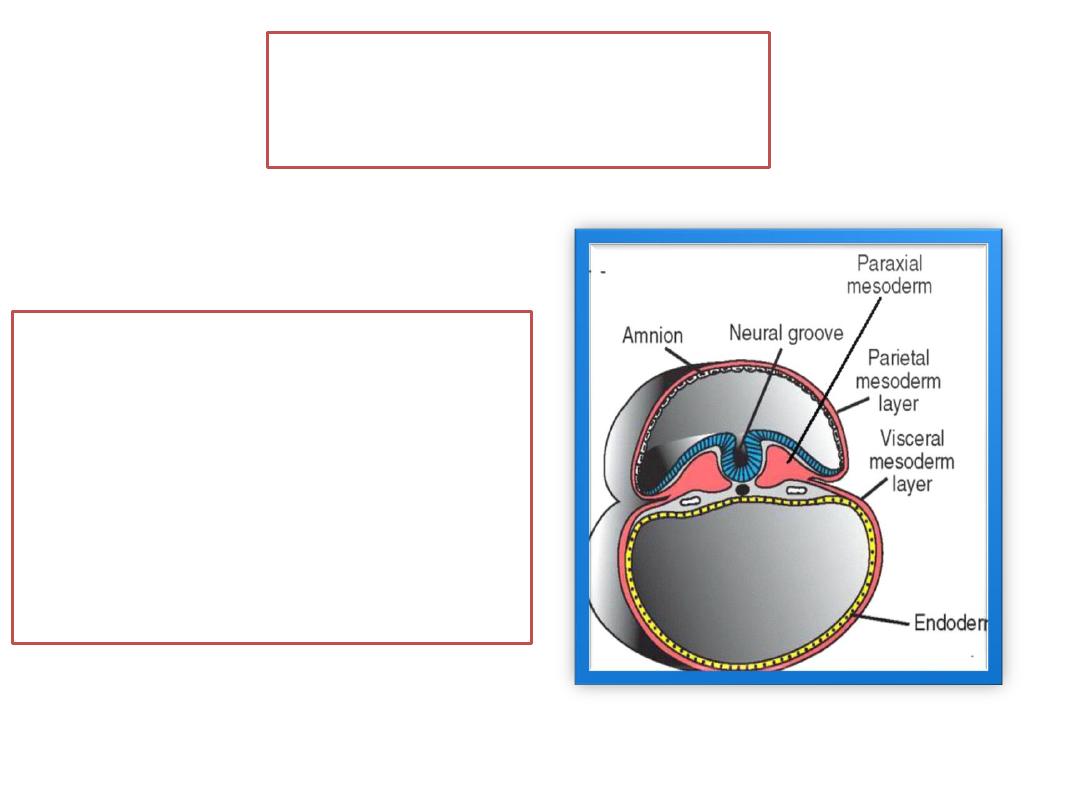
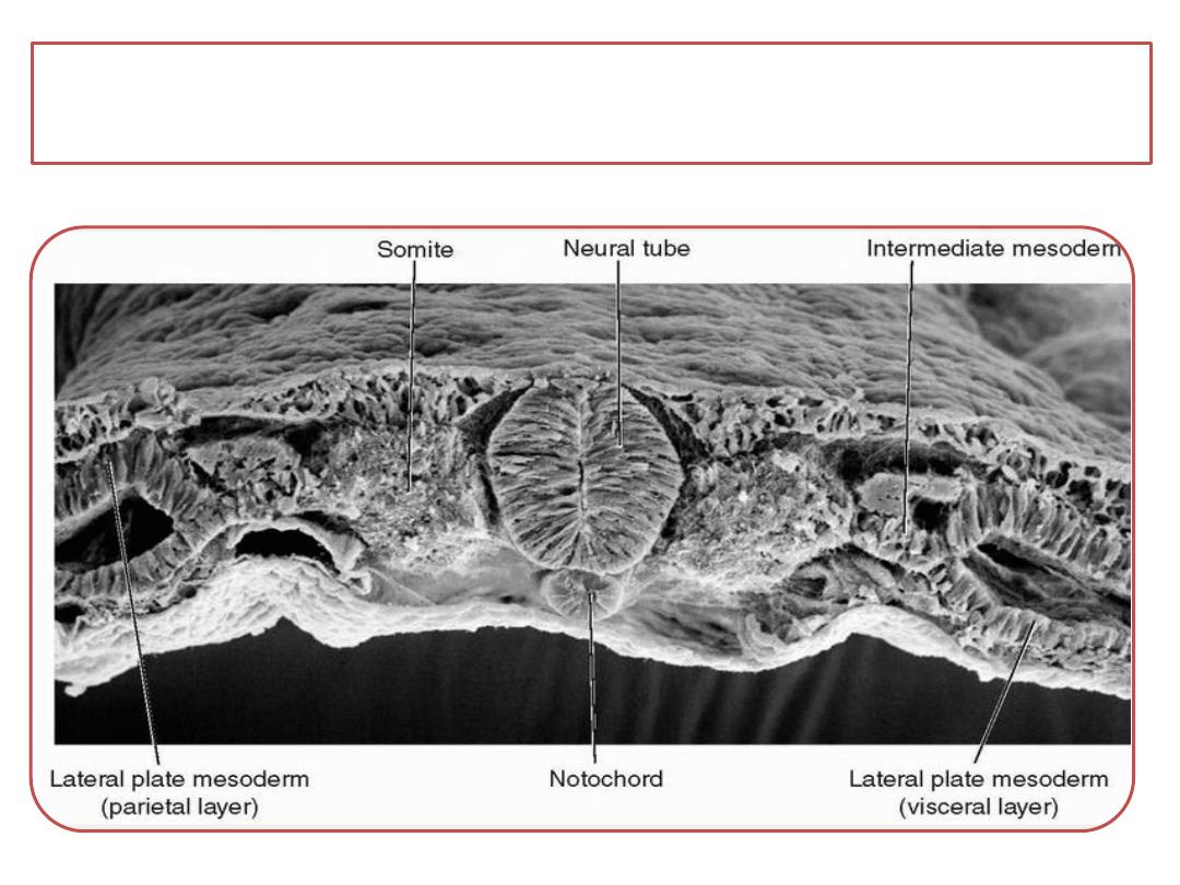
Skeletal muscle
•
is derived from paraxial
mesoderm, which forms
somites from the occipital
to the sacral regions and
somitomeres in the head.
Somitomeres
•
seven in number
•
are partially segmented
whorls of mesenchymal
cells derived from paraxial
mesoderm .
•
remain loosely organized
structures and never
segregate into sclerotome
and dermomyotome
segments.
Somites
•
initially form as
somitomeres
•
extend from the occipital
region to the tail bud.
•
Immediately after
segmentation, these
somitomeres undergo a
process of epithelization
and form a “ball” of
epithelial cells with a
small cavity in the center
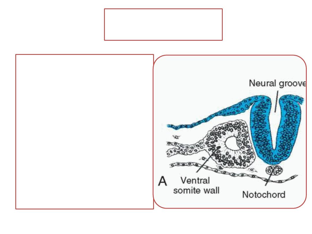
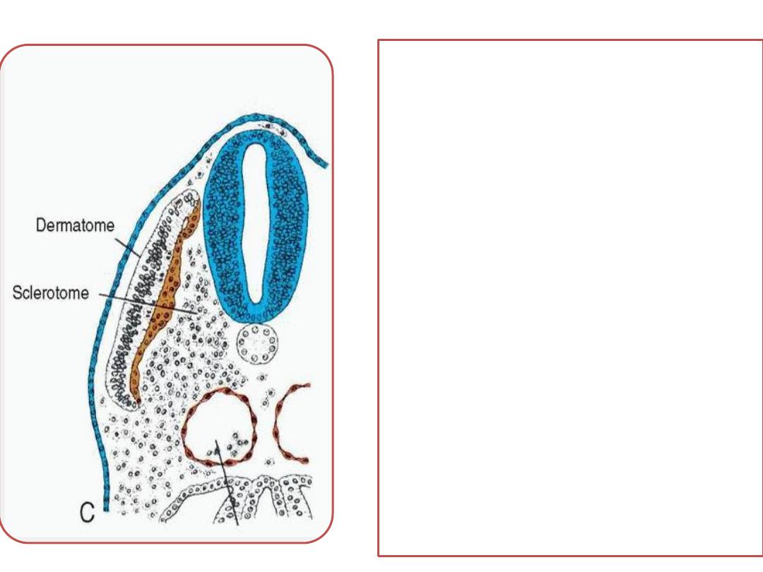
The ventral region of
each somite form
sclerotome for the
vertebrae and ribs.
Cells in the upper region
of the somite form
dermatome and
two muscle-forming
areas at the ventrolateral
(VLL) and dorsomedial
(DML) lips (or edges).
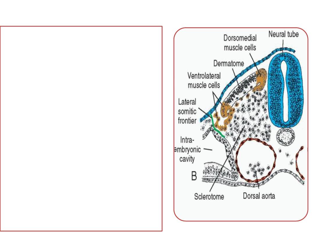
• Cells from the DML &
VLL form progenitor
muscle cells ventral to
the dermatome to form
dermomyotome
• Some cells from the
ventrolateral region
migrate into the
adjacent parietal layer
of the lateral plate
mesoderm
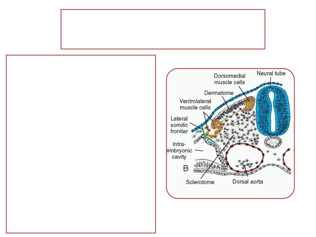
Lateral somitic frontier
• a well-defined border between
each somite and the parietal
layer of lateral plate mesoderm
• This frontier separates two
mesodermal domains in the
embryo:
(1) Primaxial domain comprises the
region around the neural tube
and contains only somite-derived
(paraxial mesoderm) cells
(2) Abaxial domain consists of the
parietal layer of lateral plate
mesoderm together with somite
cells that have migrated across
the lateral somitic frontier.

•
Abaxial domain form
infrahyoid,
abdominal wall (rectus abdominus, internal and
external oblique, and transversus abdominus),
limb muscles.
•
Primaxial domain form
muscles of the back,
shoulder girdle
intercostal muscles.
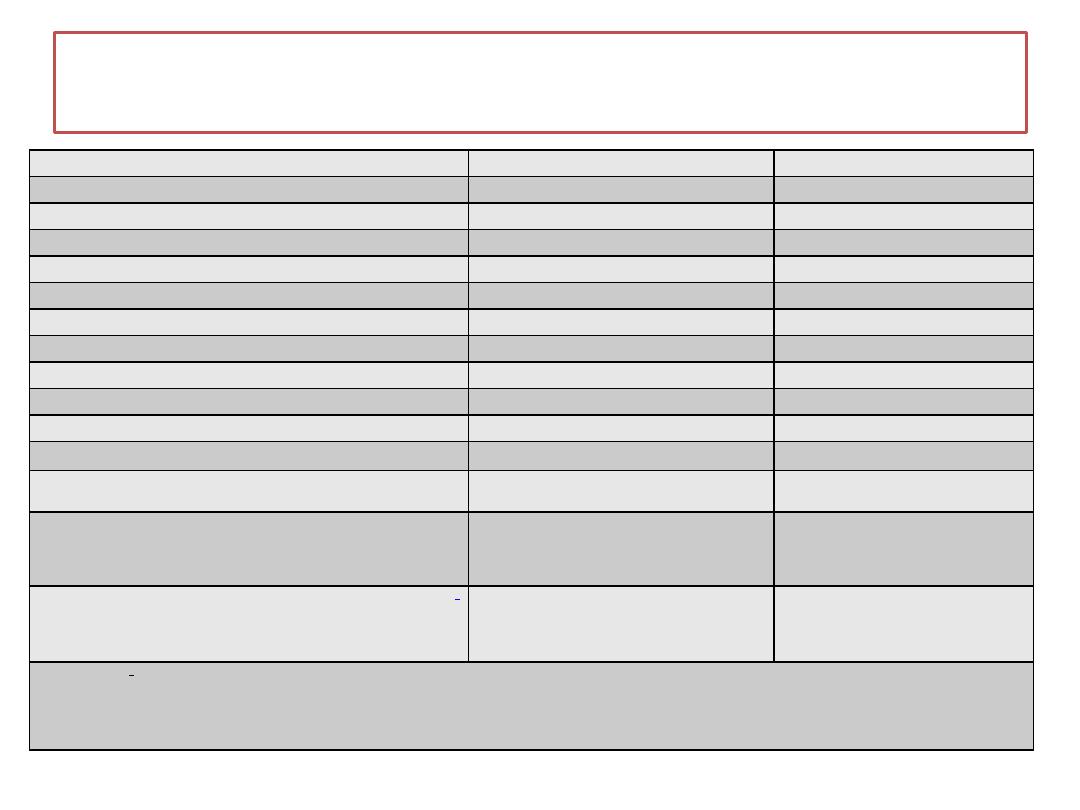
Origins of Muscles From Abaxial and Primaxial Precursors
Primaxial
Abaxial
Cervical region
Scalenes
Infrahyoid
Geniohyoid
Prevertebral
Thoracoabdominal region
Intercostals
Pectoralis major and minor
External oblique
Internal oblique
Transversus abdominus
Sternalis
Rectus abdominus
Pelvic diaphragm
Upper limb
Rhomboids
Distal limb muscles
Levator scapulae
Latissimus dorsi
Lower limb
All lower limb muscles
in origin.
abaxial
The precise origin of muscles in the pelvic region and lower limb has not been determined, but most if not all are
a
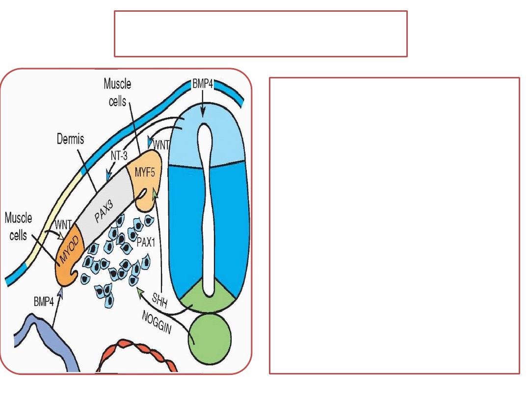
Muscle cell precursors
•
Abaxial muscle cell
precursors receive many
of their signals for
differentiation from
lateral plate mesoderm
• Primaxial muscle cell
precursors receive many
of their developmental
signals from the neural
tube and notochord.
The lateral somitic frontier
• It defines the border of dermis derived from
dermatomes in the back and dermis derived
from lateral plate mesoderm in the body wall.
•
It defines a border for rib development with
the bony components of each rib derived
from primaxial sclerotome cells and the
cartilaginous parts of those ribs derived from
abaxial sclerotome cells.

Regardless of their domain, each myotome
receives its innervation from spinal nerves
derived from the same segment as the muscle
cells.

The new description of muscle
development differs from the old concept
•
The new description of muscle
development characterized by primaxial
and abaxial domains (based on the
actual embryological origin )
•
The old concept of epimeres (back
muscles) and hypomeres (limb and body
wall muscles), which was based on a
functional definition of innervation)
•
Epimeric ( epiaxial) muscles were
innervated by dorsal primary rami;
•
hypomeric ( hypaxial) muscles by ventral
primary rami
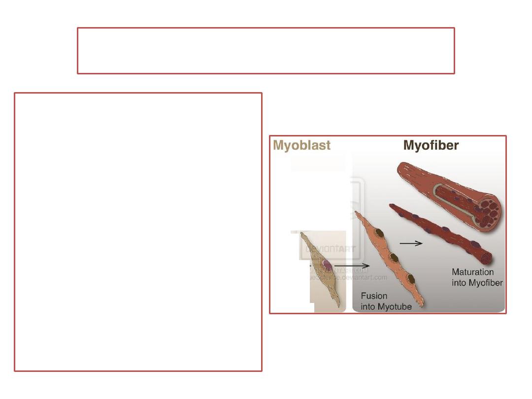
Myogenesis (muscle formation)
•
Myoblasts : primordial muscle cells
•
Myotubes is formed by fusion of
myoblasts to form elongated,
multinucleated, cylindrical structures-
myotubes.
myofilaments develop in the cytoplasm
of the myotubes.
By the end of the3rd month , cross-
striations, typical of skeletal muscle,
appear.
•
As the myotubes develop, they become
invested with external laminae
Fibroblasts produce the perimysium
and epimysium
the endomysium is formed by the
external lamina, and reticular fibers.
Tendons of muscles
• are derived from sclerotome cells lying
adjacent to myotomes at the anterior and
posterior borders of somites.

PATTERNING OF MUSCLES
are controlled by connective tissue into
which myoblasts migrate.
In the head region, these connective tissues are
derived from neural crest cells;
in cervical and occipital regions, they differentiate
from somitic mesoderm; and
in the body wall and limbs, they originate from
the parietal layer of lateral plate mesoderm.
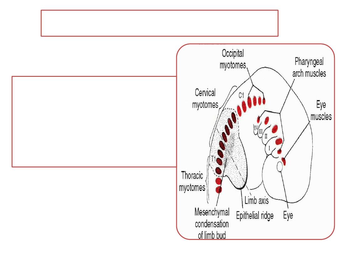
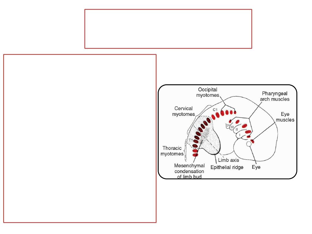
HEAD MUSCULATURE
• All voluntary muscles of
the head region are
derived from paraxial
mesoderm (somitomeres
and somites)
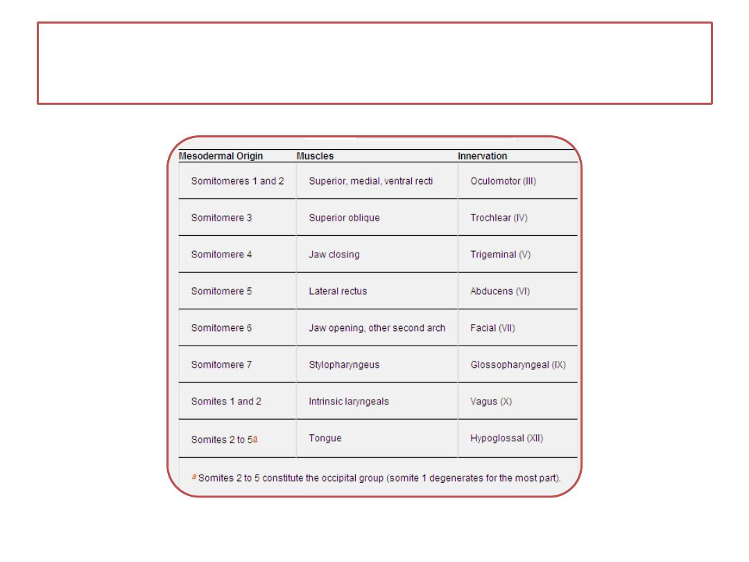
Table showing the origin of craniofacial muscles
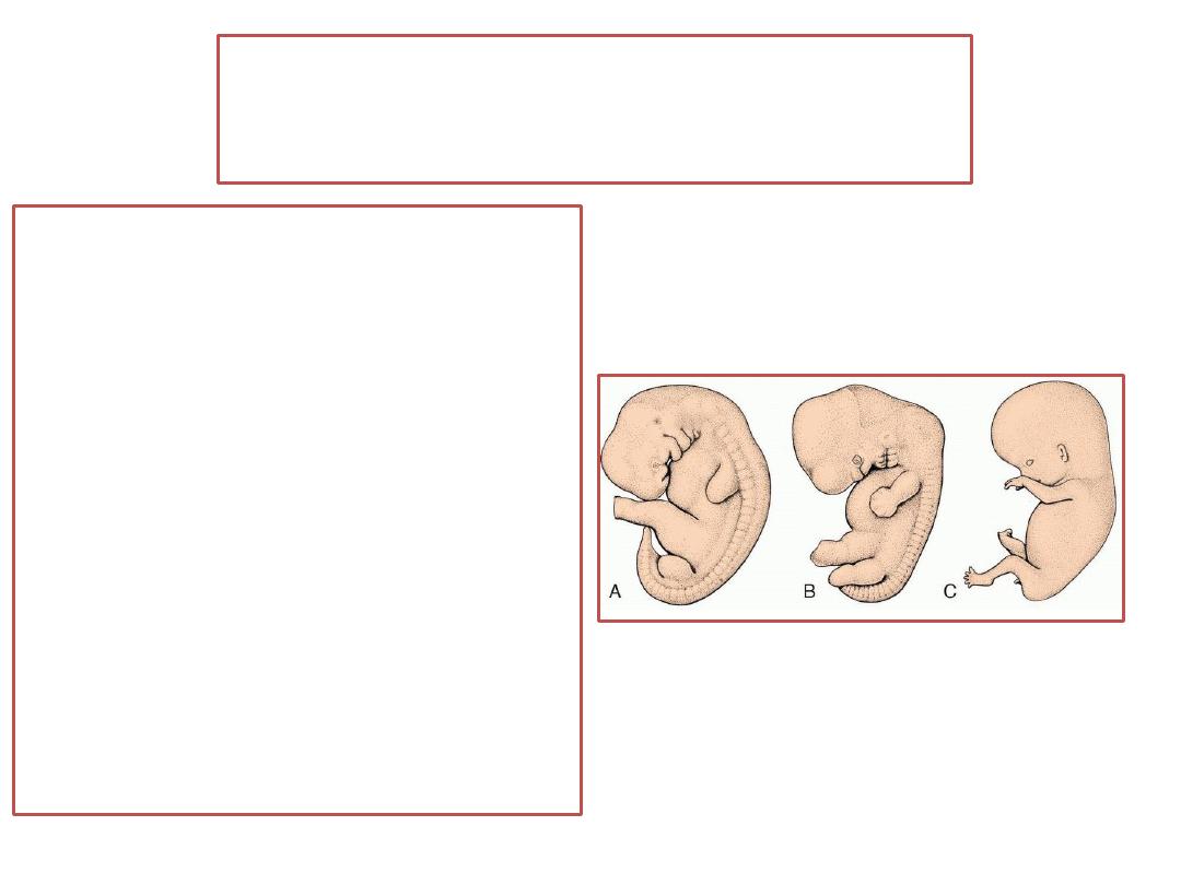
LIMB MUSCULATURE
• The first indication of limb
musculature is observed in
the 7
th
week of
development as a
condensation of
mesenchyme near the base
of the limb buds.
• The mesenchyme is derived
from dorsolateral cells of
the somites that migrate
into the limb bud to form
the muscles
CARDIAC MUSCLE
• Cardiac muscle develops from splanchnic
mesoderm surrounding the endothelial heart
tube.
• Myofibrils develop as in skeletal muscle, but
myoblasts do not fuse.
•
Late in the embryonic period, Purkinje fibers
form which are special bundles of muscle cells
develop with relatively few myofibrils and
relatively larger diameters than typical cardiac
muscle fibers.
SMOOTH MUSCLE
•
Smooth muscles in dorsal aorta and large
arteries: derived from lateral plate mesoderm
and neural crest cells.
•
Smooth muscle in coronary arteries, originates
from proepicardial cells and neural crest cells.
• Smooth muscle in the wall of the gut and gut
derivatives is derived from the splanchnic layer
of lateral plate mesoderm
• Only the sphincter and dilator muscles of the
pupil and muscle tissue in the mammary and
sweat glands are derived from ectoderm.
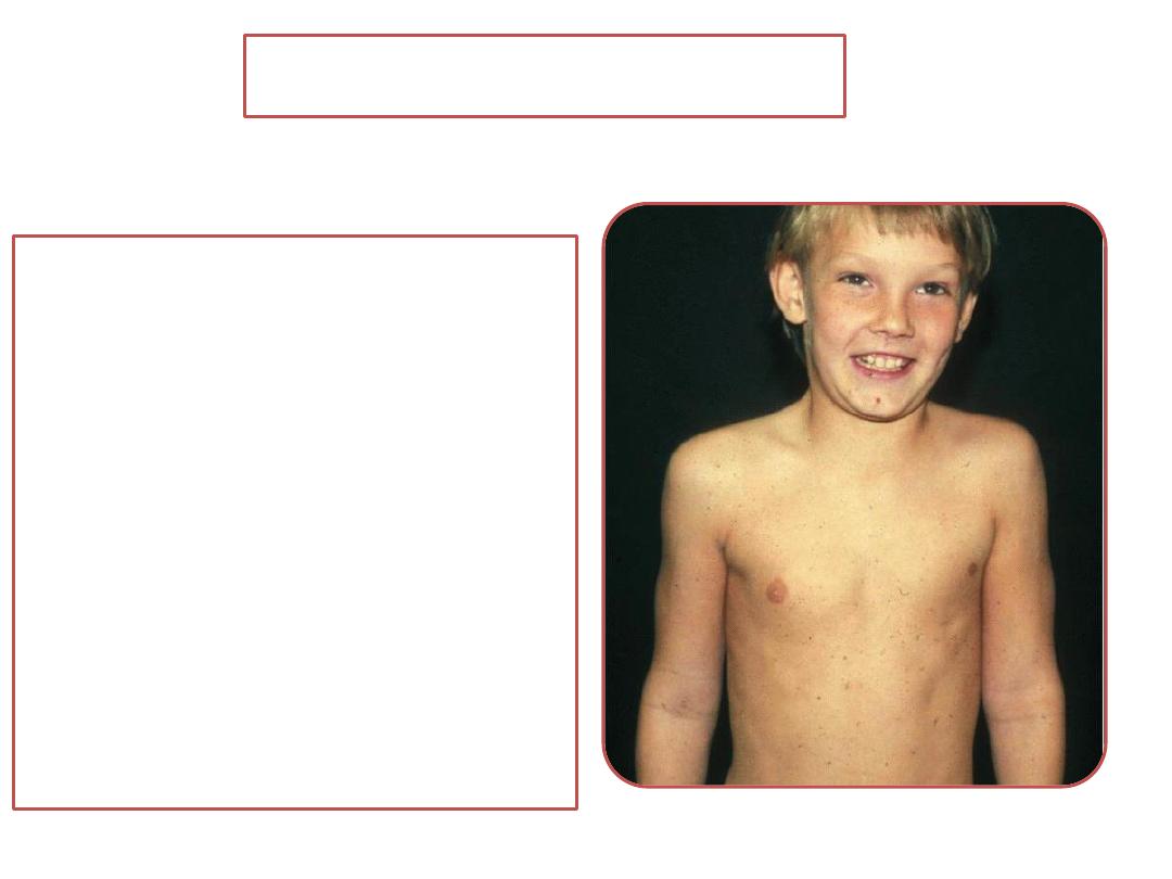
Poland sequence
• Partial or complete
absence of a muscle is
common.
• Poland
sequence is
characterized by absence
of the pectoralis minor
and partial loss of the
pectoralis major (usually
the sternal head) muscles
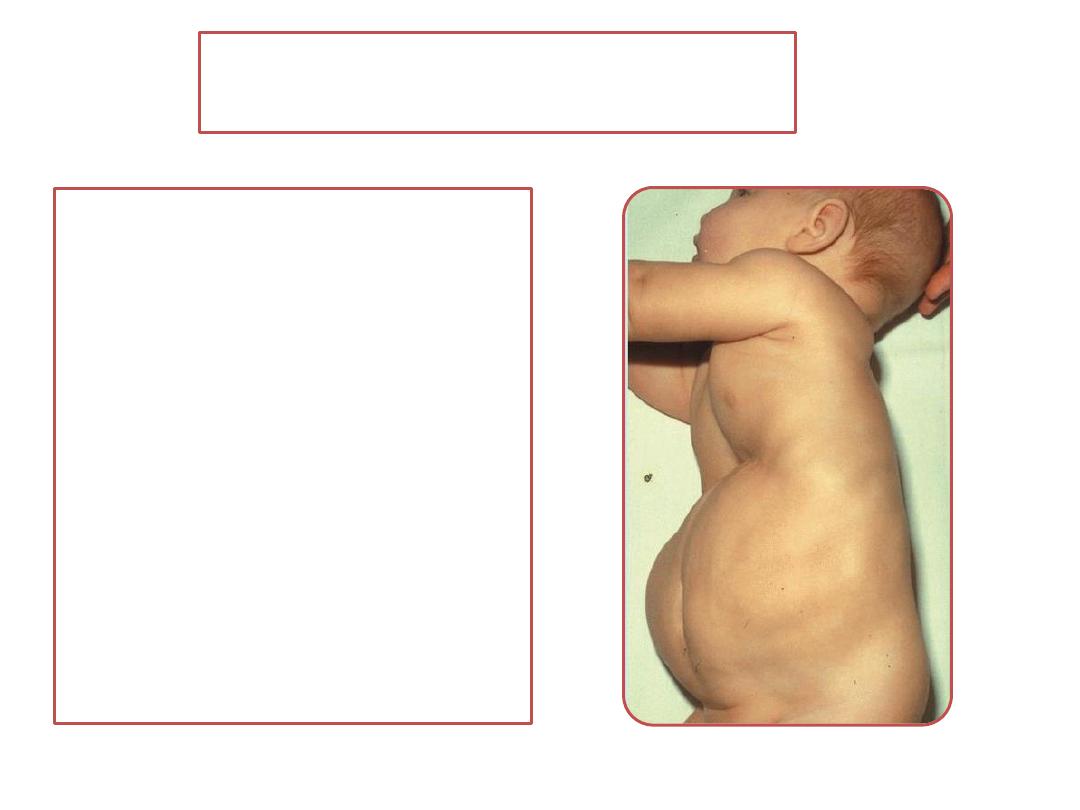
Prune belly syndrome
• Is partial or complete
absence of abdominal
musculature. Usually, the
abdominal wall is so thin
that organs are visible
and easily palpated.
• This defect is associated
with malformations of the
urinary tract and bladder,
including urethral
obstruction.
Muscular dystrophy
• a group of inherited muscle diseases that
cause progressive muscular wasting and
weakness.
• There are a large number of these types of
diseases of which Duchenne's muscular
dystrophy (DMD) is the most common. The
disease is inherited as X-linked recessive such
that males are much more often affected than
females.
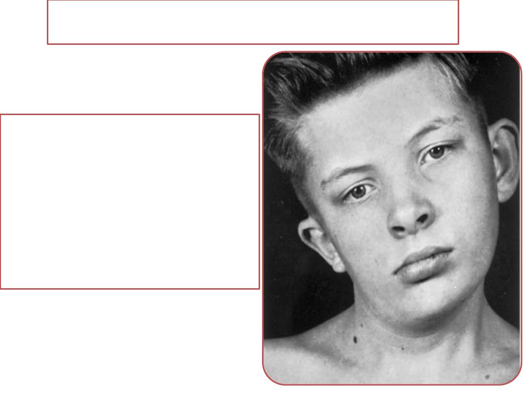
Congenital Torticollis(wry neck)
• result from tearing of fibers of
the sternocleidomastoid muscle
during childbirth.
• Shortening of the muscle
usually follows, which causes
lateral bending of the head to
the affected side
SUMMARY
With the exception of some smooth muscle tissue, the muscular
system develops from the mesodermal germ layer
Summary
•
Skeletal muscle is derived from paraxial mesoderm, which
forms somites from the occipital to the sacral regions and
somitomeres in the head
•
Smooth muscle differentiates from
somatic mesoderm which provides smooth muscle in the
walls of many blood and lymphatic vessels.
visceral splanchnic mesoderm surrounding the gut and its
derivatives
ectoderm muscles of the iris (sphincter and dilator
pupillae) and the myoepithelial cells in mammary and
sweat glands
Cardiac muscle is derived from visceral splanchnic
mesoderm surrounding the heart tube.

Thank you