
Head & neck
Prof. Dr. Malak A. Al-yawer

At the end of this lecture, the medical student will be able to:
List the embryonic origin of mesenchyme of the head region
Define ectodermal placodes and mention its significance
List the components of pharyngeal apparatus
State the fate of pharyngeal arches, pouches, clefts and
membranes
List the embryonic origin of tongue
Define the embryonic origin of the thyroid gland
State the fate of facial and nasal prominences
Define the embryonic origin of nasal cavity
Define the embryonic origin of paranasal sinuses
Learning Objectives

• (1) Paraxial mesoderm
• (2) Lateral plate mesoderm
• (3) Neural crest cells
• (4) Ectodermal placodes
Mesenchyme for formation of the
head region is derived from
(1) Paraxial mesoderm
(Somites and Somitomeres) forms
1. Large portion of the
membranous and cartilaginous
components of the
neurocranium (Skull)
2. All voluntary muscles of the
craniofacial region
3. the dermis and connective
tissues in the dorsal region of
the head
4. the meninges caudal to the
prosencephalon.
(2) Lateral plate mesoderm forms
laryngeal cartilages
(arytenoid and cricoid)
connective tissue in this
region.
(3) Neural crest cells
•
originate in the neuroectoderm of
forebrain, midbrain, and hindbrain
regions and
•
migrate ventrally into the
pharyngeal arches and rostrally
around the forebrain and optic cup
into the facial region.
•
In these locations, they form
1.
The entire viscerocranium (face)
2.
Parts of the membranous and
cartilaginous regions of the
neurocranium
3.
All other tissues in these regions
including cartilage, bone, dentin ,
tendon, dermis, pia and arachnoid,
sensory neurons and glandular
tissue

• Are thickened regions of ectoderm
• Cells from ectodermal placodes, together with
neural crest, form
neurons of the fifth, seventh, ninth, and
tenth cranial sensory ganglia.
(4)
Ectodermal placodes

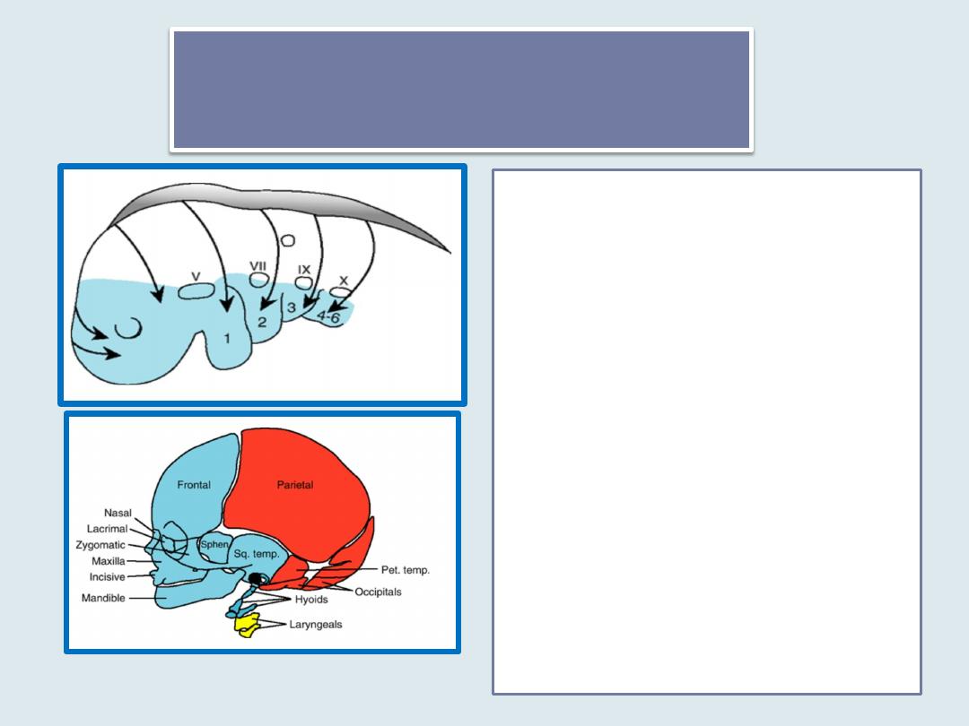
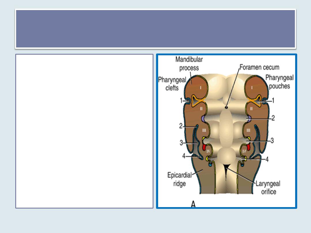
• Pharyngeal arches
• Pharyngeal pouches
• Pharyngeal clefts
• Pharyngeal membrane
Pharyngeal apparatus consists of:
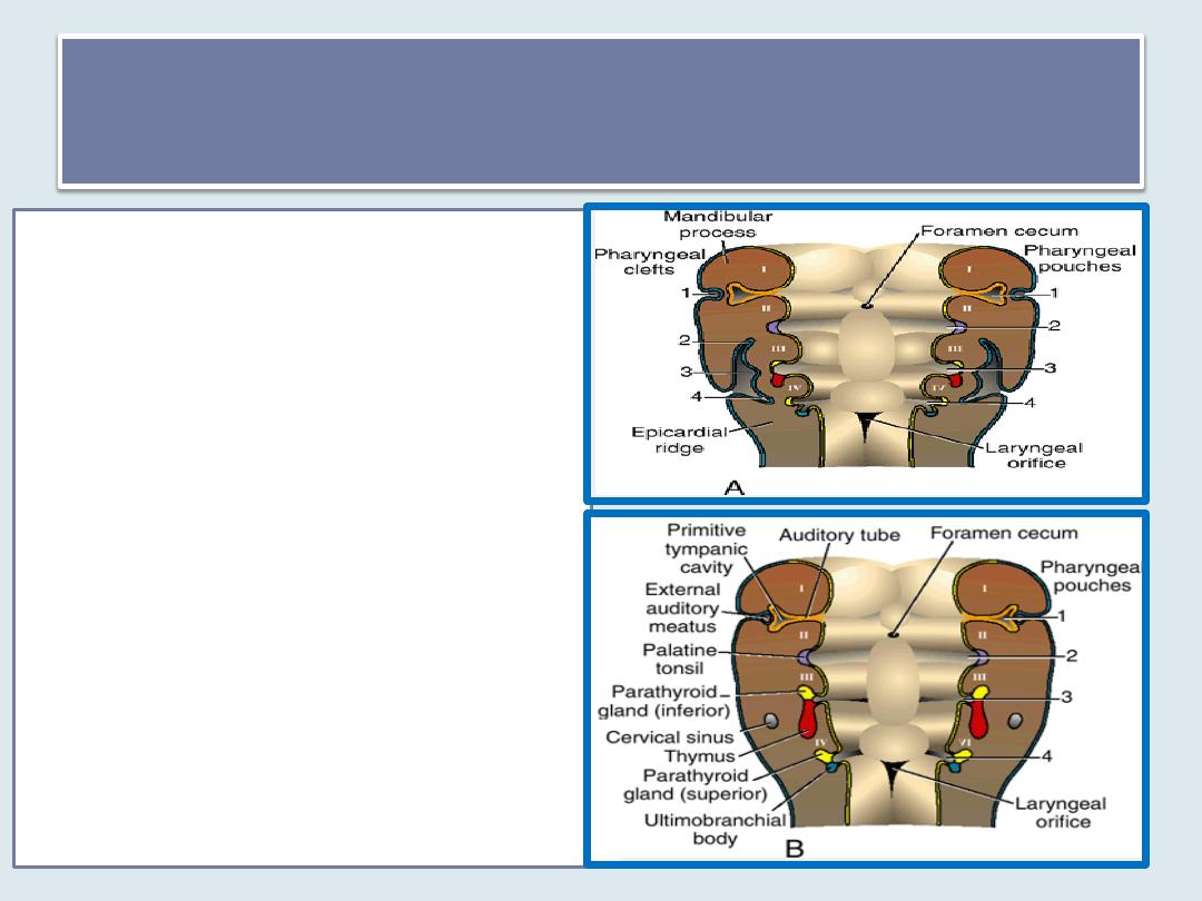
Pharyngeal arches
• The old term for these structures is brachial arches because they
somewhat resemble the gills(brachia) of a fish
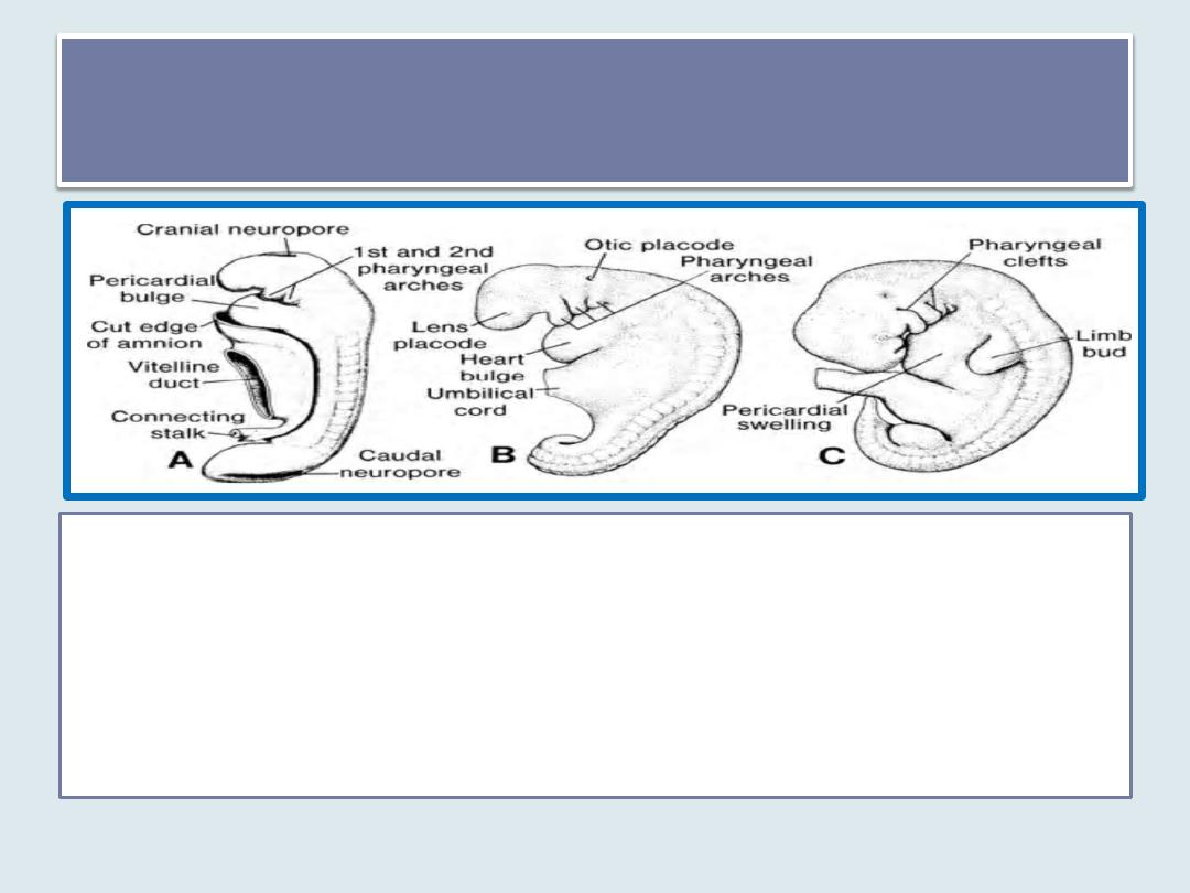
• appear in the
fourth and fifth weeks
of development and contribute to
the characteristic external appearance of the embryo. It Contribute mostly
to neck development but the first arch contributes to facial development.
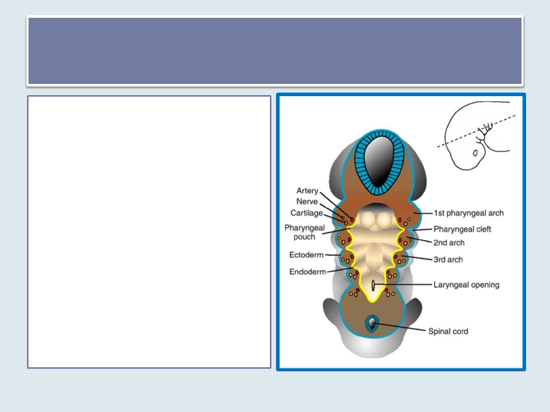
Pharyngeal arch
Each pharyngeal arch
1. consists of a
mesenchymal core
derived from mesoderm
and neural crest cells and
2. is lined internally by
endoderm and
3. externally by ectoderm.
Each arch also contains
1. an artery (one of the
aortic arches) and
2. a cranial nerve
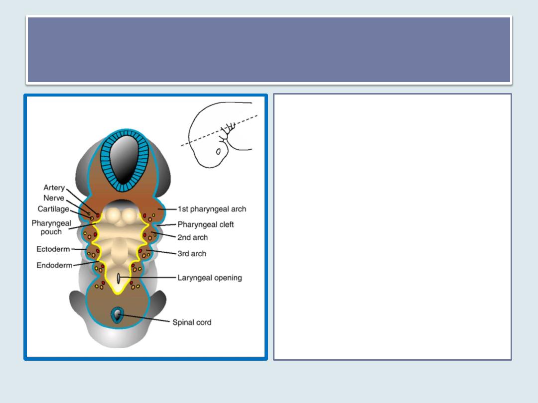
Pharyngeal arch
Each arch will
contribute specific
skeletal and muscular
components to the
head and neck.
Between the arches
are
1. pouches on the inner
surface
2. clefts externally.
1
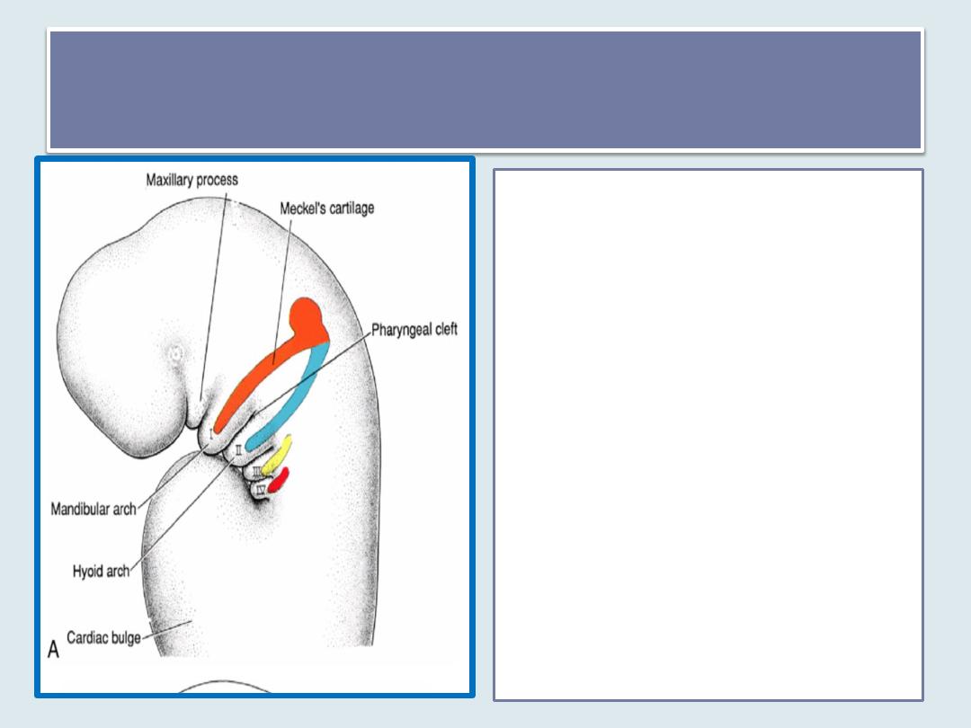
st
pharyngeal arch
• The first pharyngeal arch consists
of
1.
a dorsal portion (maxillary
process)
2.
a ventral portion(mandibular
process) which contains
Meckel's cartilage .
• During further development,
Meckel's cartilage disappears
except for two small portions at
its dorsal end that persist and
form the incus and malleus .
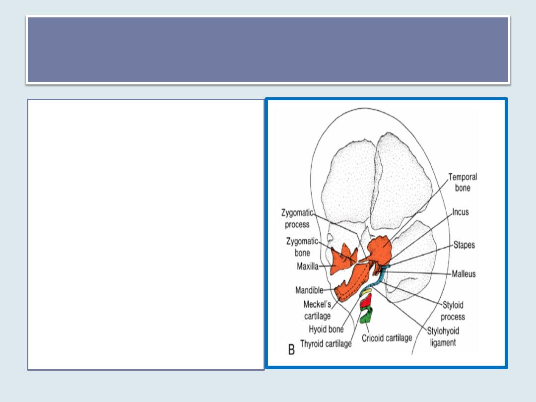
Skeleton of the 1
st
pharyngeal arches
• Mesenchyme of the maxillary
process gives rise to the
premaxilla, maxilla, zygomatic
bone, and part of the temporal
bone.
• The mandible is also formed by
membranous ossification of
mesenchymal tissue surrounding
Meckel's cartilage.
• In addition, the first arch
contributes to formation of the
bones of the middle ear( incus
and malleus), anterior ligment of
malleus and sphenomandibular
ligment.
Musculature & nerve supply of the first
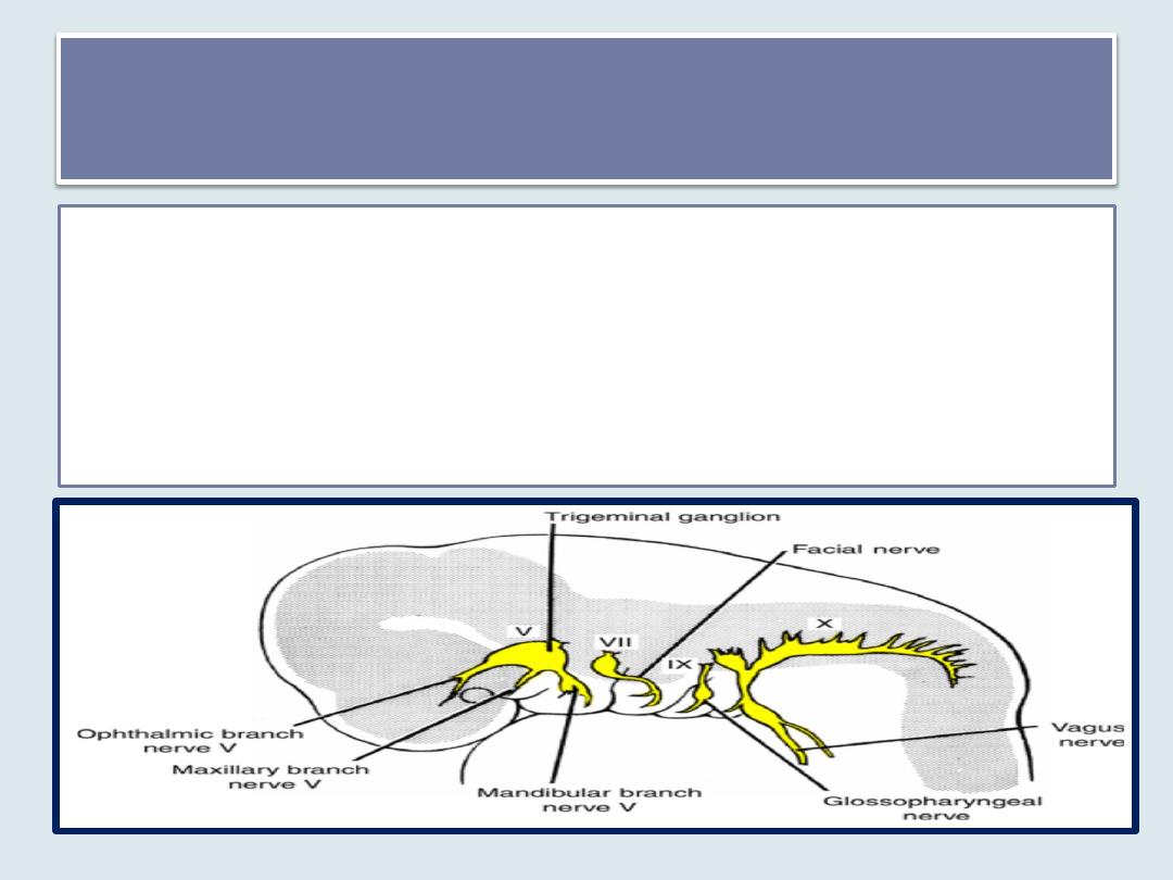
pharyngeal arch
• includes the muscles of mastication (temporalis, masseter, and
pterygoids), anterior belly of the digastric, mylohyoid, tensor tympani, and
tensor palatini.
• The nerve supply to the muscles of the first arch is provided by the
mandibular branch of the trigeminal nerve.
• Sensory supply to the skin of the face is provided by ophthalmic,
maxillary, and mandibular branches of the trigeminal nerve.
Second Pharyngeal Arch (hyoid arch)
• The cartilage of hyoid arch
(Reichert's cartilage) gives rise to
the stapes, styloid process of the
temporal bone, stylohyoid
ligament, and ventrally, the lesser
horn and upper part of the body
of the hyoid bone.
• Muscles of the hyoid arch are
the stapedius, stylohyoid,
posterior belly of the digastric,
auricular, and muscles of facial
expression.
• The facial nerve, the nerve of the
second arch, supplies all of these
muscles.
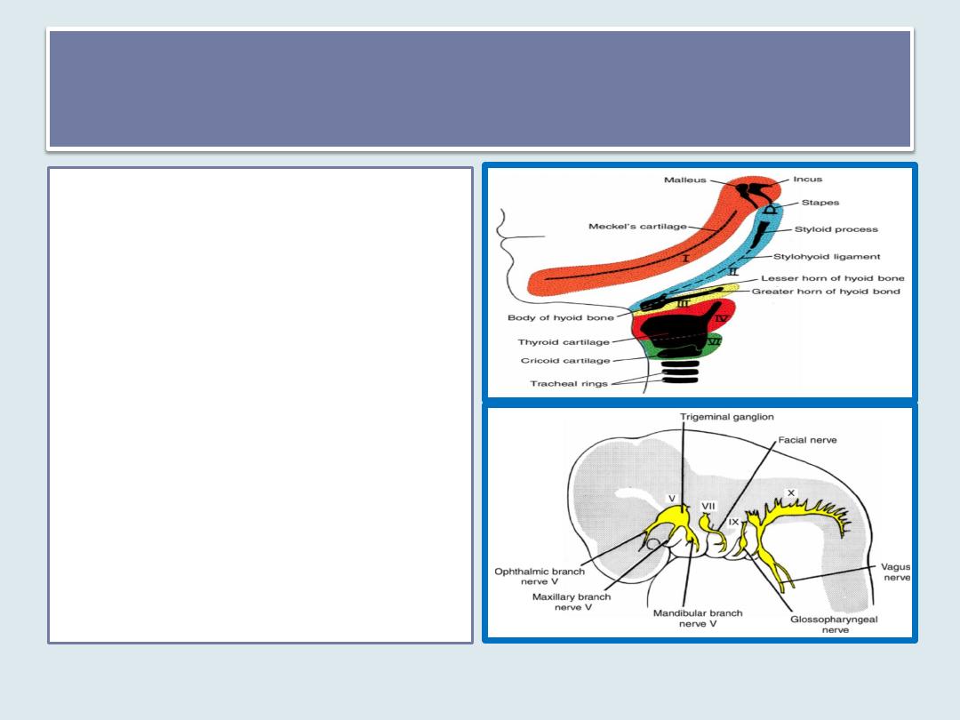
Third Pharyngeal arch
• produces the lower part of
the body and greater horn
of the hyoid bone .
• The musculature is limited
to the stylopharyngeus
muscles.
• These muscles are
innervated by the
glossopharyngeal nerve
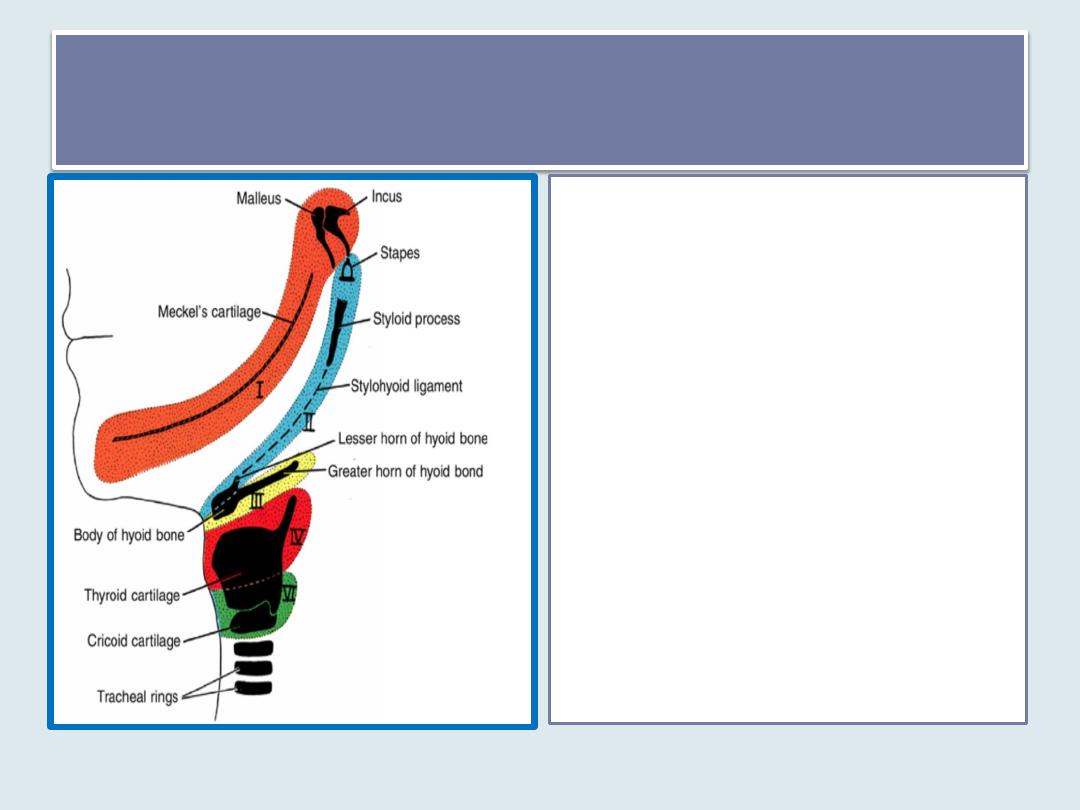
Fourth and Sixth Pharyngeal Arches
Cartilaginous components of the
fourth and sixth pharyngeal
arches fuse to form the thyroid,
cricoid, arytenoid, corniculate,
and cuneiform cartilages of the
larynx .
Muscles of the fourth arch
(cricothyroid, levator palatini,
and constrictors of the pharynx)
are innervated by the superior
laryngeal branch of the vagus.
Muscles of the 6
th
arch (Intrinsic
muscles of the larynx and
striated muscles of esophagus )
are supplied by the recurrent
laryngeal branch of the vagus.
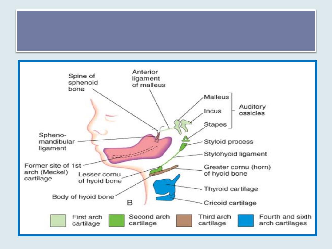
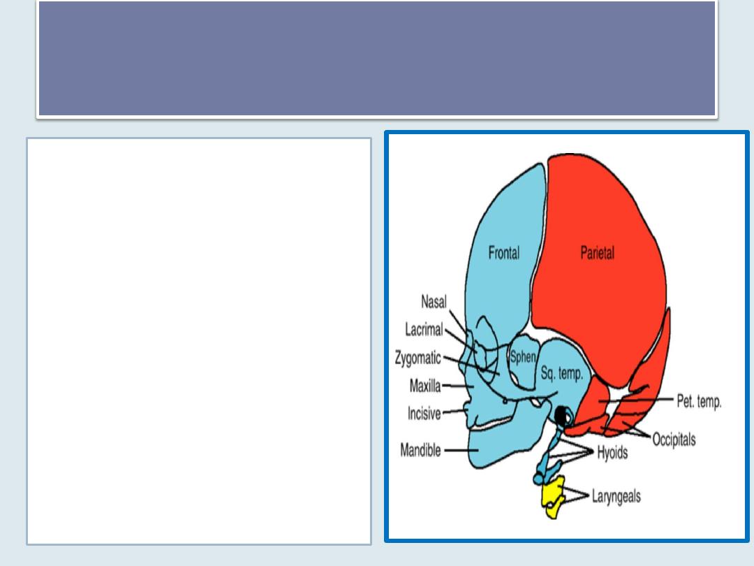
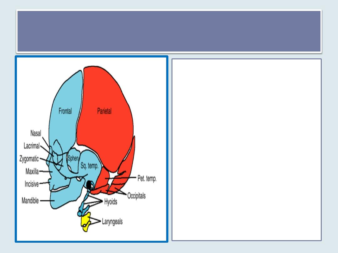
View of a 24-week fetus illustrating the adult derivatives of the
arch cartilages
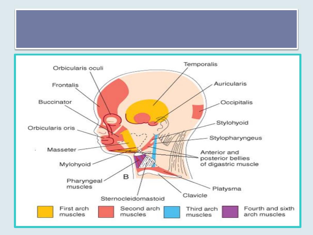
Sketch of the head and neck regions of a 20-week fetus, dissected to show the
muscles derived from the pharyngeal arches.
Parts of the platysma and sternocleidomastoid muscles have been removed to show the
deeper muscles

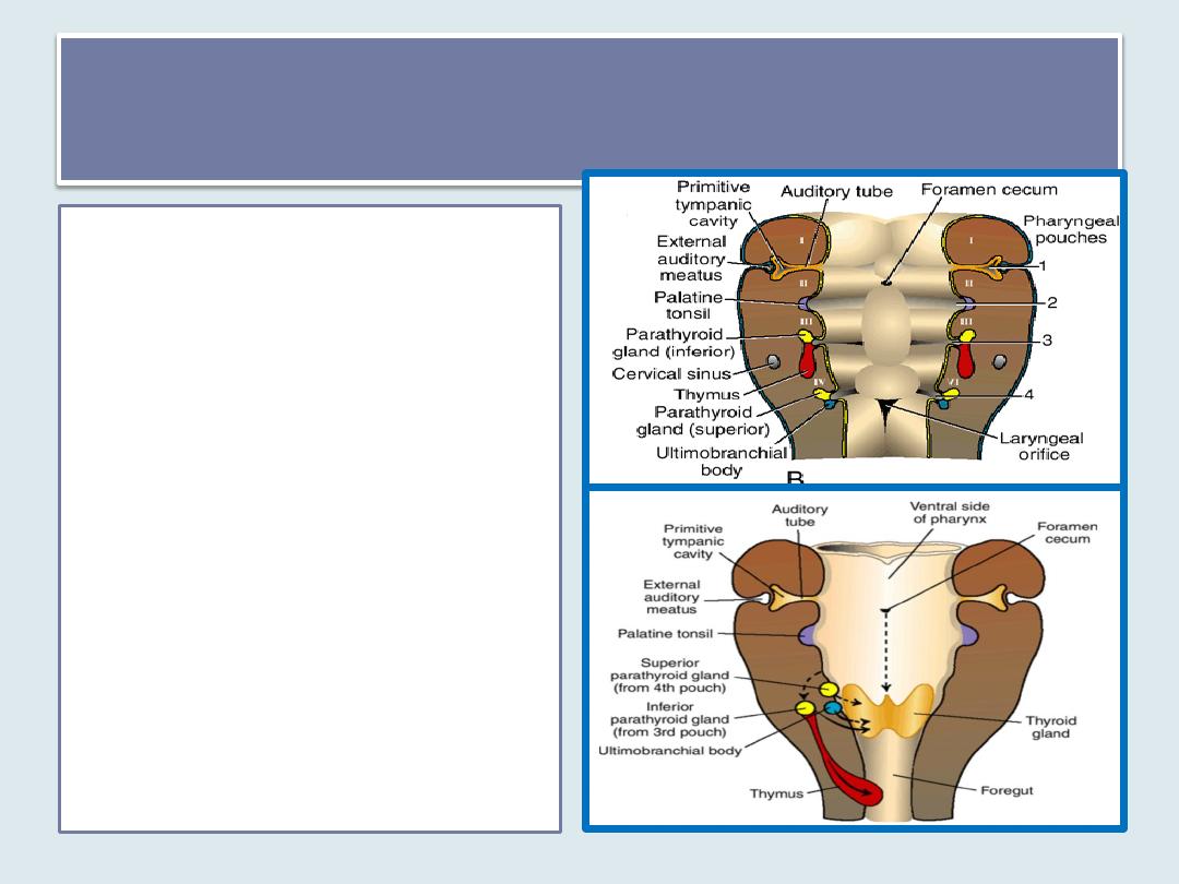
Pharyngeal Pouches
Four pairs of pharyngeal pouches.
The fifth is rudimentary
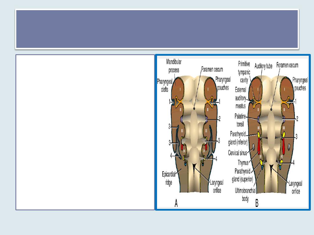
First Pharyngeal Pouch
• forms a stalk-like diverticulum,
the tubotympanic recess, which
comes in contact with the
epithelial lining of the first
pharyngeal cleft, the future
external auditory meatus.
• The distal portion of the
diverticulum widens into a saclike
structure, the primitive tympanic
or middle ear cavity, and the
proximal part remains narrow,
forming the auditory
(eustachian) tube.
• The lining of the tympanic cavity
later aids in formation of the
tympanic membrane or eardrum
Second Pharyngeal Pouch
• The epithelial lining of the
second pharyngeal pouch
proliferates and forms buds
that penetrate into the
surrounding mesenchyme,
forming the primordium of
the palatine tonsil .
• Part of the pouch remains
and is found in the adult as
the tonsillar fossa
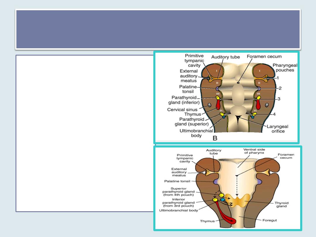
Third Pharyngeal Pouch
• eithelium of the dorsal wing
of the third pouch
differentiates into the
inferior parathyroid gland,
while the ventral wing
forms the thymus
• Both gland primordia lose
their connection with the
pharyngeal wall, and the
thymus then migrates in a
caudal and a medial
direction, pulling the
inferior parathyroid with it.
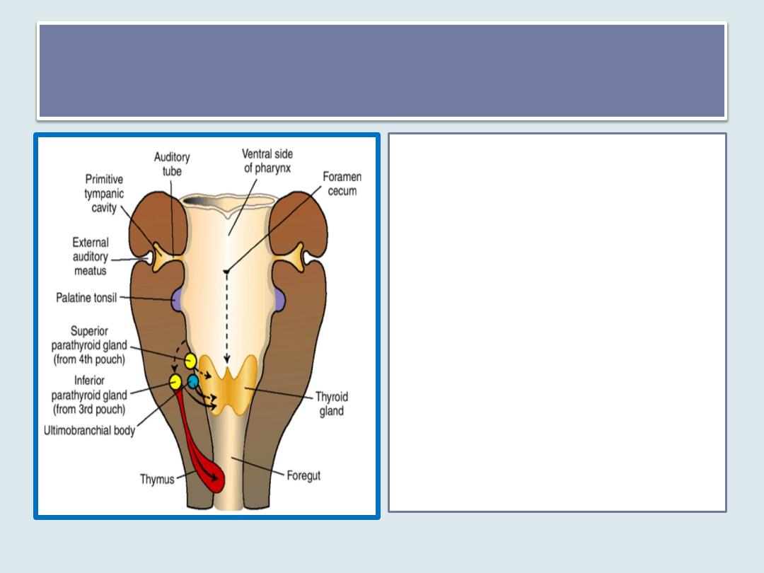
Fourth Pharyngeal Pouch
• Epithelium of the dorsal wing of
the fourth pharyngeal pouch
forms the superior parathyroid
gland.
• Epithelium of the ventral wing of
the fourth pharyngeal pouch
gives rise to the ultimobranchial
body, which is later incorporated
into the thyroid gland. Cells of the
ultimobranchial body give rise to
the parafollicular, or C, cells of
the thyroid gland. These cells
secrete calcitonin, a hormone
involved in regulation of the
calcium level in the blood.

Pharyngeal Clefts
•Four pairs of pharyngeal clefts
•Only one of them contributes to the
definitive structure of the embryo
First pharyngeal cleft
• The dorsal part of the
first cleft penetrates
the underlying
mesenchyme and gives
rise to the external
auditory meatus.
• The epithelial lining at
the bottom of the
meatus participates in
formation of the
eardrum
2
nd
,3
rd
& 4
th
pharyngeal clefts
• Active proliferation of
mesenchymal tissue in the
second arch causes it to
overlap the third and fourth
arches. Finally, it merges with
the epicardial ridge in the
lower part of the neck , and
the second, third, and fourth
clefts lose contact with the
outside .
• The clefts form a cavity lined
with ectodermal epithelium,
the cervical sinus, but with
further development, this
sinus disappears
.

• it is not unusual for
accessory glands or
remnants of tissue to
persist along the
pathway.
• This is true particularly
for thymic tissue, which
may remain in the neck,
and for the parathyroid
glands.
Ectopic Thymic and Parathyroid Tissue
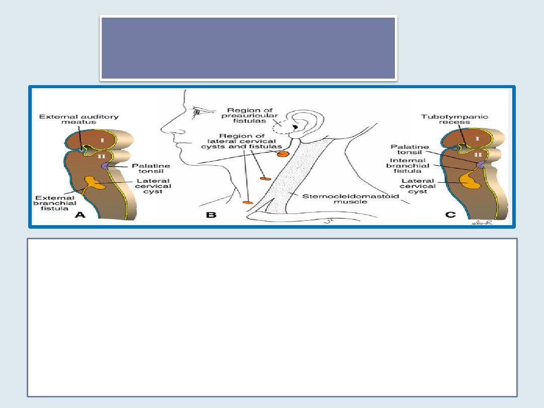
Branchial Fistulas
• occur when the second pharyngeal arch fails to grow caudally over the
third and fourth arches, leaving remnants of the second, third, and fourth
clefts in contact with the surface by a narrow canal
• A. Lateral cervical cyst opening at the side of the neck by way of a fistula.
• B. Lateral cervical cysts and fistulas in front of the sternocleidomastoid
muscle. Note also the region of preauricular fistulas.
• C. A lateral cervical cyst opening into the pharynx at the level of the
palatine tonsil.

• These membranes form where the epithelia of the
clefts and pouches approach each other
• The endoderm of the pouches and ectoderm of the
grooves are soon separated by mesenchyme
• Only first pharyngeal membrane becomes the
tympanic membrane, others obliterate
Pharyngeal Membranes
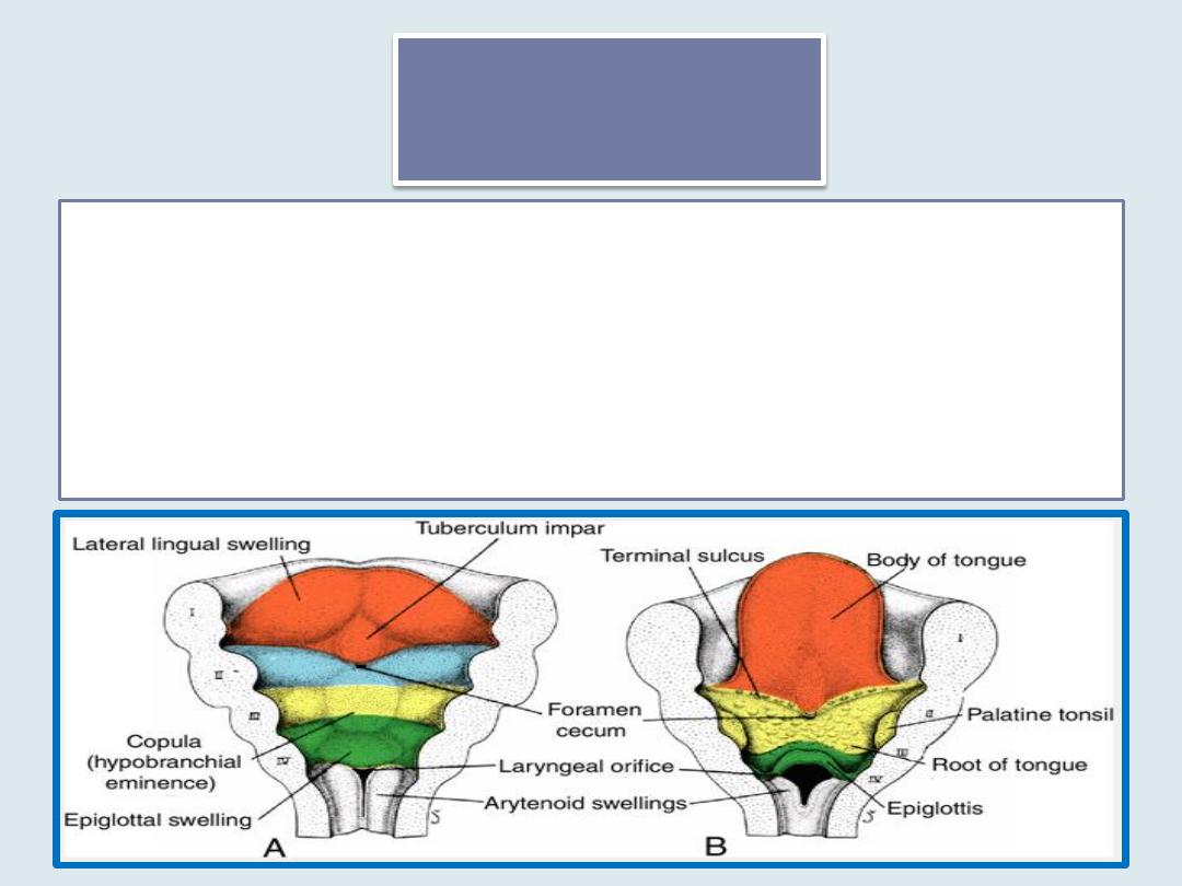
Tongue
•
Anterior two-thirds, or body, of the tongue derived from two lateral lingual
swellings and one median swelling (tuberculum impar) (first pharyngeal arch).
•
The posterior part, or root, of the tongue derived from a second median swelling (
copula, or hypobranchial eminence) is formed by mesoderm of the second, third,
and part of the fourth arch---------
•
Third median swelling, formed by the posterior part of the fourth arch, marks
development of the epiglottis.
•
Immediately behind this swelling is the laryngeal orifice, which is flanked by the
arytenoid swellings
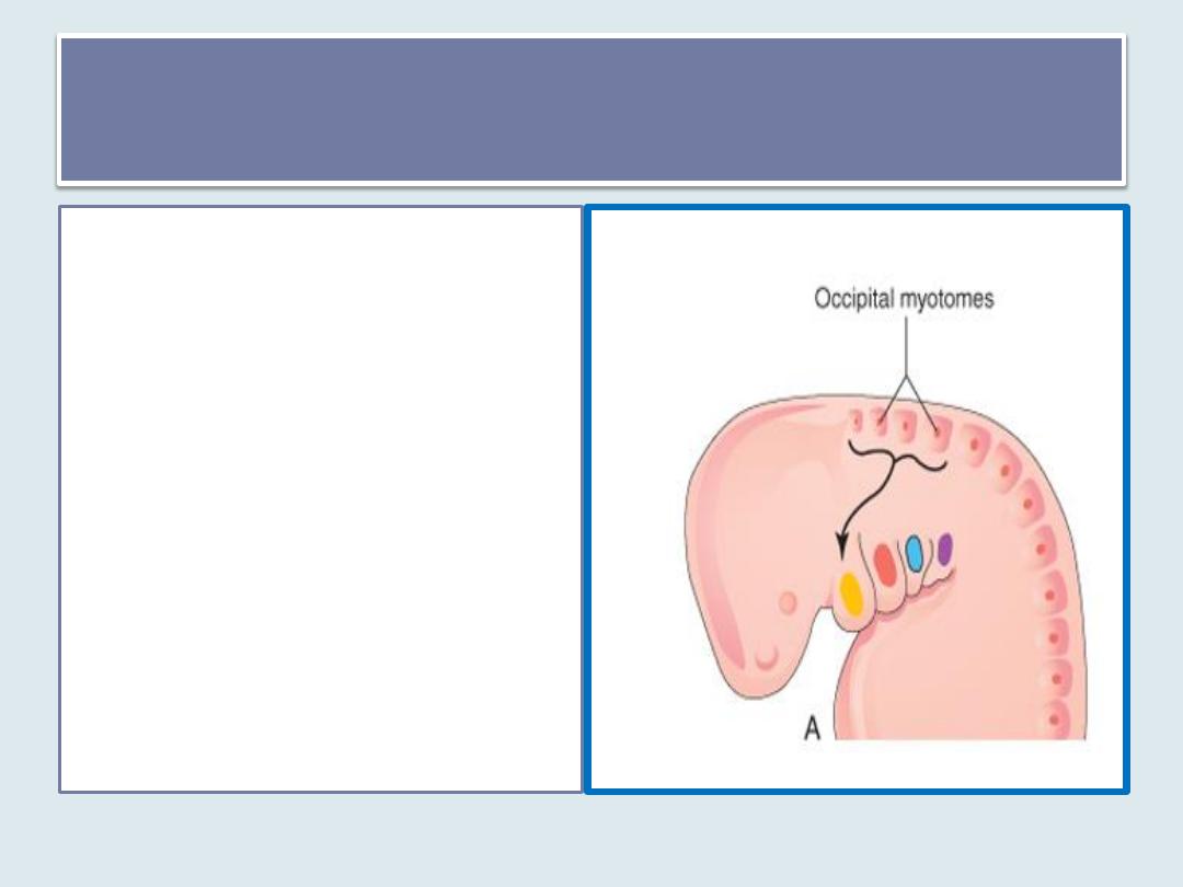
Muscular innervation of the tongue
• Some of the tongue muscles
probably differentiate in
situ, but most are derived
from myoblasts originating
in occipital somites. Thus,
tongue musculature is
innervated by the
hypoglossal nerve.
• The arrow shows the
pathway taken by myoblasts
from the occipital
myotomes to form the
tongue musculature.
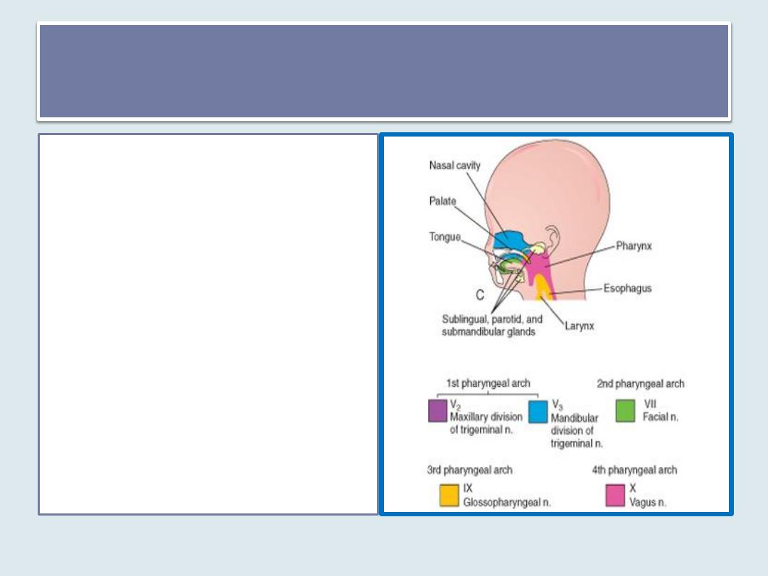
General sensory innervation
•
Since the mucosa covering the body
of the tongue originates from the
first pharyngeal arch, sensory
innervation to this area is by the
mandibular branch of the trigeminal
nerve.
•
The posterior part, or root, of the
tongue originates from the second,
third, and part of the fourth
pharyngeal arch. The fact that
sensory innervation to this part of
the tongue is supplied by the
glossopharyngeal nerve indicates
that tissue of the third arch
overgrows that of the second.
•
The epiglottis and the extreme
posterior part of the tongue are
innervated by the superior laryngeal
nerve, reflecting their development
from the fourth arch.

• the anterior two-thirds of the tongue is
provided by the chorda tympani branch of the
facial nerve.
• while the posterior third is supplied by the
glossopharyngeal nerve.
Special sensory innervation (taste)
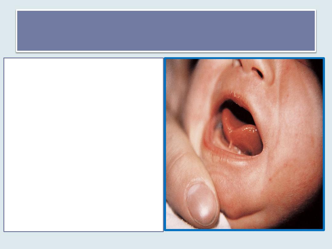
Tongue-tie
• The frenulum is the only
tissue that anchors the tongue
to the floor of the mouth.
• In
ankyloglossia
(tongue-tie)
the tongue is not freed from
the floor of the mouth.
• In the most common form of
ankyloglossia, the frenulum
extends to the tip of the
tongue.
Thyroid Gland
• The thyroid gland
appears as an epithelial
proliferation in the floor
of the pharynx between
the tuberculum impar
and the copula at a point
later indicated by the
Foramen cecum .
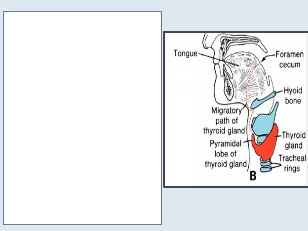
• the thyroid descends in front of
the pharyngeal gut as a bilobed
diverticulum .
• During this migration, the thyroid
remains connected to the tongue
by a narrow canal( thyroglossal
duct). This duct later disappears.
• With further development, the
thyroid gland descends in front
of the hyoid bone and the
laryngeal cartilages. It reaches its
final position in front of the
trachea in the 7
th
week
• The thyroid begins to function at
approximately the end of the 3
rd
month, at which time the first
follicles containing colloid
become visible
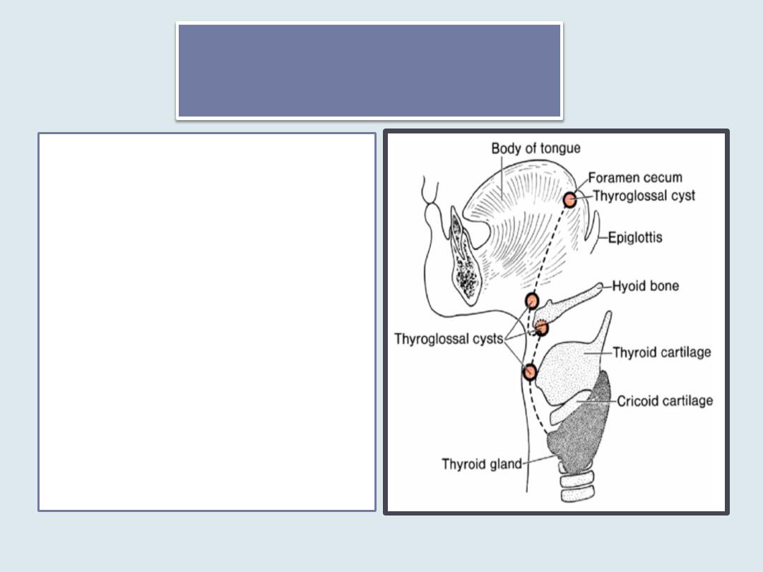
Thyroglossal cyst
• it is a cystic remnant of the
thyroglossal duct,
• may lie at any point along
the migratory pathway of
the thyroid gland but is
always near or in the
midline of the neck.
• approximately 50% of these
cysts are close to or just
inferior to the body of the
hyoid bone
• they may also be found at
the base of the tongue or
close to the thyroid
cartilage.
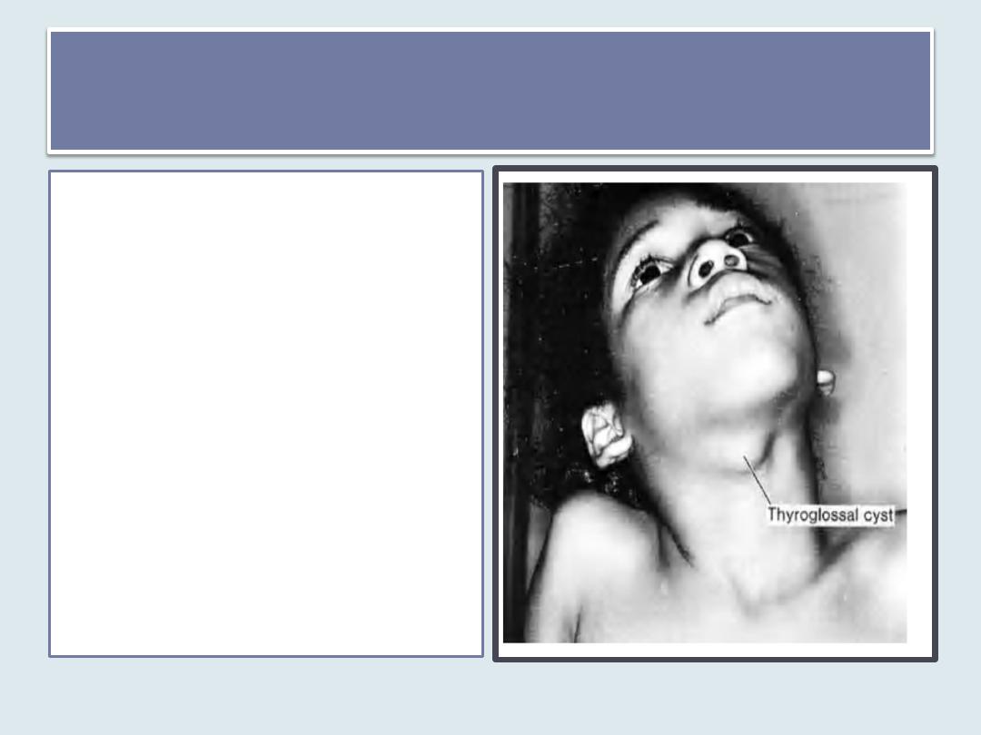
Thyroglossal cyst
•
They are commonly found behind
the arch of the hyoid bone.
•
An important diagnostic
characteristic is their midline
location
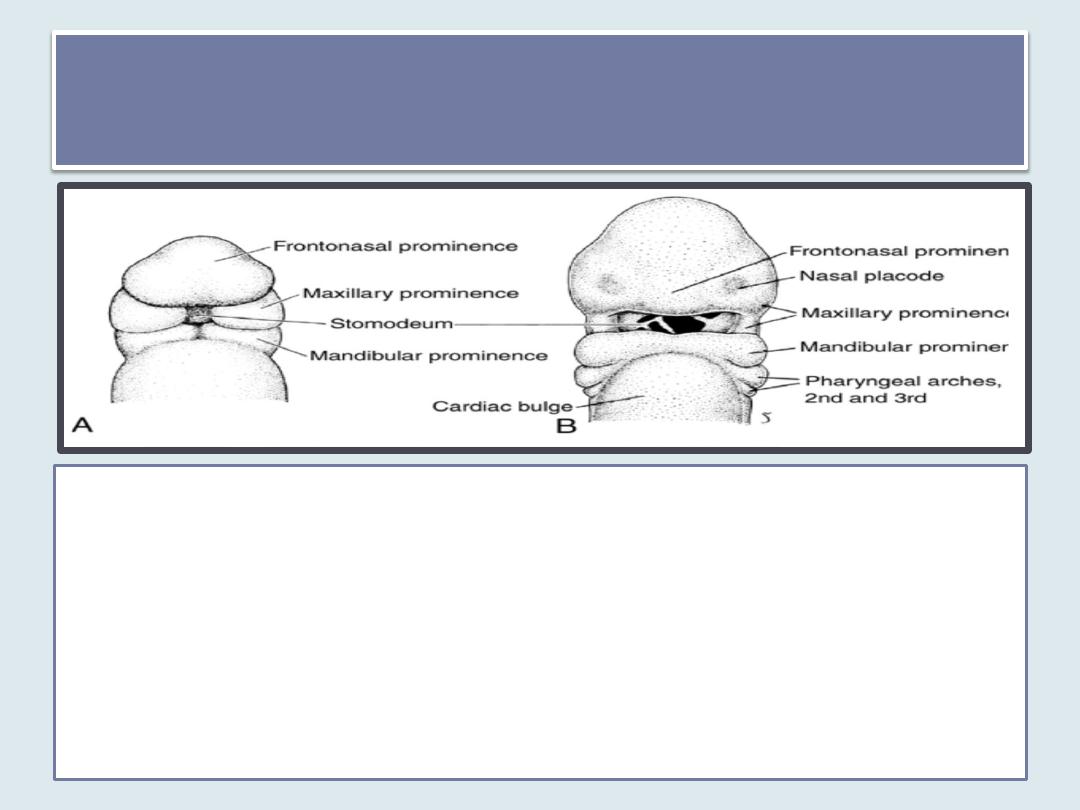
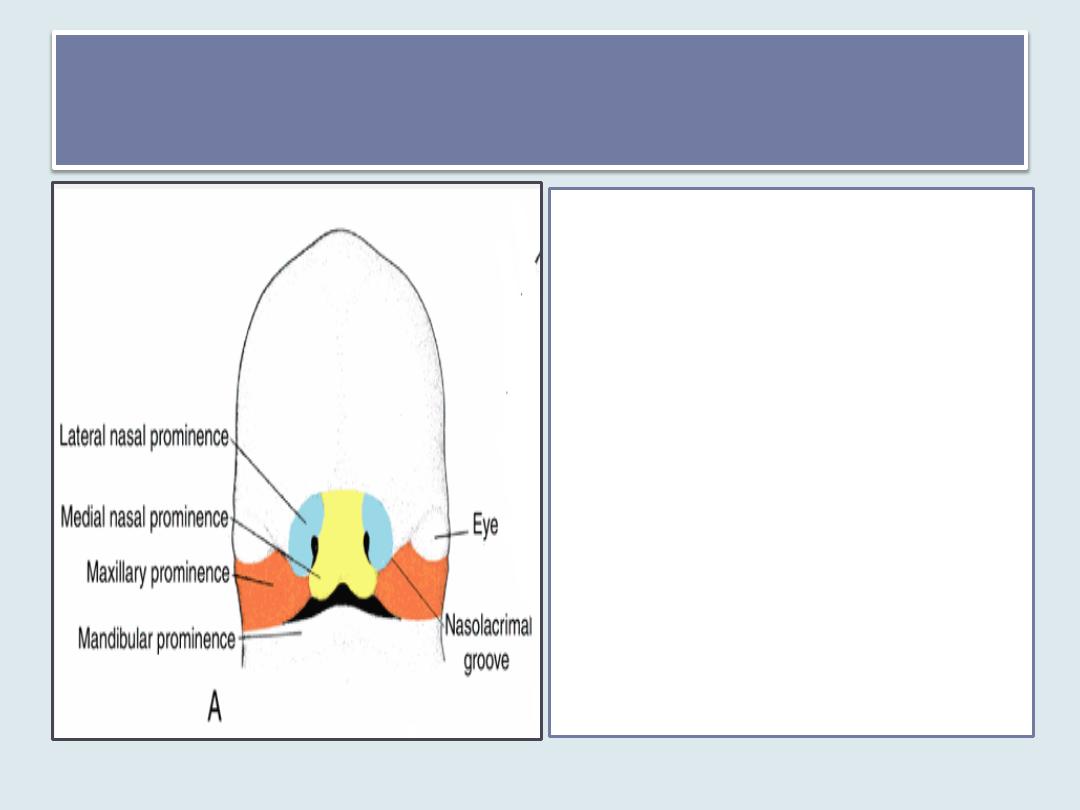
Facial prominences
At the end of the fourth week, facial prominences consisting primarily of
neural crest-derived mesenchyme and formed mainly by the first pair of
pharyngeal arches .
Maxillary prominences can be distinguished lateral to the stomodeum,
Mandibular prominences can be distinguished caudal to this structure
Frontonasal prominence constitutes the upper border of the stomodeum.
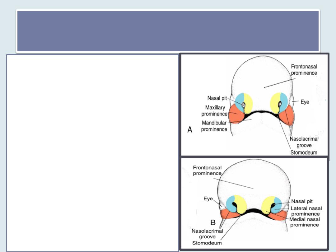
Nasal prominences
• On both sides of the frontonasal
prominence, local thickenings of
the surface ectoderm, the nasal
(olfactory) placodes, originate
under inductive influence of the
ventral portion of the forebrain
• During the fifth week, the nasal
placodes invaginate to form
nasal pits.
• In so doing, they create a ridge of
tissue that surrounds each pit
and forms the nasal
prominences:
1. Lateral nasal prominences on
the outer edge of the pits;
2. Medial nasal prominences on
the inner edge of pits
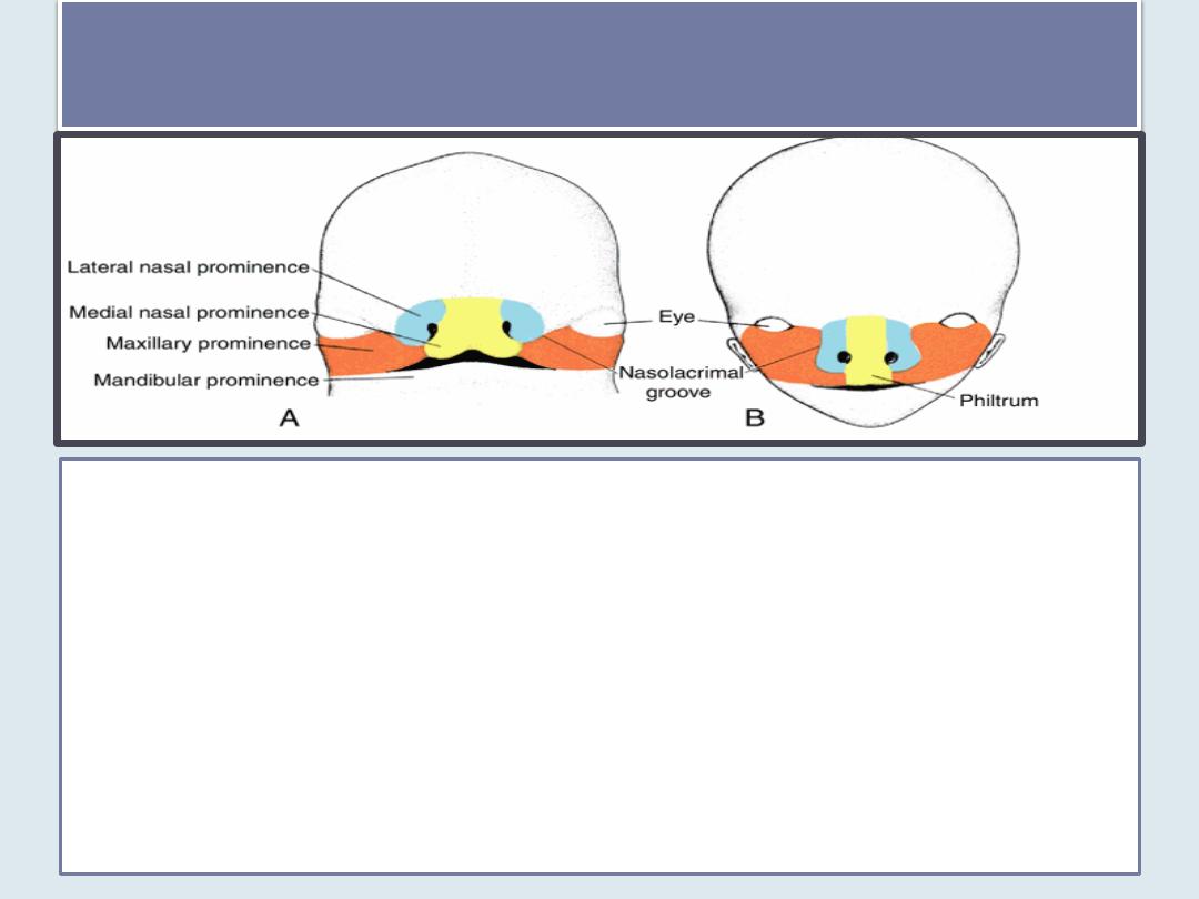
upper lip& lower lip development
During the following 2 weeks, the maxillary prominences grow medially,
compressing the medial nasal prominences toward the midline. Subsequently,
the cleft between the medial nasal prominence and the maxillary prominence
is lost, and the two fuse
• Hence, the upper lip is formed by the
two medial nasal prominences and the
two maxillary prominences.
• The lateral nasal prominences do not participate in formation of the upper
lip
• The lower lip and jaw form from the mandibular prominences that merge
across the midline.
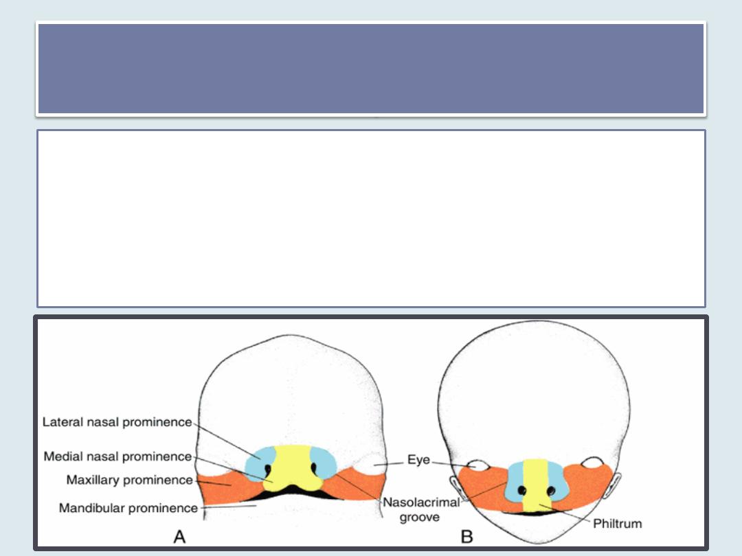
Nasolacrimal duct& cheeks
development
•
Initially, the maxillary and lateral nasal prominences are separated by a deep furrow,
the nasolacrimal groove.
•
Ectoderm in the floor of this groove forms a solid epithelial cord that detaches from
the overlying ectoderm. After canalization, the cord forms the nasolacrimal duct; its
upper end widens to form the lacrimal sac.
•
Following detachment of the cord, the maxillary and lateral nasal prominences merge
with each other.
•
The nasolacrimal duct then runs from the medial corner of the eye to the inferior
meatus of the nasal cavity, and the maxillary prominences enlarge to form the cheeks
and maxillae.
The nose
•
is formed from five facial
prominences:
1. the frontal prominence
gives rise to the bridge;
2. the merged medial nasal
prominences provide the
crest and tip;
3. the lateral nasal
prominences form the
sides (alae)
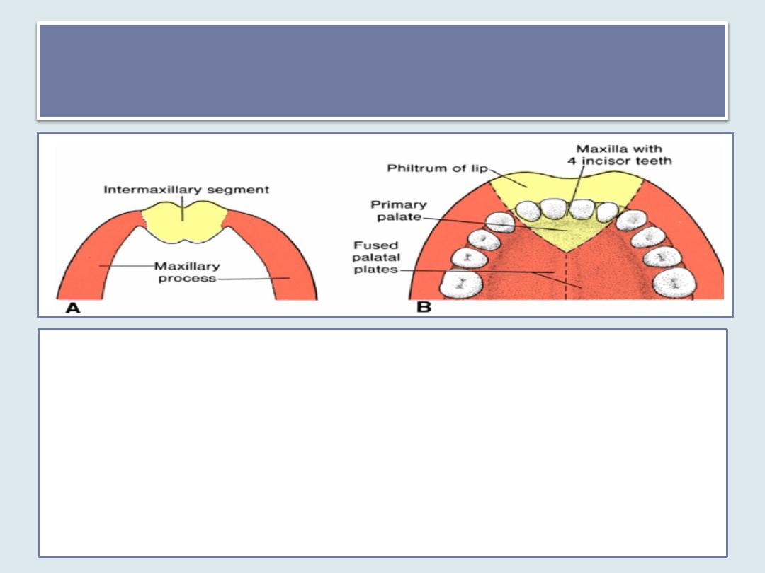
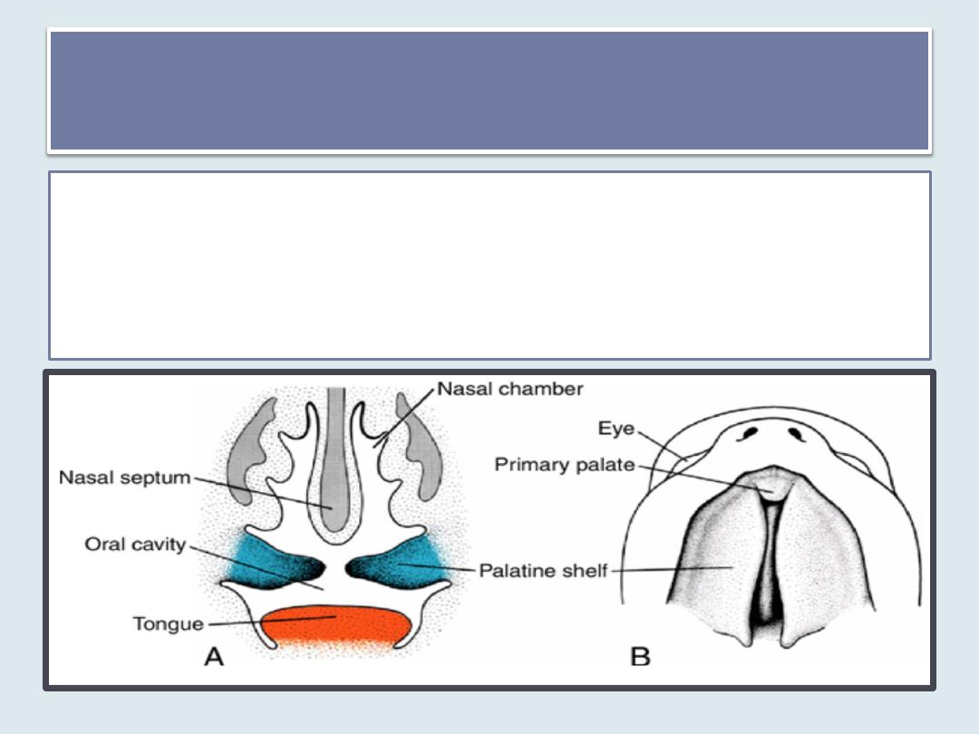
Intermaxillary Segment
• As a result of medial growth of the maxillary prominences, the two medial
nasal prominences merge not only at the surface but also at a deeper
level. The structure formed by the two merged prominences is the
intermaxillary segment. It is composed of
1. a labial component, which forms the philtrum of the upper lip;
2. an upper jaw component, which carries the four incisor teeth; and
3. a palatal component, which forms the triangular primary palate .
• The intermaxillary segment is continuous with the rostral portion of the
nasal septum, which is formed by the frontal prominence.
Secondary Palate
• The main part of the definitive palate is formed by two shelf-
like outgrowths from the maxillary prominences.
• These outgrowths, the palatine shelves, appear in the sixth
week of development and are directed obliquely downward
on each side of the tongue
Secondary Palate
• In the
seventh week
, the palatine shelves ascend to
attain a horizontal position above the tongue and
fuse, forming the Secondary palate
• Anteriorly, the shelves fuse with the triangular primary palate, and
the incisive foramen is the midline landmark between the primary
and secondary palates.
• At the same time as the palatine shelves fuse, the nasal septum
grows down and joins with the cephalic aspect of the newly
formed palate

• Cleft lip and cleft palate are common defects.
• The incisive foramen is considered the
dividing landmark between the anterior and
posterior cleft deformities.
Facial Clefts

Those anterior to the
incisive foramen include
1. lateral cleft lip
2. cleft upper jaw
3. cleft between the primary
and secondary palates .
Such defects are due to
• a partial or
• complete lack of fusion of
the maxillary prominence
with the medial nasal
prominence on one or both
sides.
Anterior cleft deformity
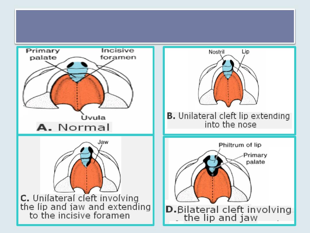
Anterior cleft deformity
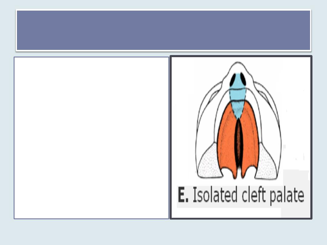
Posterior cleft deformity
Those that lie posterior to
the incisive foramen
include
1. cleft (secondary) palate
2. cleft uvula .
•
Cleft palate results from
a lack of fusion of the
palatine shelves.

The frequency of isolated cleft palate is much lower than that
of cleft lip ,
occurs more often in females (67%) than in males, and
is not related to maternal age.
In females, the palatal shelves fuse approximately 1 week
later than in males. This difference may explain why isolated
cleft palate occurs more frequently in females than in males.
may be caused by hereditary factors and anticonvulsant
drugs
Cleft palate
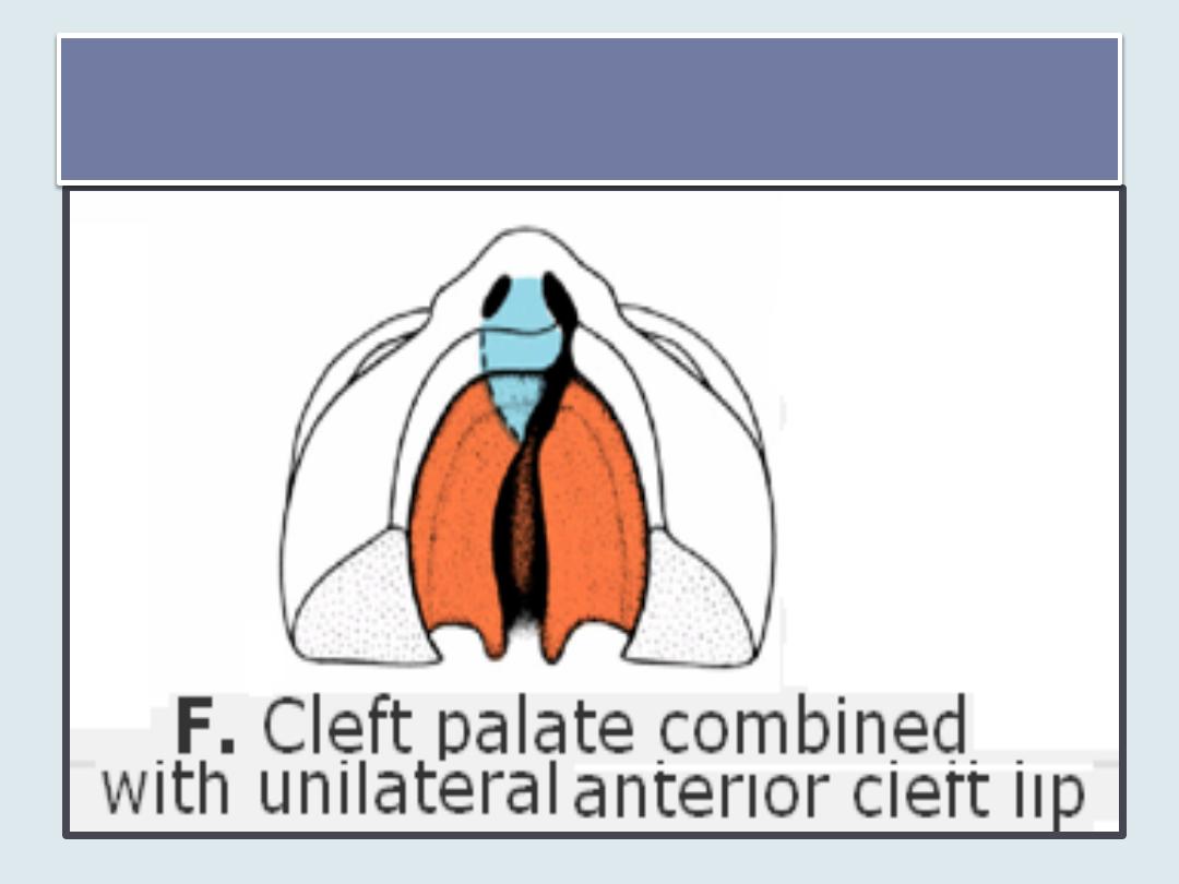
combination of anterior and posterior
clefts
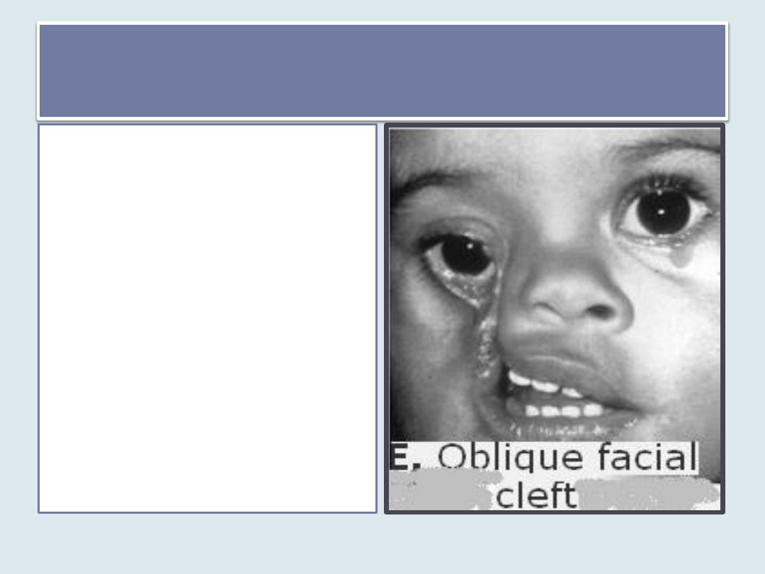
Oblique facial clefts
• are produced by failure
of the maxillary
prominence to merge
with its corresponding
lateral nasal
prominence.
• When this occurs, the
nasolacrimal duct is
usually exposed to the
surface
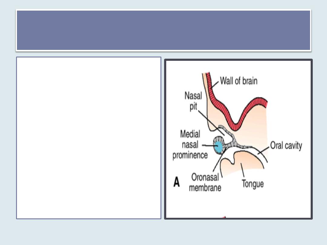
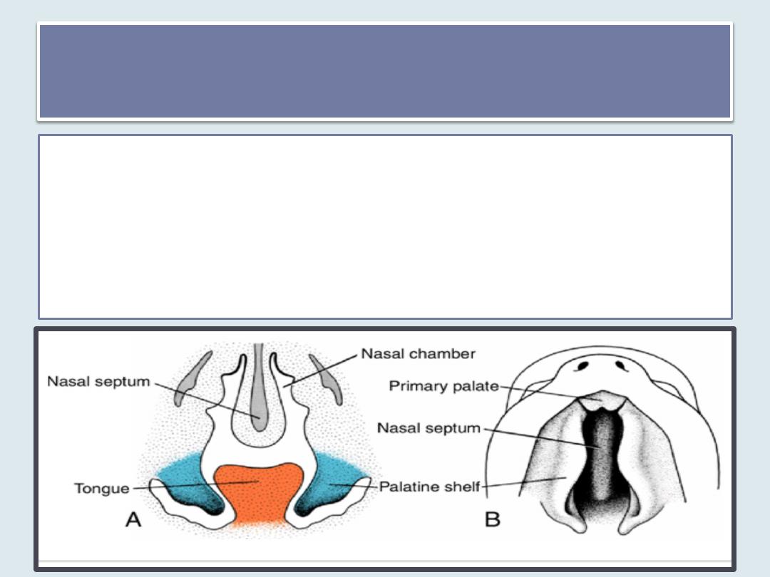
Nasal Cavities
•
During the sixth week, the
nasal pits deepen,
partly because of growth
of the surrounding nasal
prominences and
partly because of their
penetration into the
underlying mesenchyme.
•
At first, the oronasal
membrane separates the
pits from the primitive
oral cavity
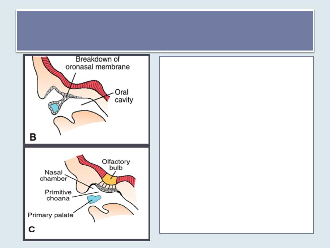
primitive choanae
• Breakdown of oronasal
membrane result in the
formation of the
primitive choanae
• These choanae lie on
each side of the midline
and immediately behind
the primary palate.
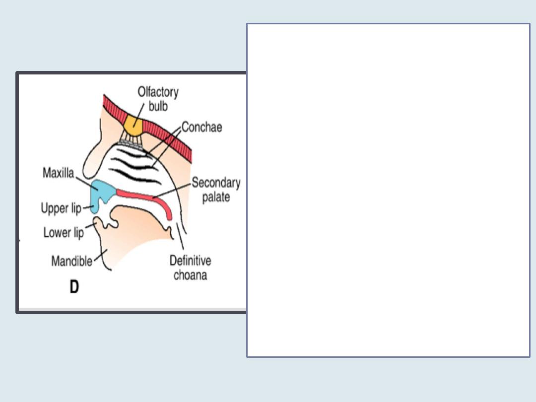
• Later, the definitive choanae
lie at the junction of the
nasal cavity and the pharynx
due to
1. The formation of the
secondary palate and
2. further development of the
primitive nasal chambers ,

• the ectodermal epithelium
in the roof of each nasal
cavity becomes specialized
to form the olfactory
epithelium.
• Some epithelial cells
differentiate into olfactory
receptor cells (neurons).
• The axons of these cells
constitute the olfactory
nerves, which grow into the
olfactory bulbs of the brain
• the superior, middle, and
inferior nasal conchae
develop as elevations of the
lateral walls of the nasal
cavities.

• develop as diverticula of the lateral nasal wall
and extend into the maxilla, ethmoid, frontal,
and sphenoid bones.
• They reach their maximum size during puberty
and contribute to the definitive shape of the
face.
Paranasal air sinuses

is influenced by development of
1. paranasal sinuses
2. nasal conchae
3. teeth .
The adult form of the face

Summary
• Mesenchyme for formation of the head region is derived from paraxial
mesoderm, lateral plate mesoderm, neural crest cells and ectodermal
placodes
• Pharyngeal apparatus consists of pharyngeal arches, pouches, clefts and
membranes
• Pharyngeal arches Contribute mostly to neck development but the first
arch contributes to facial development
• The thyroid gland originates from an epithelial proliferation in the floor of
the tongue and descends to its level in front of the tracheal rings
• The paired maxillary and mandibular prominences and the frontonasal
prominence are the first prominences of the facial region.
• The nasal placodes invaginate to form nasal pits creating the medial and
lateral nasal prominences
• Upper lip, lower lip, cheek, nose, primary palate and secondary palates
derived from these prominences

Thank you
Next Lecture: Urinary System