
Eye
Prof. Dr. Malak A. Al-yawer

Learning objectives
• At the end of this lecture, the medical student will be
able to
• List the embryonic origin of the different components
of the eye
• Describe how the optic vesicle gives rise to retina, iris
and ciliary body
• Describe how the lens originates from lens placode
• State the embryonic origin of choroid, sclera , cornea
and vitreous body
• Describe how optic stalk transforms into optic nerve
• State some clinical correlates
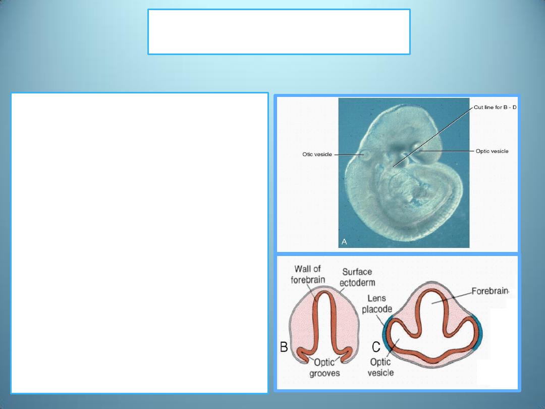
The optic vesicles.
• The developing eye appears in
the 22-day embryo as a pair of
shallow grooves on the sides
of the forebrain.
• With closure of the neural
tube, these grooves form
outpocketings of the
forebrain, the optic vesicles.
• These vesicles subsequently
come in contact with the
surface ectoderm and induce
changes in the ectoderm
necessary for lens formation
.
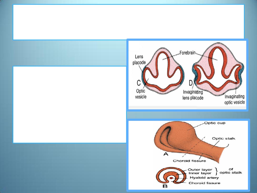
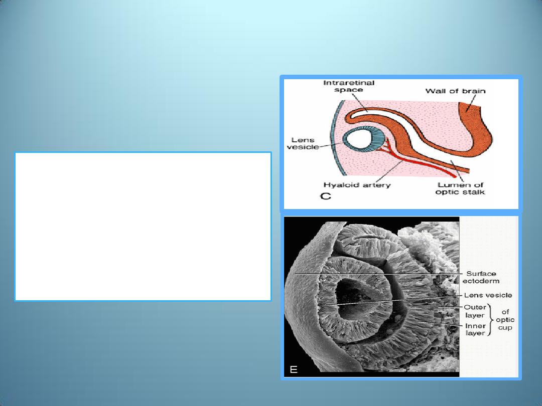
Double-walled optic cup
• Shortly thereafter the
optic vesicle begins to
invaginate and forms
the double-walled optic
cup .
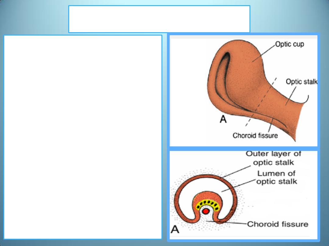
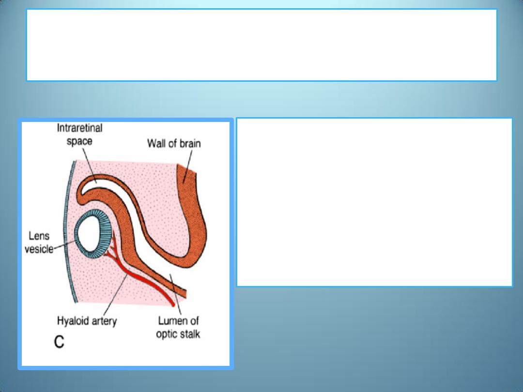
The choroid fissure
•
Invagination is not restricted
to the central portion of the
cup but also involves a part
of the inferior surface that
forms the choroid fissure.
•
Formation of this fissure
allows the hyaloid artery to
reach the inner chamber of
the eye .
•
During the 7
th
week,
1. the lips of the choroid fissure
fuse, and
2. the mouth of the optic cup
becomes a round opening,
the future pupil .
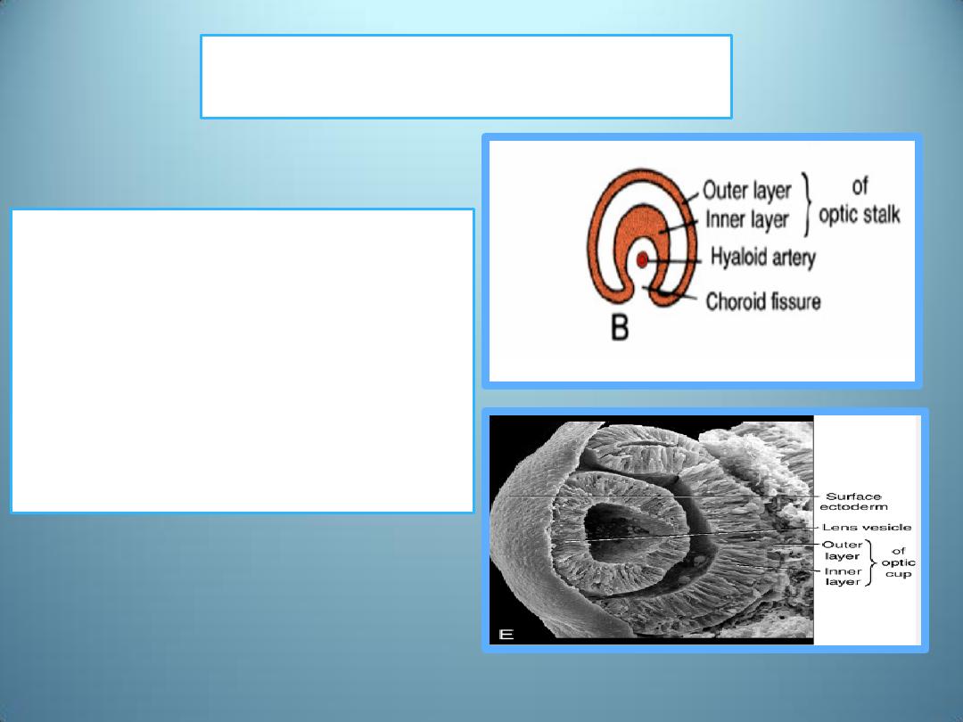
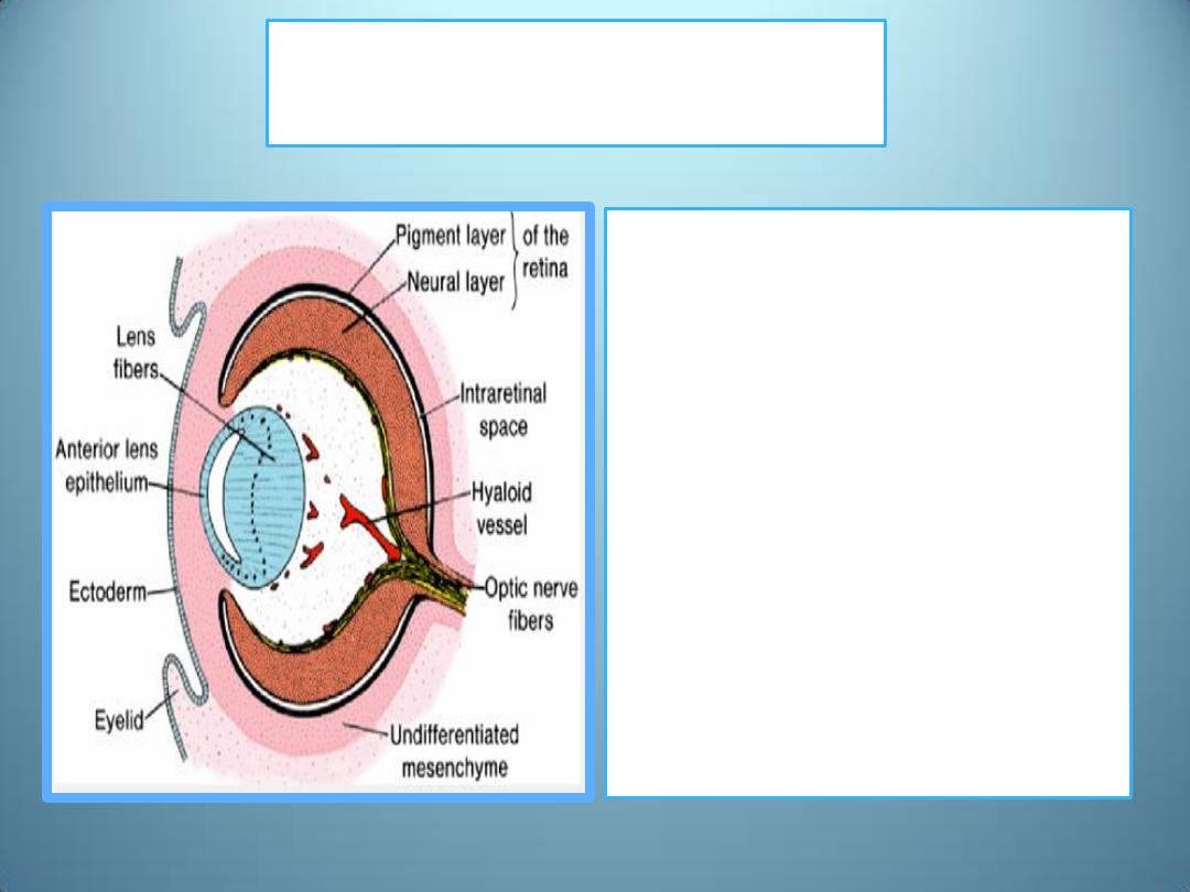
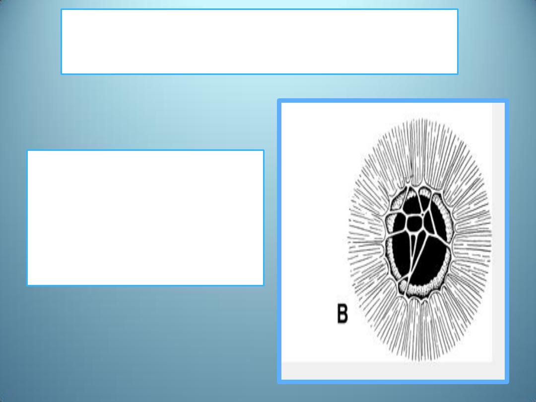
Intraretinal space
• The inner and outer layers of
this cup are initially separated
by a lumen, the intraretinal
space (B)
• but soon this lumen
disappears, and the two layers
appose each other
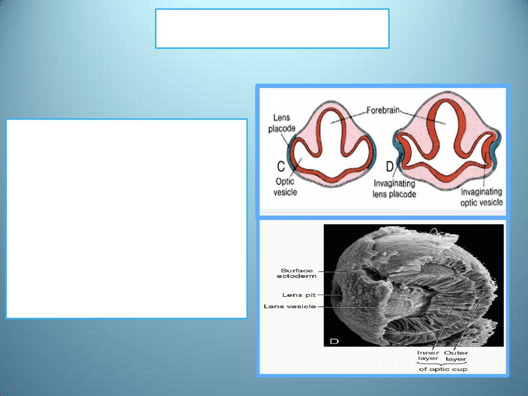
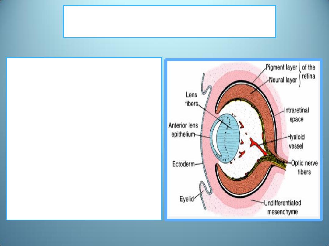
Lens vesicle
• During these events, cells of
the surface ectoderm,
initially in contact with the
optic vesicle, begin to
elongate and form the lens
placode .
• This placode subsequently
invaginates and develops
into the lens vesicle.
• During the 5
th
week, the
lens vesicle loses contact
with the surface
ectoderm and lies in the
mouth of the optic cup
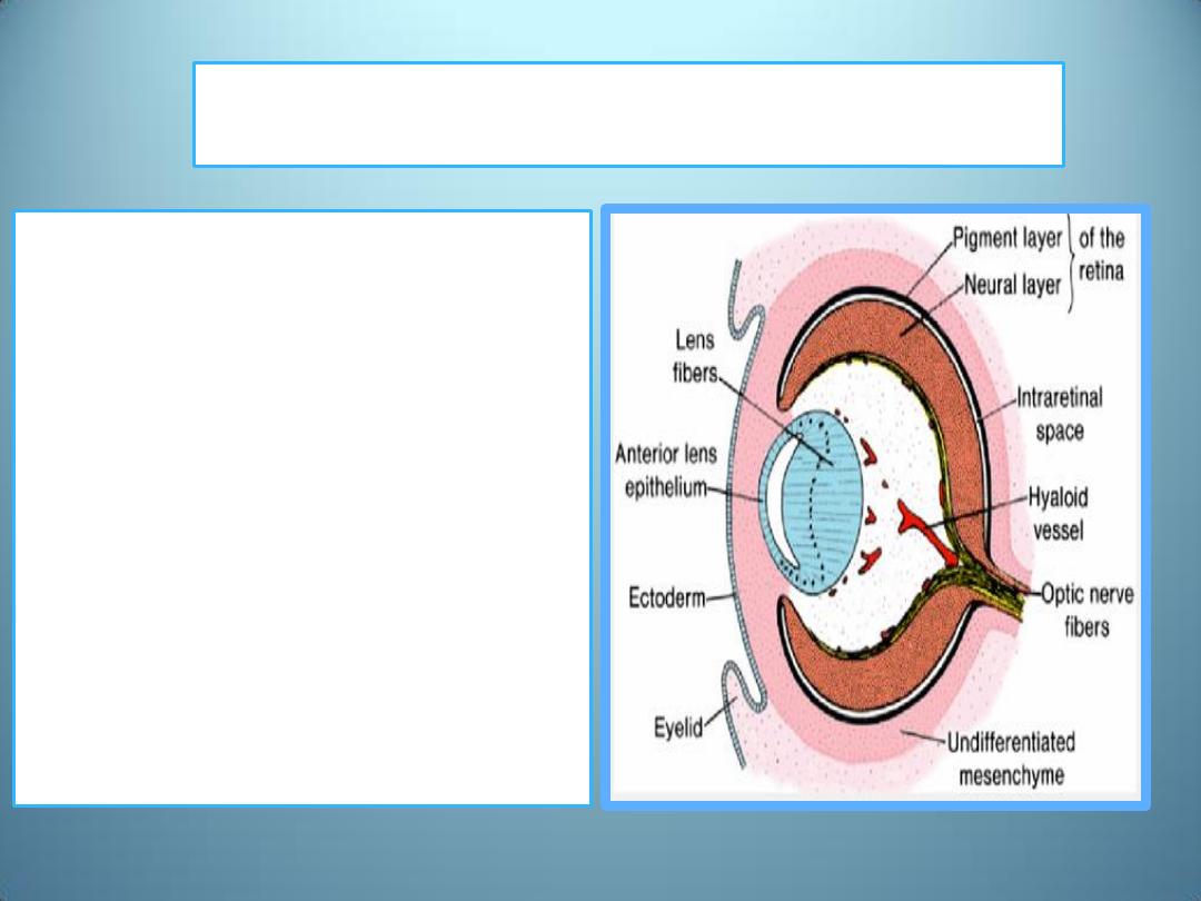
Pigmented
layer of the retina
• The outer layer of the
optic cup, which is
characterized by small
pigment granules, is
known as the pigmented
layer of the retina.

Neural layer of the optic cup
•
Development of the inner (neural) layer of the
optic cup is more complicated.
1. Pars optica retinae :The posterior four-fifths of
the neural layer (the photoreceptive layer,
mantle layer, fibrous layer)
2. pars ceca retinae : It is the anterior fifth of the
inner layer. It divides into pars iridica retinae
retina which forms the inner layer of the iris
and pars ciliaris retinae which participates in
formation of the ciliary body.
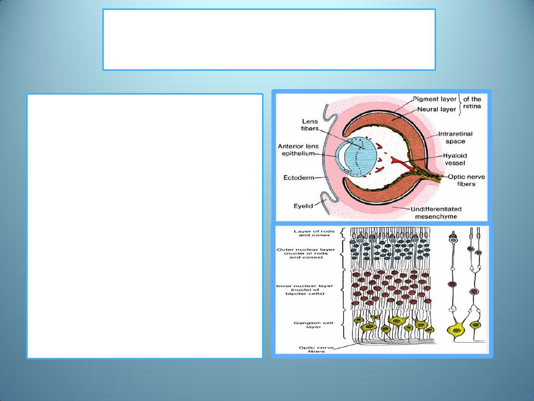
Pars optica retinae
1.
photoreceptive layer
• contains cells bordering
the intraretinal space
that differentiate into
light-receptive
elements ,rods and
cones
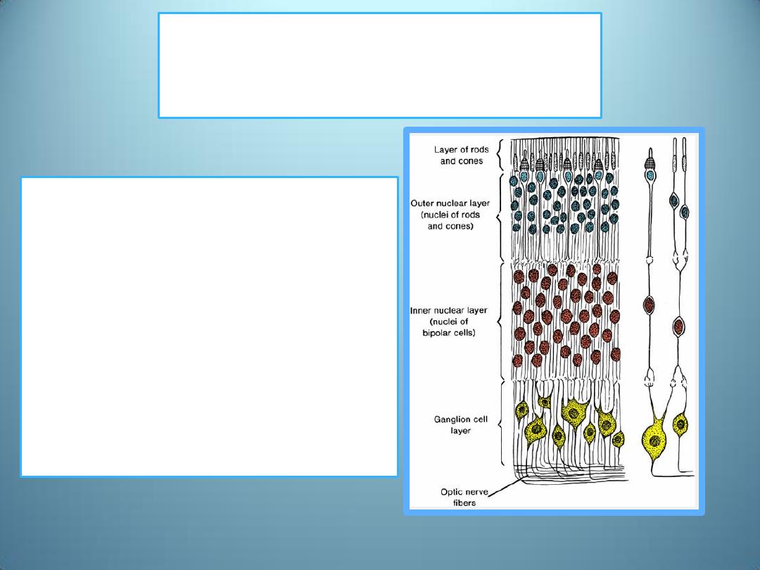
Pars optica retinae
2.
the mantle layer
•
Adjacent to this
photoreceptive layer is the
mantle layer, which, as in the
brain, gives rise to neurons
and supporting cells, including
1. the outer nuclear layer
2. inner nuclear layer
3. ganglion cell layer
Pars optica retinae
3. Fibrous layer
• On the surface is a fibrous
layer that contains axons of
nerve cells of the deeper
layers.
• Nerve fibers in this zone
converge toward the optic
stalk, which develops into
the optic nerve .
• Hence, light impulses pass
through most layers of the
retina before they reach the
rods and cones .
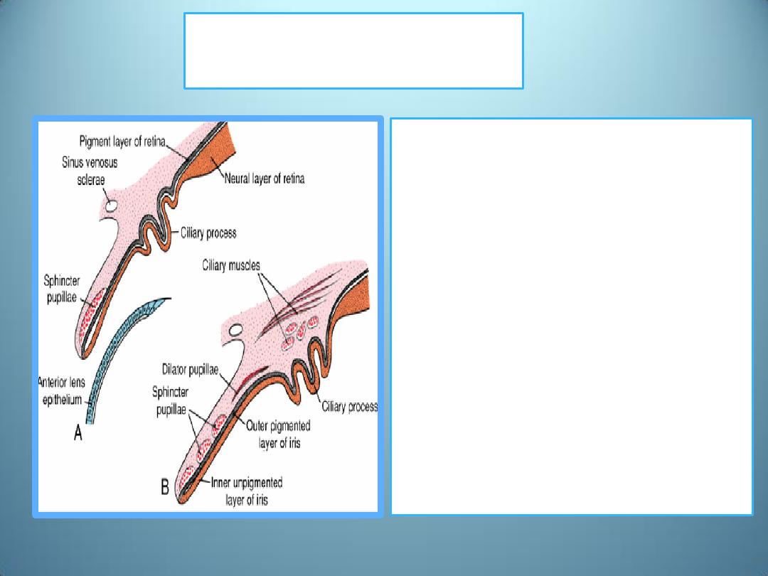
The iris
•
In the adult, is formed by
1. the pigment-containing
external layer,
2. the unpigmented internal
layer of the optic cup, and
3. a layer of richly vascularized
connective tissue that
contains the pupillary
muscles (the sphincter and
dilator pupillae muscles
develop from the underlying
ectoderm of the optic cup).
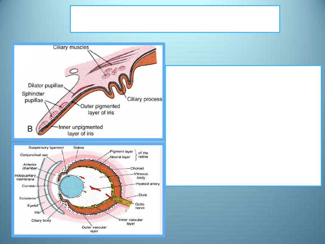
The pars ciliaris retinae
• is easily recognized by its
marked folding and
• Externally it is covered by a
layer of mesenchyme that
forms the ciliary muscle ;
• on the inside it is connected to
the lens by a network of elastic
fibers, the suspensory
ligament or zonula
Lens
Shortly after formation of the lens vesicle
•
Cells of the posterior wall
1. begin to elongate anteriorly
and
2. form long fibers that
gradually fill the lumen of
the vesicle
Growth of the lens
• By the end of the 7
th
week,
these primary lens fibers
reach the anterior wall of the
lens vesicle.
• Growth of the lens is not
finished at this stage since
new (secondary) lens fibers
are continuously added to the
central core.
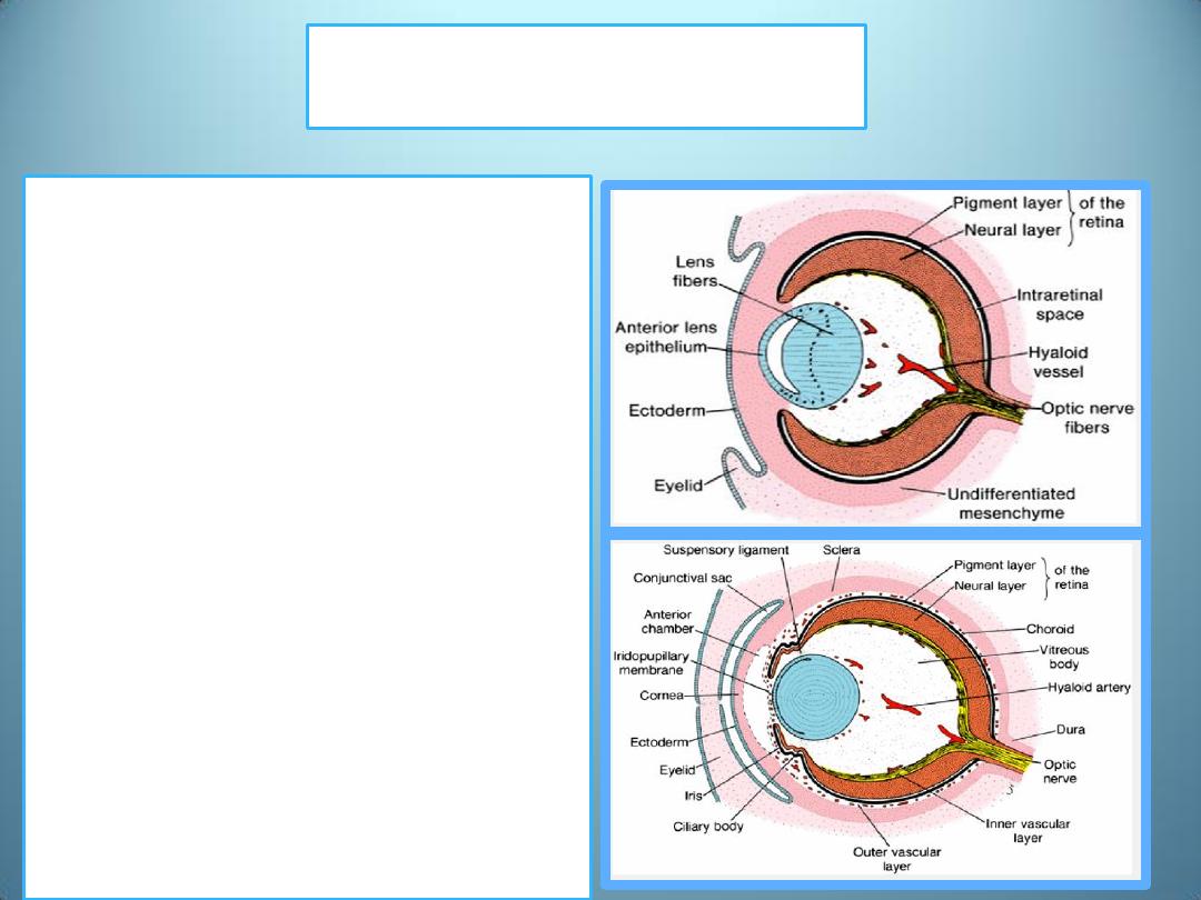
Choroid and Sclera
• At the end of the 5
th
week,
the eye primordium is
completely surrounded by
loose mesenchyme.
• This tissue soon differentiates
into
1. an inner layer comparable
with the pia mater of the
brain ;it later forms a highly
vascularized pigmented layer
known as the choroid
2. an outer layer comparable
with the dura mater; develops
into the sclera and is
continuous with the dura
mater around the optic nerve.
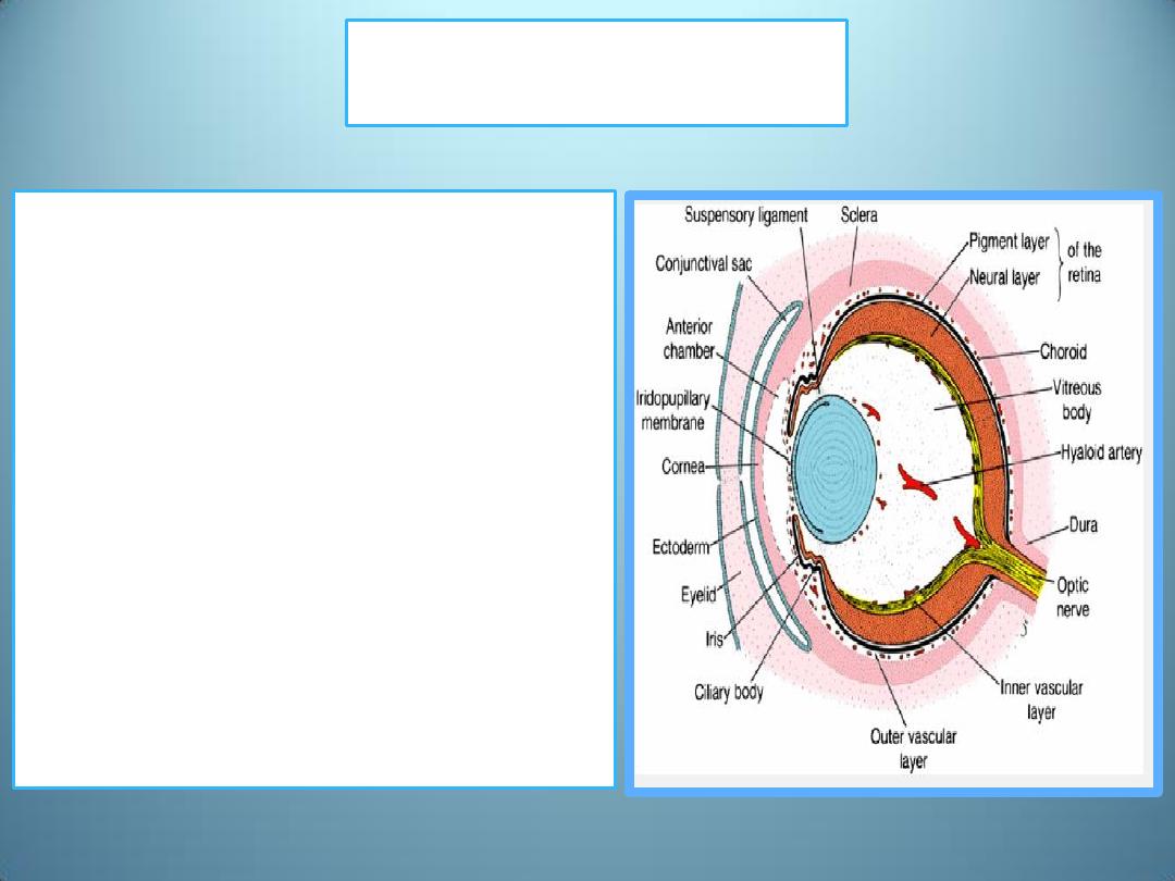
Cornea
Differentiation of mesenchymal
layers overlying the anterior aspect
of the eye is different
The anterior chamber forms
through vacuolization and splits the
mesenchyme into
1.
an inner layer in front of the lens
and iris, the iridopupillary
membrane ,and
2.
an outer layer continuous with
the sclera, the substantia propria
of the cornea
The anterior chamber itself is lined
by flattened mesenchymal cells.
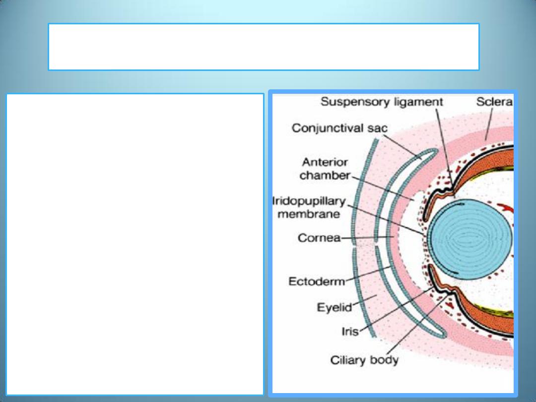
Hence, the cornea is formed by:
(a) an epithelial layer derived
from the surface ectoderm ,
)b) the substantia propria or
Stroma ,which is continuous with
the sclera, and
(c) an epithelial layer, which
borders the anterior chamber.
• The iridopupillary membrane
in front of the lens disappears
completely, providing
communication between the
anterior and posterior eye
chambers.
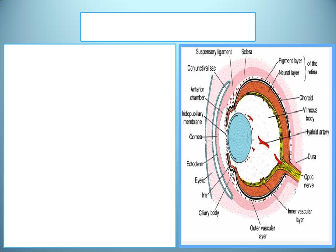
Vitreous Body
Mesenchyme not only surrounds the
eye primordium from the outside but
also invades the inside of the optic
cup by way of the choroid fissure.
Here it forms
1. the hyaloid vessels, which during
intrauterine life supply the lens and
form the vascular layer on the inner
surface of the retina
2. a delicate network of fibers
between the lens and retina.
The interstitial spaces of this network
later fill with a transparent gelatinous
substance, forming the vitreous body
The hyaloid vessels in this region are
obliterated and disappear during fetal
life, leaving behind the hyaloid canal.
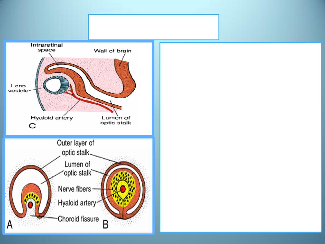
Optic Nerve
• The optic cup is connected to
the brain by the optic stalk,
which has a groove, the
choroid fissure ,on its ventral
surface.In this groove are the
hyaloid vessels.
• The nerve fibers of the retina
returning to the brain lie
among cells of the inner wall
of the stalk.
• During the 7
th
week, the
choroid fissure closes, and a
narrow tunnel forms inside
the optic stalk.
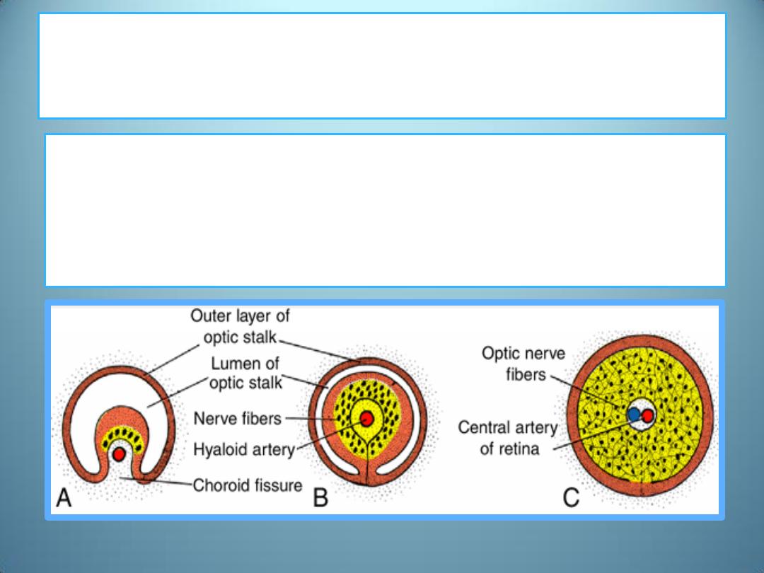
As a result of the continuously increasing number of
nerve fibers
,
• the inner wall of the stalk grows, and
• the inside and outside walls of the stalk fuse.
• cells of the inner layer provide a network of neuroglia that
support the optic nerve fibers.
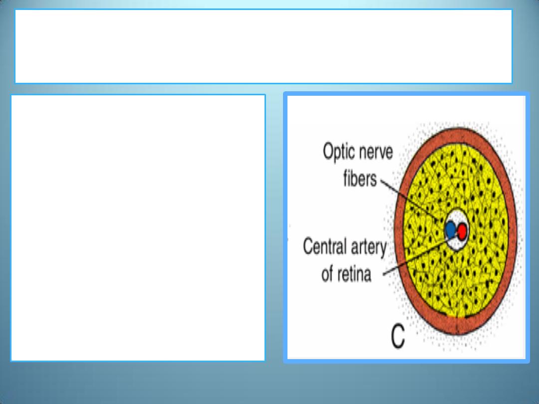
The optic stalk is thus transformed into the
optic nerve
• Its center contains a portion
of the hyaloid artery, later
called the central artery of
the retina.
• On the outside, a
continuation of the choroid
and sclera, the pia, arachnoid
and dura layer of the nerve,
respectively, surround the
optic nerve.
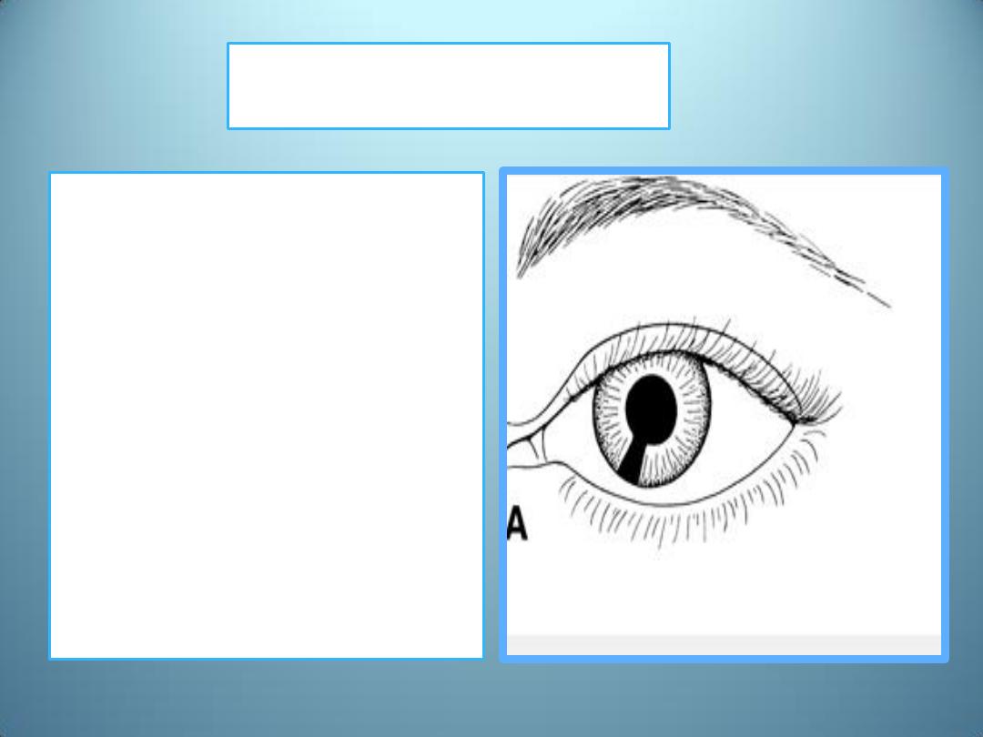
Coloboma
• may occur if the choroid
fissure fails to close.
• Although such a cleft is
usually in the iris only—
coloboma iridis—it may
extend into the ciliary body,
the retina, the choroid, and
the optic nerve.
• Coloboma is a common eye
abnormality frequently
associated with other eye
defects.
• Colobomas (clefts) of the
eyelids may also occur.
The iridopupillary membrane
• may persist instead of
being resorbed during
formation of the
anterior chamber.

Congenital cataracts
the lens becomes opaque
during intrauterine life.
this anomaly is usually
genetically determined
many children of mothers
who have had German
measles (rubella) between
the fourth and seventh
weeks of pregnancy have
cataracts.
If the mother is infected
after the seventh week of
pregnancy, the lens escapes
damage, but the child may
be deaf as a result of
abnormalities of the
cochlea.

The hyaloid artery
• may persist to form a cord or
cyst.
• Normally, the distal portion of
this vessel degenerates,
leaving the proximal part to
form the central artery of the
retina .
Congenital aphakia
(absence of the lens) and
• )
aniridia
( absence of the
iris)
are rare anomalies

Microphthalmia
the eye is too small;
• the eyeball may be only
two-thirds of its normal
volume.
• Usually associated with
other ocular abnormalities,
• microphthalmia frequently
results from intrauterine
infections such as
cytomegalovirus and
toxoplasmosis.
Anophthalmia
• is absence of the eye.
• In some cases, histological
analysis reveals some ocular
tissue.
• The defect is usually
accompanied by severe
cranial abnormalities.

Cyclopia & synophthalmia
•
Cyclopia single eye and synophthalmia fusion of
the eyes
•
comprise a spectrum of defects in which the eyes
are partially or completely fused
•
The defects are due to a loss of midline tissue that
may occur
1. as early as days 19 to 21 of gestation or
2. at later stages when facial development is initiated .

Summary
• Optic vesicles are derived from a pair of shallow grooves on each side of
the forebrain at the end of the 4
th
week of development
• The optic vesicles come in contact with the surface ectoderm and induce
lens formation.
• The optic vesicle begins to invaginate to form doubled wall optic cup
• the pigment and neural layers of the retina are derived from the outer and
inner layers of the optic cup respectively
• Mesenchyme surrounding eye primordium differentiate into choroid and
sclera
• As a result of anterior chamber formation, mesenchyme infront the eye
differentiate into iridopupillary membrane and substantia propria of the
cornea
• Mesenchyme inside the eye differentiate into choroid vessels and vitreous
body
• Optic nerve is derived from optic stalk