Histology
Lec.1 Dr. Faraid
THE DIGESTIVE SYSTEM
The digestive system consists of the digestive tract oral cavity,
esophagus, stomach, small and large intestines, rectum, and anus” and
its associated glands ”salivary glands, liver, and pancreas”.
The first step in the complex process known as digestion occurs in the
mouth, where food is moistened by saliva and ground by the teeth into
smaller pieces; saliva also initiates the digestion of carbohydrates.
Digestion continues in the stomach and small intestine, where the food
”transformed into its basic components (eg , amino acids,
monosaccharides, free fatty acids, monoglycerides)”is absorbed. Water
absorption occurs in the large intestine, causing the undigested
contents to become semisolid.
THE ORAL CAVITY
The oral cavity is lined with a protective, stratified squamous
epithelium, keratinized or nonkeratinized depending on the region. The

keratin layer protetcts the oral mucosa from damage and is present
mostly in the hard palate and gingiva (gum). Nonkeratinized squamous
epithelium covers the soft palate, lips, cheeks, and the floor of the
mouth.
The oral cavity is divided into:
1- The vestibule: is just internal to the lips and cheeks, extending as
far as the teeth.
2- The oral cavity proper: is behind the teeth, having a roof
comprised of the hard and soft palates and a floor from which the
tongue projects.
Posteriorly, the oral cavity opens into the oropharynx.
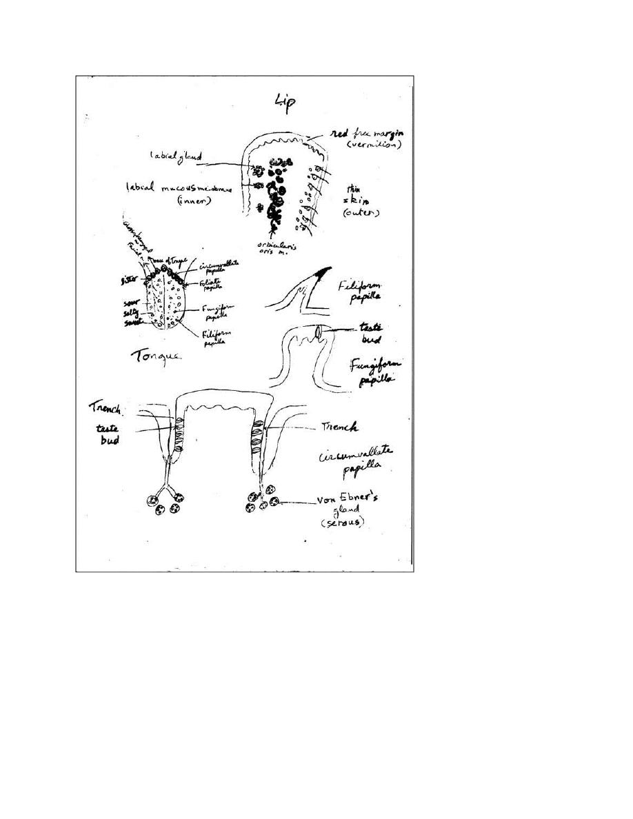
The lips
:
The outer surface of each lip is covered with skin that contains hair
follicles, sebaceous glands, and sweat glands.
The red free margin (vermilion) of the lip is covered with a modified
skin which represents a transition from skin to mucous membrane. The
connective tissue (c.t.) papillae of the dermis beneath it are numerous,
high and vascular and as a result, the blood in their capillaries readily
shows through the transparent epidermis to make the lips appear red.
The inner surface of the lip is lined by mucous membrane. The
epithelium of this surface is thicker than the epidermis covering the
outer surface of the lip and is of the stratified squamous non-
keratinized type. High papillae of the c.t. lamina propria extend into it.
Small clusters of labial glands are embedded in the lamina propria and
connected with the surface by means of little ducts.

The substance of the lips consists of striated muscle fibers (the
orbicularis oris muscle).
The tongue
:
The tongue is a mass of striated muscle covered by mucous
membrane .The muscle is arranged in bundles running in the vertical,
transverse and longitudinal directions, and crossing one another at
right angles. This arrangement gives the tongue a great mobility. The
mucous membrane consists of a thick stratified squamous epithelium
and underlying lamina propria containing many blood vessels,
lymphatics and nerve fibers. The mucous membrane on the lower
surface of the tongue is smooth, whereas its dorsal surface is irregular,
and covered anteriorly with numerous epithelial elevations called
papillae. The posterior one-third of the dorsal surface is separated from
anterior two-thirds by a V-shape line (Sulcus Terminalis). Behind this V-
line, the dorsal surface present eminences composed mainly of
aggregations of lymphatic nodules in the lamina propria beneath the
epithelium (Lingual tonsils).
Papillae: are elevations of the oral epithelium and lamina propria (are
mucosal projections).
Types of lingual papillae:
1-Filiform papillae: they have an elongated, conical shape. They are the
most numerous and distributed over the dorsum of the anterior two
thirds of the tongue. Each papilla has a thin core of c.t. lamina propria
and is covered by a pointed cap of stratified squamous epithelium

which is cornified (keratinized). These papillae do not contain taste
buds.
2-Fungiform papillae: are fewer and larger than the filiform and are
scattered irregularly among them. They have narrow stalk (base) and a
dilated upper part with smooth surface in the shape of mushroom.
They frequently contain taste buds on their upper surfaces, and they
are very vascular.
3-Circumvallate papillae: are the largest and least numerous; only 7 to
12 in number and are arranged along the V-line. They are large circular
papillae whose flattened surfaces extend above the other papillae.
They are surrounded by a deep groove (trench).They have many taste
buds on their lateral surfaces. The ducts of von Ebner's glands (serous
gland) open into the bottom of trench and maintain a continuous flow
of fluid over the taste buds and this is important in removing food
particles from the vicinity of the taste buds so that, they can receive
and process new gustatory stimuli. Von Ebner's glands secrete a lipase
that probably prevents the formation of hydrophobic layer over the
taste buds that would hinder their function. Lingual lipase is active in
the stomach and can digest up to 30% of dietary triglyceride.
4-Foliate papillae: are rudimentary in human .They consist of two or
more parallel ridges and furrows. They are located on the dorsolateral
surface of the tongue. Ducts from serous glands drain into the bases of
the furrows. Numerous taste buds are present in the walls of the
furrows.

Taste Buds:-
They are specialized sensory receptors (chemoreceptors). They are
located in the epithelium of the tongue (on the apical surfaces of
fungiform papillae and on the lateral surfaces of circumvallate and
foliate papillae). They are also present on the soft palate, pharynx &
epiglottis. The taste buds are oval in shape, pale in staining (as
compared to surrounding epithelium) & they are intra-epithelial &
traverse the whole thickness of epithelium (extend from basement
membrane to the surface). At the apical part, it opens in a very
small/minute opening called ″Taste pore″ or ″Gustatory pore″. There
are 50-100 cells in each bud. The following types of cells can be
distinguished in the taste bud:
1- Supporting cells
Their function is supporting for taste buds. They are slender cells
extending from the basal lamina to the taste pore where they have
numerous microvilli. They are dark because they contain large number
of fine filaments in the cytoplasm. Also they have granules in the apical
part contain glycosaminoglycans.
2-Taste cells (gustatory cells)
They are light because their cytoplasm contains few filaments. Also
the cells have apical microvilli. They are characterized by the presence
of numerous synaptic vesicles in their basal cytoplasm. Dendritic
processes of sensory nerves are found in close proximity to these
vesicles.

3-Undifferentiated basal cells
They are found in the base of the taste buds. They are probably the
precursor of the other cells.
Turnover of these cells is rapid, every 10-14 days.
Lingual glands:-
are glands of the tongue (minor salivary glands).
They can be divided into 3 main groups according to their structure &
location.
1-A paired group of mixed mucous & serous glands:- are located in
the anterior part of the tongue near the apex. They are embedded in
the muscle but are closer to the ventral than to the dorsal surface; they
have several ducts which open on the ventral surface.
2- Von Ebner’s glands:- are located in the region of the circumvallate
papillae. They are pure serous glands. Their ducts open into the
trenches of circumvallate papillae.
ال
3-Mucous glands of the root of the tongue:- are the most numerous.
They lie in the posterior third of the tongue. Their ducts open into the
crypts of lingual tonsils and into the depressions between the tonsils.
Teeth:-
In adult human, the 32 permanent teeth are disposed in two
bilaterally symmetric arches in the maxillary & mandibular bones. There
are 8 teeth in each quadrant: 2 incisors, 1 canine, 2 premolars & 3
permanent molars. Twenty of the permanent teeth are preceded by

deciduous (baby) teeth. The permanent molars have no deciduous
precursors.
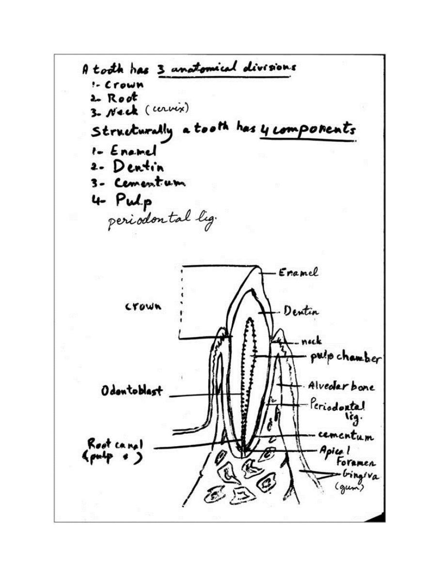
A tooth has 3 anatomical divisions:-
1. Crown: - the portion that projects above the gingiva (gum).
2. Root(s):- one or more below gingiva that hold the teeth in bony
sockets called alveoli, one for each tooth.
3. Neck (cervix):- where the crown & the root meet.
Structurally, a tooth has the following components:-
1. Enamel:- is the hardest substance in the body. It consists of about
95% ca salts (mainly hydroxyapatite). Structurally it is composed
of enamel rods (or prisms) that are bound together by interrod
enamel. Both interrod enamel & enamel rods are formed of
hydroxyapatite crystals; they differ only in the orientation of the
crystals. Each rod extends through the entire thickness of the
enamel layer. Enamel is produced by cells of ectodermal origin
(ameloblasts), whereas most of the other structures of the teeth
derive from mesodermal or neutral crest cells. Ameloblasts secrete
the enamel matrix. Ameloblasts degenerate when the tooth erupts,
after which time the enamel cannot be replaced by new synthesis.
The enamel is acellular after tooth eruption & therefore cannot
repair itself.
2. Dentin
(
e
)
:- is a calcified tissue similar to bone but harder because
of its higher content of ca salts (70% of dry weight) in the form of
crystals of hydroxyapatite. It forms the bulk of the tooth & gives the
main strength to it. It differs from bone in that it contains no cells &

no lacunae but has only processes of cells (odontoblasts) whose
bodies lie adjacent to the dentin in the pulp cavity. Odontoblasts
have slender branched cytoplasmic extensions that penetrate
perpendicularly through the width of the dentin. These odontoblasts
processes are present in small canals called dentinal tubules.
Odontoblasts produce the organic matrix of dentin only at the
dentinal surface. Dentin, unlike enamel, forms throughout the life. In
contrast to bone, dentin persists as a mineralized tissue for a long
time after destruction of the odontoblasts.
3. Cementum:- is a bone-like tissue secreted by cells of the periodontal
ligament which lines the tooth socket. The cementum forms a
protective covering over the dentin & serves to attach the tooth to the
surrounding structures. This tissue covers the dentin of the root & is
similar in composition to bone. It is thicker in the apical region of the
root & in this area; there are cells with the appearance of the
osteocytes, the cementocytes. Like osteocytes, they are encased in
lacunae; unlike those cells, however, cementocytes do not
communicate through canaliculi; & their nourishment comes from the
periodontal ligament. Like bone tissue, cementum is labile & reacts by
resorption or production of a new tissue according to the stresses to
which it is subjected. Continuous production of the cementum in the
apex compensates for the physiologic wear of the teeth & maintains
close contact between the roots of the teeth & their sockets. When the
periodontal ligament is destroyed the cementum undergoes necrosis &
may be resorbed.

4. Pulp:- the pulp fills the pulp cavity. The shape of the pulp cavity is
quite similar to that of the tooth in which it occurs. It consists of an
expanded pulp chamber & a narrow pulp canal or root canal in the
each root. A root canal communicates with periodontal tissues through
the apical foramen. The dental pulp is essential to the nourishment &
vitality of the tooth. Dental pulp consists of a loose c.t. Its main
components are odontoblasts, fibroblast, macrophages, thin collagen
fibrils & a ground substance containing glycosaminoglycans. Pulp is a
highly innervated & vascularized tissue. It contains both myelinated
& unmyelinated nerve fibers.
5. Periodontal ligament:- is composed of a special type of dense c.t.
whose fibers penetrate the cementum of the tooth & bind it to the
bony walls of the socket, permitting limited movement of the tooth. It
serves as the periosteum of the alveolar bone. In the periodontal
ligament, there are fibroblasts, osteoblasts & cementoblasts. It has a
high protein turnover rate & high rate of the collagen renewal. Protein
or vitamin C deficiency (scurvy) may cause atrophy of this ligament,
resulting in the loosening or loss of teeth.
Periodontium:-
The periodontium comprises the structures responsible for
maintaining the teeth in the maxillary & mandibular bone.
It consists of:-
1- Cementum

2- Periodontal ligament (membrane)
3- Alveolar bone
4-Gingiva (gum)
Alveolar bone:- this portion of bone is in immediate contact with the
periodontal ligament. It is an immature type of bone
(primary=woven bone) in which the collagen fibers are not arranged
in the typical lamellar pattern of adult bone. Many of the collagen
fibers of the periodontal ligament are arranged in bundles that
penetrate this bone & the cementum, forming a connecting bridge
between these structures.
Gingiva(gum):- is a mucous membrane firmly bound to the
periosteum of the maxillary or mandibular bone. It is composed of
stratified squamous epithelium & lamina propria containing
numerous connective tissue papillae.
Surface of the tongue on the region close to its V-shaped boundary,
between the anterior and posterior portions. Note the lymphoid
nodules (lingual tonsil), glands, and papillae.
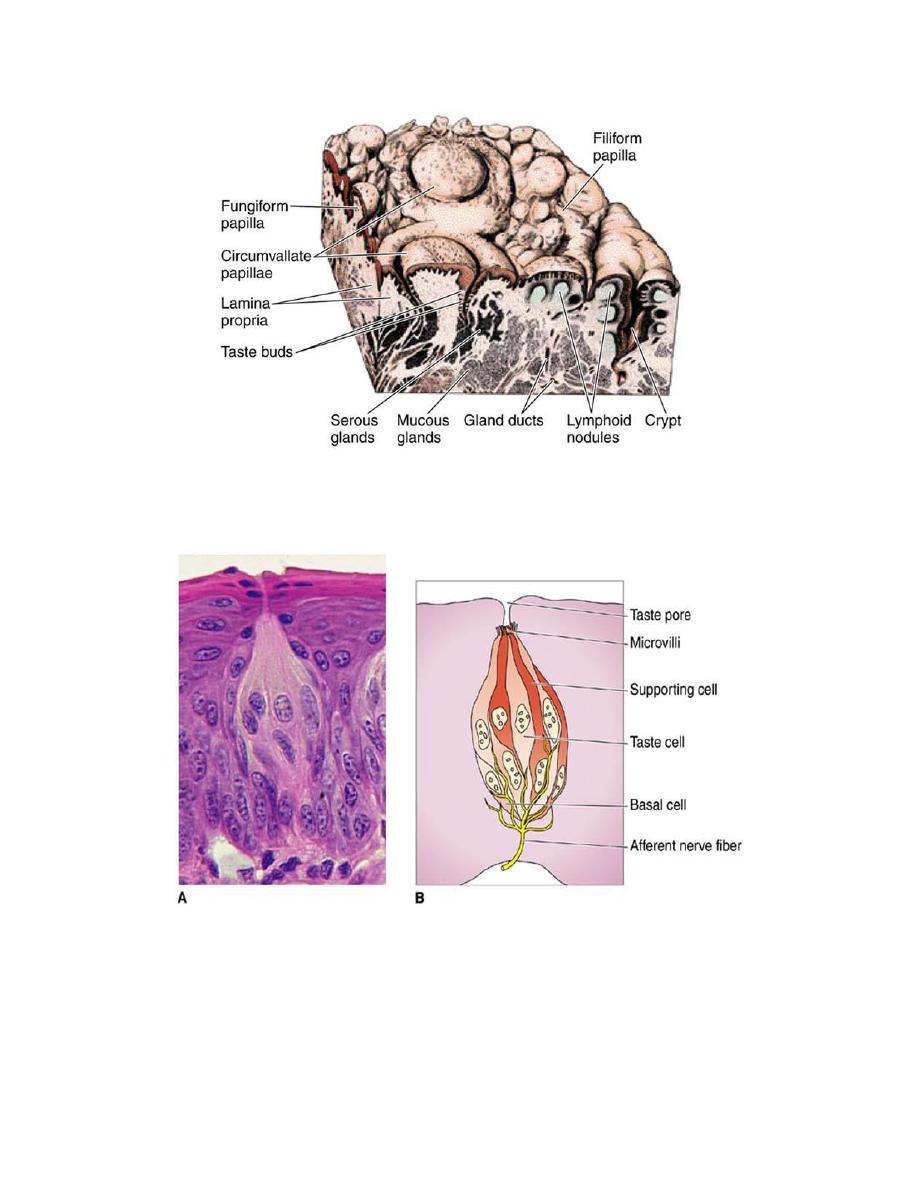
Photomicrograph (A) and drawing (B) of a taste bud, showing the taste cells and the taste
pore. The drawing also illustrates several cell types (basal, taste, and supporting) and afferent
nerve fibers that, upon stimulation, will transmit the sensory information to the central
gustatory neurons. A: Hematoxylin and eosin (H&E) stain.
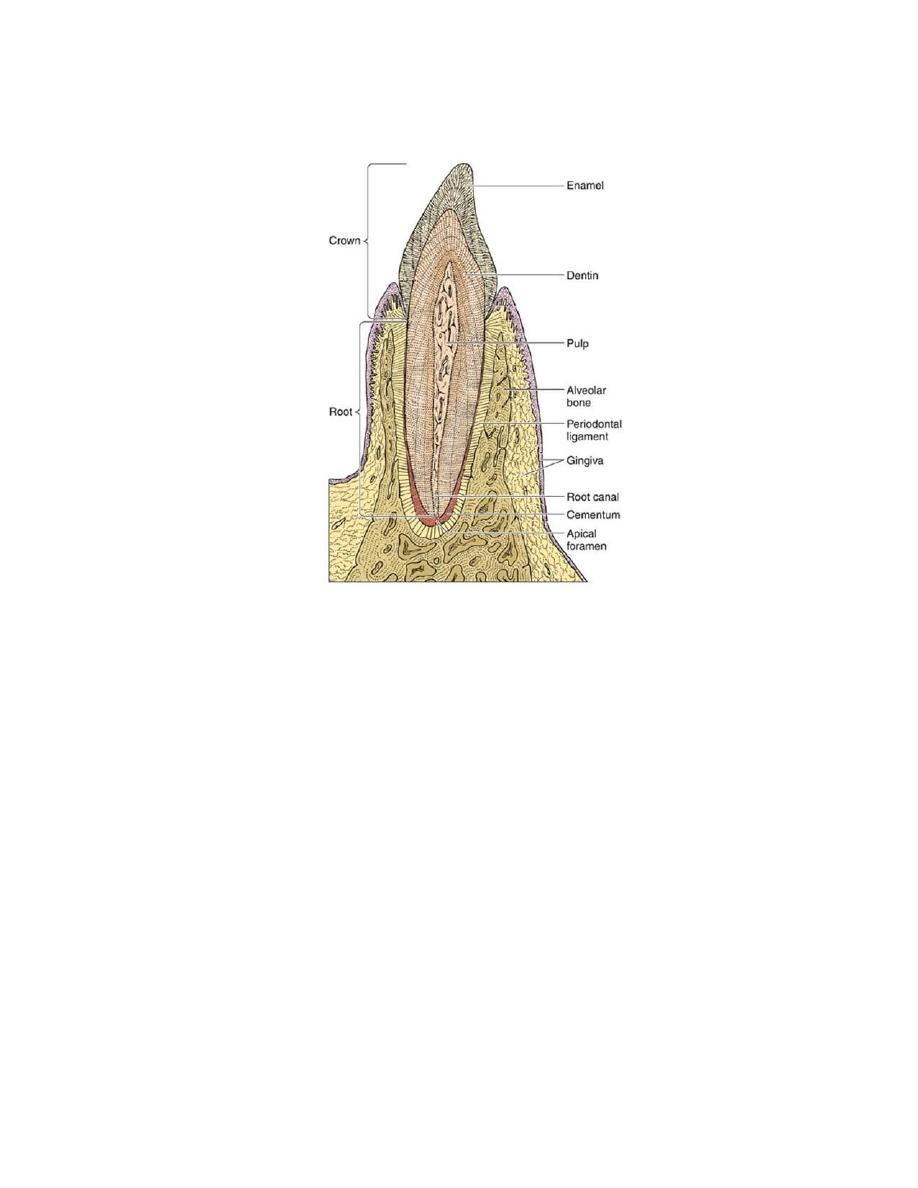
Diagram of a sagittal section from an incisor tooth in position in the
mandibular bone. (Redrawn and reproduced, with permission, from
Leeson TS, Leeson CR: Histology, 2nd ed. Saunders, 1970.)