
4
HISTOLOGY
Prof. Dr. Huda Al-Khateeb
2013-2014
Lec.2
General structure of blood vessels (BV):
Blood vessels (BV) have three basic types of tissues. These are:
1. Endothelium.
2. Smooth muscle cells
3. Connective tissue
The amount and the arrangement of these three types of tissues,
within BV wall, are influenced by:
1. Mechanical factor – represented primarily by blood pressure.
2. Metabolic factor – reflects the local needs of tissue.
VASCULAR ENDOTHELIUM
It is a special type of simple squamous epithelium. It serves many functions,
these are:
1. It forms a semipermeable barrier between plasma and interstitial
fluid.
2. It converts angiotensin I to angiotensin II. The latter increase blood
pressure.
3. It converts bredykinin, serotonin, prostaglandins and noradrenaline
------------etc. to biologically inert compounds.
4. It enhances lipolysis leading to formation of triglycerides and
cholesterol.
5. It produces endothelin, which is a vasoconstrictive factor.
6. It produces nitric oxide, which is a relaxing agent.

5
7. It produces vascular endothelial growth factors (VEGFs). In embryo,
VEGFs is responsible for the formation of vascular system, while in
adults, they regulate capillary growth in normal and pathological
conditions.
8. It has anti-thrombogenic action (preventing blood coagulation).
Damage to endothelium will uncover connective tissue and induce
platelates aggregation, thus cause thrombus and emboli formation.
VASCULAR SMOOTH MUSCLE CELLS
Found in all vessels, except capillaries and venules.
They are arranged in helical layers in tunica media.
Each muscle is enclosed by basal lamina and connective tissue (both
are secreted by the smooth muscle).
VASCULAR CONNECTIVE TISSUE
Variable amount of connective tissue present in different BV.
Collagen fibers type I found in tunica adventitia, type III in tunica
media and type IV in the basement membrane.
Elastic fibers are responsible for shrinkage of the expanded vascular
wall. They are predominant in large arteries.
Ground substance is composed of hetrogenous gel in extracellular
spaces of the vessel wall. It affects the diffusion and permeability
across the vessel wall.
Aging causes conformational changes in collagen and elastin with
deposition of lipoproteins and calcium, in addition to atherosclerosis.
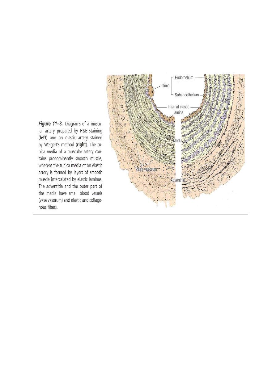
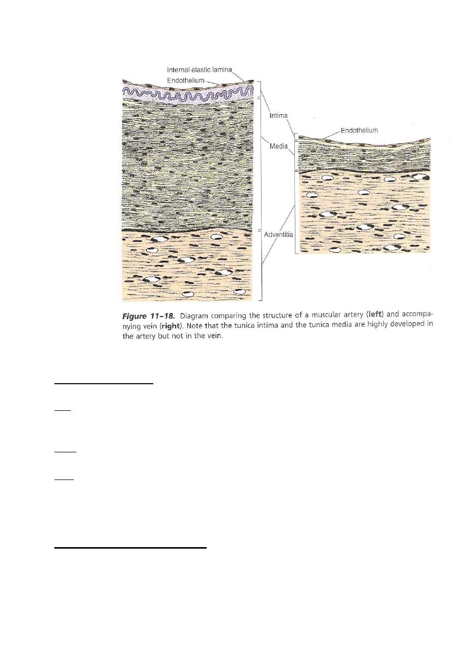
LAYERS OF WALL OF BV
Generally, each BV is composed of 3 layers; these are from inside to
outside:
1-Tunica Intima (TI) – subdivided into:
A- Endothelium- (simple sq. epith.)
B- Subendothelium- loose con.t.

6
C- Int. elastic lamina – composed of elastin that has gaps (for
diffusion of substances to nourish cells deep in the vessel wall). It is
found in arteries only.
2-tunica media (TM) – composed of:
A- Circular smooth m. fibers
B- Elastic fibers.
C- Reticular fibers (collagen fibers type III)
D- Fibroblast
E- Ground substance (proteoglycan and glycoprotein)
F- in arteries, TM has external elastic lamina that separate it from
TA
3-tunica adventitia (TA) – consist of connective tissue (collagen type I
and elastic fibers.
The structure & relative thickness of each layer vary according to the
type & size of the vessel.
Classification of a.s. :
1-large-size a.-(elastic a.)
2-medium-size a. and small- size a. -(muscular a.)
3-arteriol
In arteries, TM is usually the thickest layer, while in veins, TA is
commonly the thickest layer.
large-size a. (elastic a.) (conducting a.)
includes aorta & its largest branches.
TI – relatively well developed
-the int. elastic lamina is present but difficult to be diagnosed because It
is usually mixed with the elastic lamina of TM
TM – it is the thickest layer in this type of a.s
-characterized by a distinct elastic laminae (40-70 in no., usually increase
with age) arranged in a concentrical pattern.
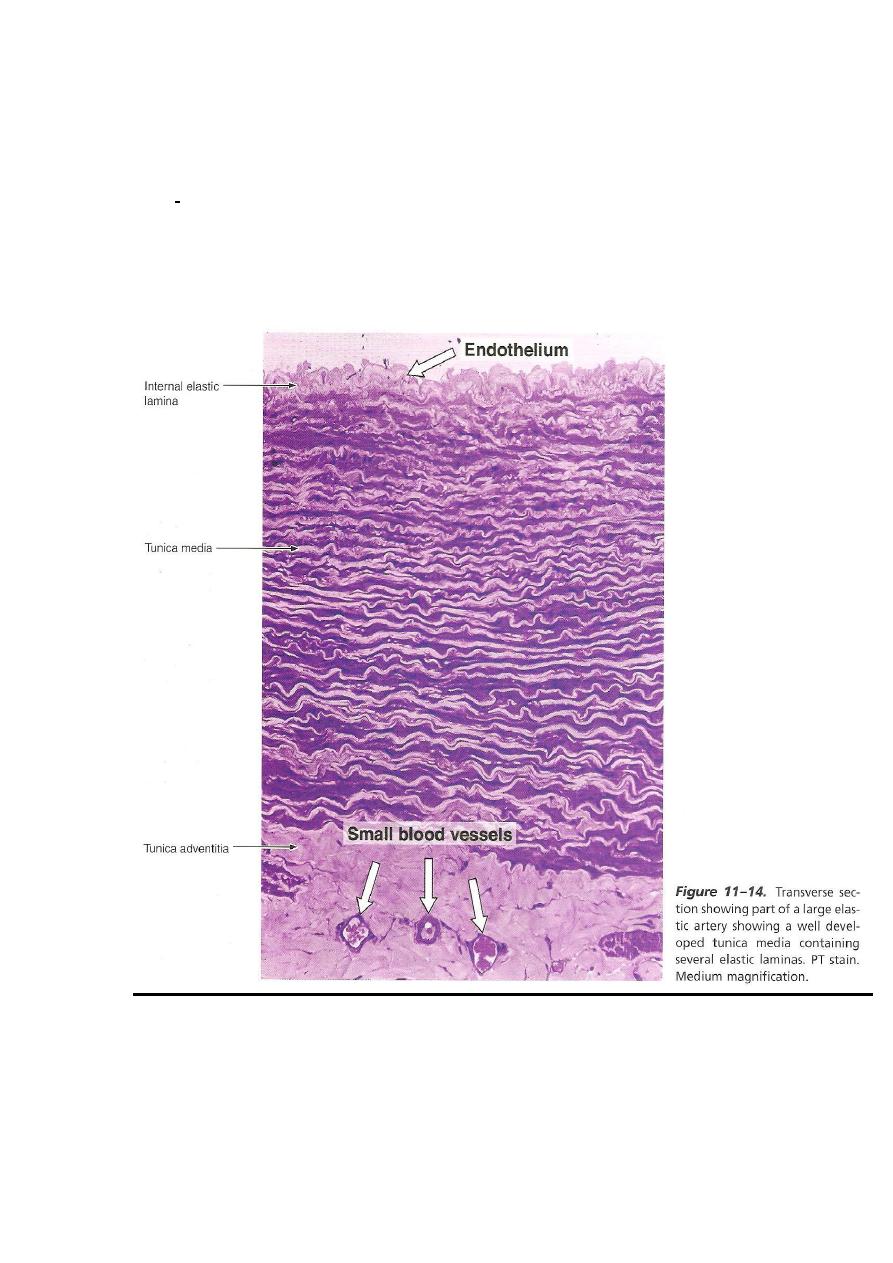
7
-interspaces between the elastic membranes are occupied by fibroblasts ,
amorphous ground substance, fine elastic net work and smooth m. cells.
TA - Loose connective tissue

8
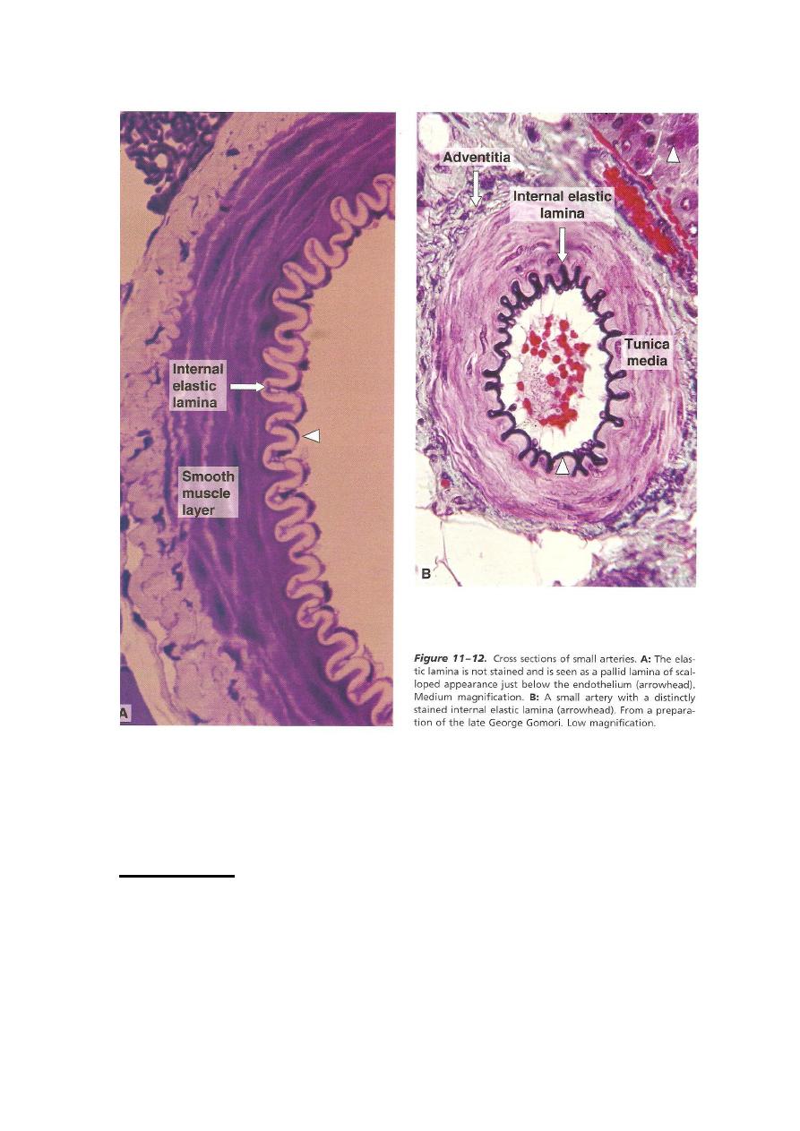
Medium- & small- size a. (Muscular a.)
(Distributing a.)
Characterized by thick wall & narrow lumen, when compared with
elastic a.
TI - prominent thick int. elastic lamina
TM
-consists of about 40 layers of circularly arranged smooth muscle fibers.
-between smooth muscle fibers, there are small amount of con.t. that
contains elastic & reticular fibers & few fibro blasts.
-ext. elastic lamina is Prominent & composed of many layers
TA - loose connective tissue
9
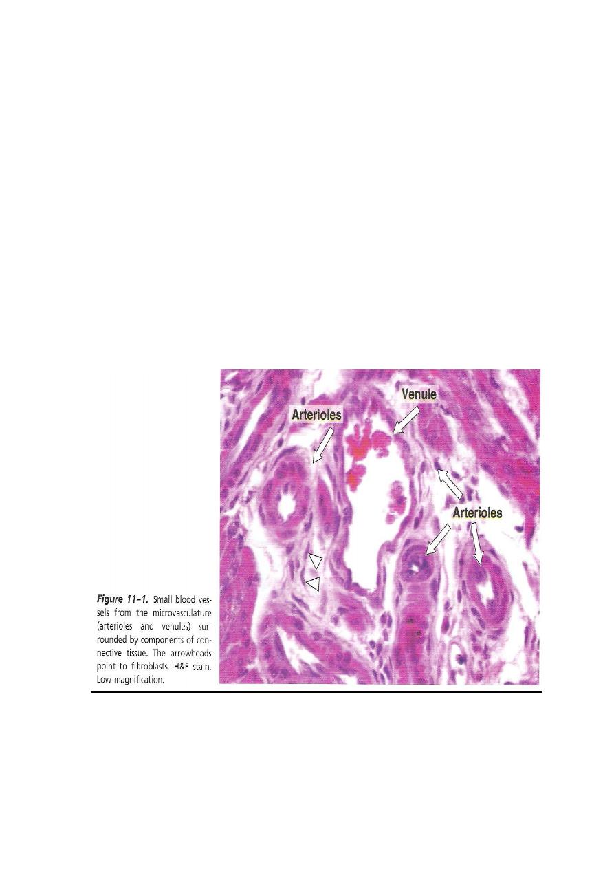
Arterioles
generally less than 0.5 mm in diameter
have relatively thick wall & narrow lumen (when compared with
venule).
TI
*Int. elastic membrane present in big arterioles only and disappear in
smaller arterioles.
01
*has no subendothelial con.t.
TM
*composed of (1-5) layers of circularly arranged smooth m. fibers among
which scattered elastic fibers
*the no. of smooth m. layers ↓ as diameter ↓. it becomes single layer at
about 20μm diameter arteriole.
*it has no external elastic lamina.
TA - very thin
00
Arterial capillary (pre-capillary) (met-arteriole)
TI
- composed of endothelium only
TM - circularly oriented scattered smooth m. fibers that have branching
processes.
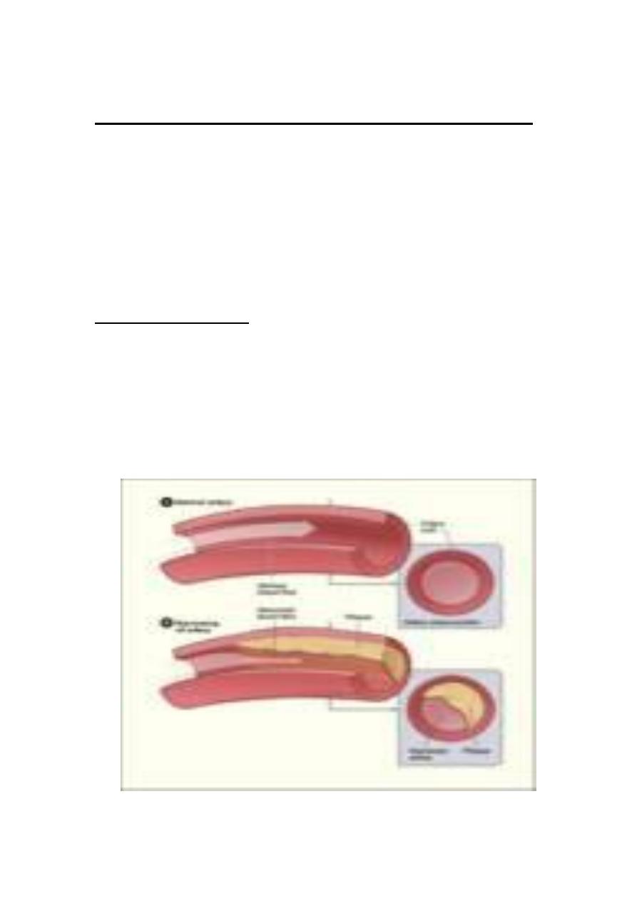
ATHEROSCLEROSIS:
It is accumulation of cholesterol in the T.I. of arteries. Grossly, the
artery contains fatty streaks and plaques on its internal surface. If these
fatty thickenings become great, they will occlude the vessel. Coronary
arteries are most arteries predisposed to atherosclerosis, which
sometimes leads to infarction (necrosis and tissue death) and other times
it is overcomed by arterial anastomosis.
01
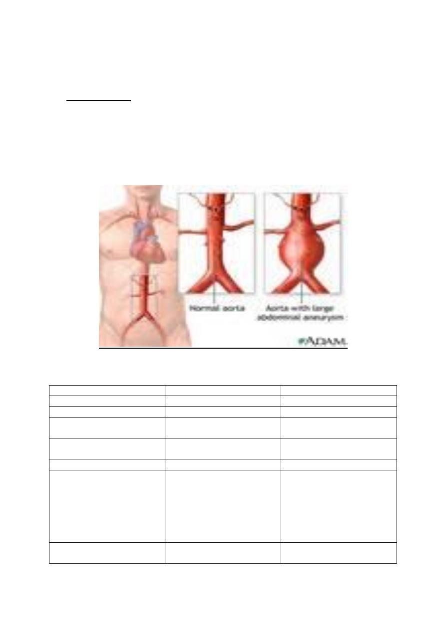
ANEURYSM:
It is a pathological dilatation of an artery, which is due to embryonic
defect, disease or lesion in its TM (that leads to its weakness in TM and
dilatation in the artery). Rapture of aneurysm brings sever consequence
and may cause death.
Elastic artery
Muscular artery
1. diameter of lumen
Wider
Narrower
2. thickness of the wall
Thinner
Thicker
3. TM is mainly composed
of
40-70 elastic lamina
40 layers of smooth m.
fibers
4. Internal and external
elastic membranes
Can't be easily diagnosed
Very prominent and
diagnosed easily
5. TI is
Thicker
Thinner
6. function
Stabilize arterial blood
pressure (during systole,
elastic lamina stretch and
reduce the increased blood
pressure. During diastole,
the elastic rebound increase
the reduced blood pressure)
Control the amount of
blood flow to organs by
contracting or relaxing its
smooth m.
7. example
Aorta and its large
branches
Coronary artery
01
04
HISTOLOGY
Prof. Dr. Huda Al-khateeb
2013-2014
Lec. 3
Capillaries (cap.):
- total length of human cap.s = 9600 km, which = 60000 mile
-connects venous to arterial sides
-(7-9) μm in diameter = diameter of a single RBC
-form a network that varies in size & shape
ex: in
lung }
liver }
kidneys } have large mesh of cap. network
glands }
mucous membranes }
skeletal muscles }
gray matter of brain }
tendon }
nerve } have spares cap. network
smooth muscles }
serous membranes }

05
Structure of cap.:
L.M.
+transverse section of cap. contains (1-2) endothelial cells, whose nuclei bulge
into the cap. Lumen.
E.M.
1-endothelium
-cytoplasm is thick opposite nucleus & thin elsewhere
-organelles usu. lie in the perinuclear area
-these are:
+small Golgi complex
+few mitochondria
+free rrbosomes
+RER
+filaments → may be related to the contractility of endothelium
2-Basal lamina- which is a product of endothelial cells.
3-thin sheath of delicate collagen & elastic fibers.
4-pericytes- slender, elongated, highly branched cells, whose cytoplasm
contain filaments (myosin, actin and tropomyosin), which are responsible for
its contractile function. They are located between endothelial cells and their
basal lamina.
* After tissue injury, pericytes proliferate and differentiate to form new blood
vessels and connective tissue cells, thus participating in tissue repair process.
06
Classification of cap.s
Cap. Are classified according to their ultrastructure into:
1-continuous cap.
2-fenestrated cap.
3-sinusoidal (or discontinuous) cap.
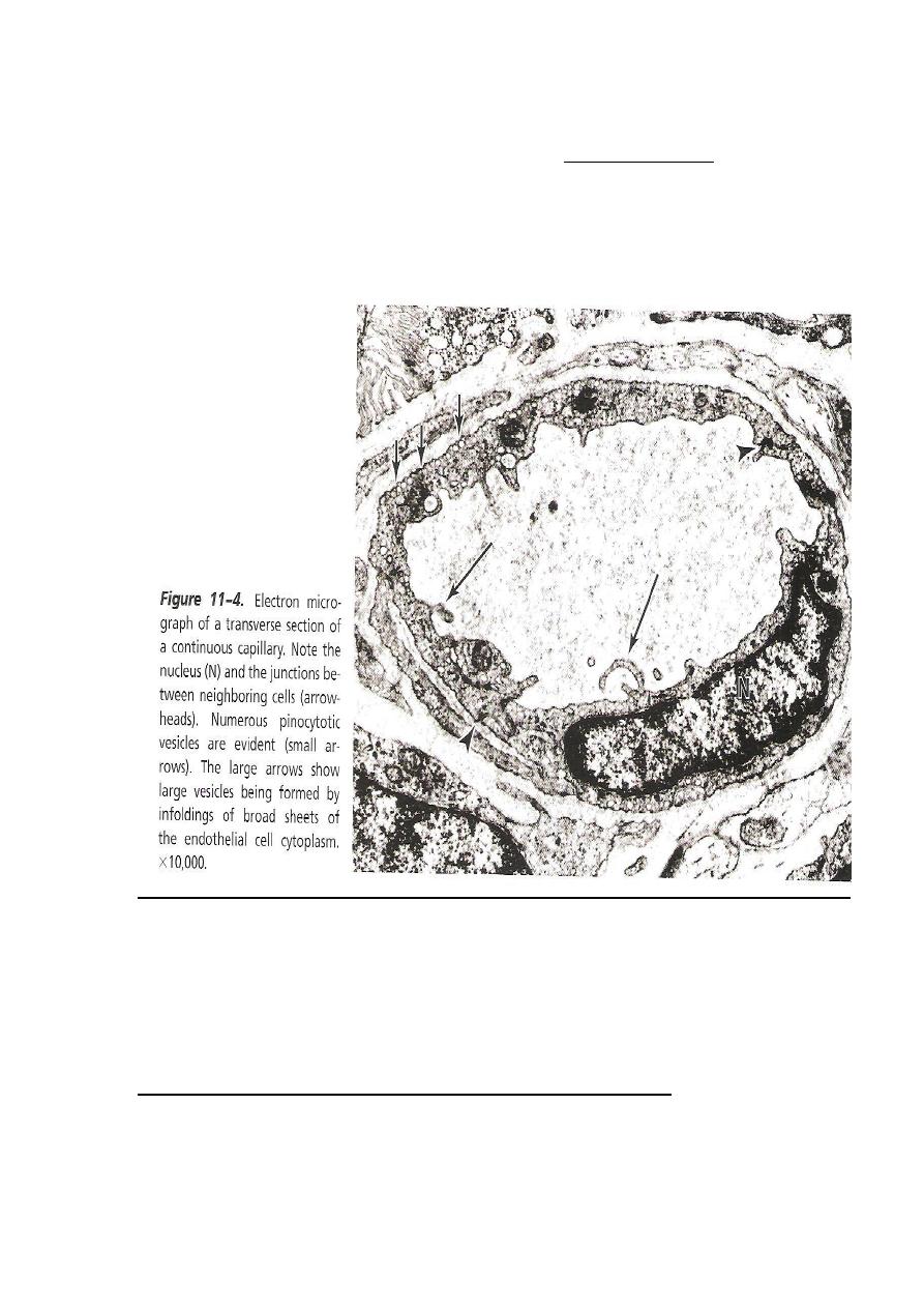
continuous (type I) cap.
*found in: muscle tissue
lung
CNS
Skin
Connective tissue
Exocrine gland
*the endothelial cytoplasm contains no. of small vesicles (pinocytotic vesicles)
of (50-70) nm in diameter.
Functionally, it appears that they are involved in the transport of fluid &
macromolecules across the cap. wall.
07
*few or no pinocytotic vesicles are encountered in continuous cap.s of nervous
system. this feature accounts for the existence of B. Brain Barrier.
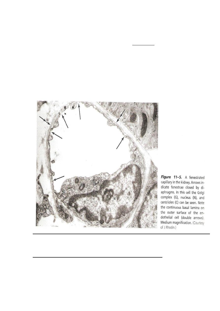
fenestrated (type ІІ) cap.s
*found in: intestinal mucosa
endocrine glands
renal Glomerulus
08
pancreas
*peripheral cytoplasm of endothelium is perforated at intervals by “pores”
ranging (30-50) nm. the pores are closed by thin diaphragm, except in cap. of
renal glomeruli.(pores have no diaphragm)
Sinusoidal cap.s (sinusoids):
*the cap. lumen is greater than other types of cap.s (30-40) μm In diameter
*the wall of sinusoid is composed of discontinuous layer of endothelial cells
* cytoplasm of endothelial cells shows multiple fenestration without
diaphragms.

09
*macrophages are closely associated with the endothelial cells. both within
& around the sinusoidal wall.
*basal lamina is incomplete.
*sinusoids are found in liver and haemopoietic organs (ex. Bone marrow &
spleen).
Venus cap.s (post-cap.s)
-diameter up to 30μm.
-wall consists of:
1-endothelium
2-thin con.t. coat contains pericytes (slender, elongated, highly branched cells
found in cap.s & postcap.s surrounding endothelium). they are greater in no.
in post cap. than that in cap.s.
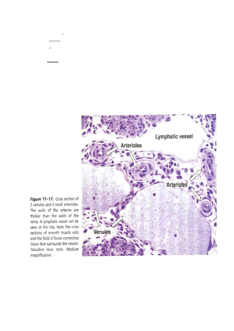
classification of v.s
1- venules
2- small to medium size v.s
3- large size v.s
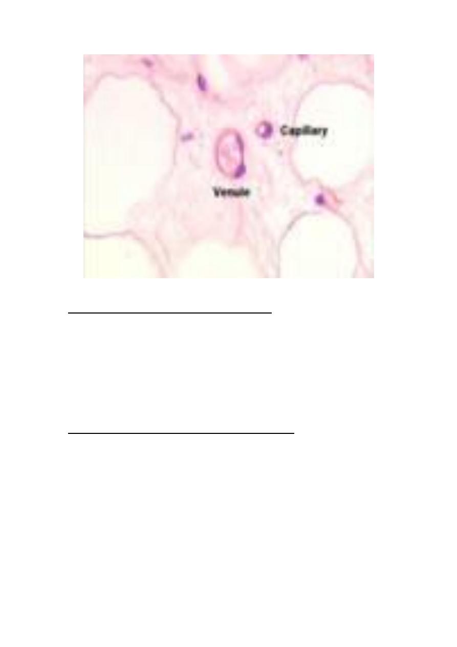
Venules
*the smallest venule (40μm in diameter) has
-T.I. that possess endothelium with basal lamina
-T.A.- outer thin sheath of collagenous fibers
*in venules of 50μm diameter smooth m. fibers appear between
endothelium & con.t. (i.e. T.M. appears).
*in 200μm diameter venule- the circular muscle fibers form a
continuous layer (T.M.)(1-3) cells thick.
-T.A. is thicker & consists of longitudinally oriented collagenous fibers
, scattered elastic fibers & fibroblasts .
Small & medium size v.s
-diameter ranges (1-9)mm
11
-T.M. has no elastic membrane
T.M.
-thin
*best developed in v.s of lower limb.
T.A.
-thickest layer
-thick longitudinal collagenous bundles and frequently few smooth
m. fibers which are arranged longitudinally along the vessels .
10
Large v.s
-includes the sup. & inf. Vena cava & their main tributaries.
T.I.
-same as medium sized v.
-it may be thicker.
T.M.
-poorly developed or absent
T.A.
-thickest composed of longitudinal coarse collagen fibers & smooth m.s
venous valves
- found in medium size v.s especially of lower limb are provided with valves
that prevent the flow of b. away from the heart.
-valves are folding of the T.I.

11
BV of BV (vasa vasorum)
-a.s & v.s of diameter over 1 mm are supplied by small , nutrient BV that is
called vasa vasorum .
-vasa vasorum usually inters T.A. & terminates in a dense cap. network which
penetrate as far as the deepest layer of the T.M.
-generally no cap.s are found in T.I. However , in some large v.s, cap.s penetrate
as far as T.I. (probably because of low venous pressure & O
2
tension) .
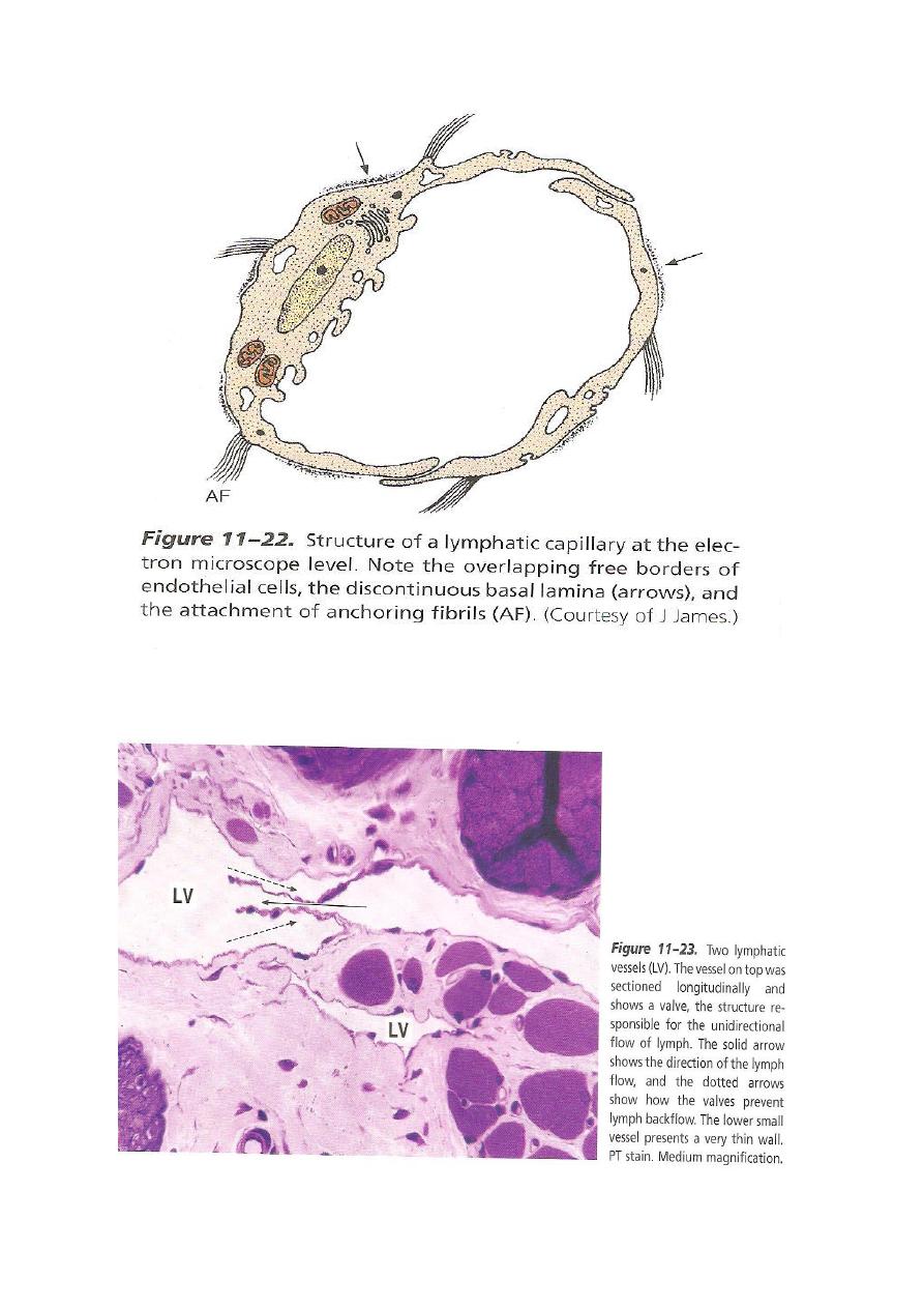
Lymph vascular system
Consists of :
1-lymphatic vessels
their structure is usu. Similar to the structure of the corresponding size v.s
2-organs → lymph nodes
11