
1
URINARY SYSTEM Dr.Firdous
Objectives:
1- Describe the structure of the kidney; both cortex and medulla.
2- Describe the functional unit of the kidney; the nephron, and its parts; renal
corpuscle, proximal convoluted tubule, distal convoluted tubule, loop of Henle. The
structure of these parts should be studied both by light and electron microscope.
3- Explain the role of glomerulus in the process of membrane filtration, and the
structure of juxtaglomerular apparatus.
4- Explain the arrangement of blood vessels in the kidney, and its blood supply.
5- Describe the structure of ureters, bladder, and urethra.
The urinary system consists of paired kidneys, ureters, and a single bladder and
urethra.
Function:
1. Maintenance of homeostasis by production of urine through filtration,
absorption, and secretion.
2. Regulation of fluid and electrolytes balance of the body.
3. Production of Renin (important in regulation of blood pressure).
4. Production of Erythropoietin (stimulates the production of erythrocytes).
KIDNEY
The kidney is
about 11cm, 6cm, 3cm in dimensions. Each kidney weights about
150 gm. It has a concave medial border; the hilum, where the nerves enter, blood
and lymphatic vessels enter and leave, and the ureter exits, and a lateral convex
side.
Each kidney is surrounded by a connective tissue capsule that is surrounded by
a mass of peri renal adipose tissue. The capsule consists of two distinct layers; an
outer layer of fibroblasts and collagen fibers, and an inner layer of
myofibroblasts. The capsule pass inward at the hilum covering the sinus, and
become continuous with the connective tissue forming the walls of calyces and
renal pelvis.
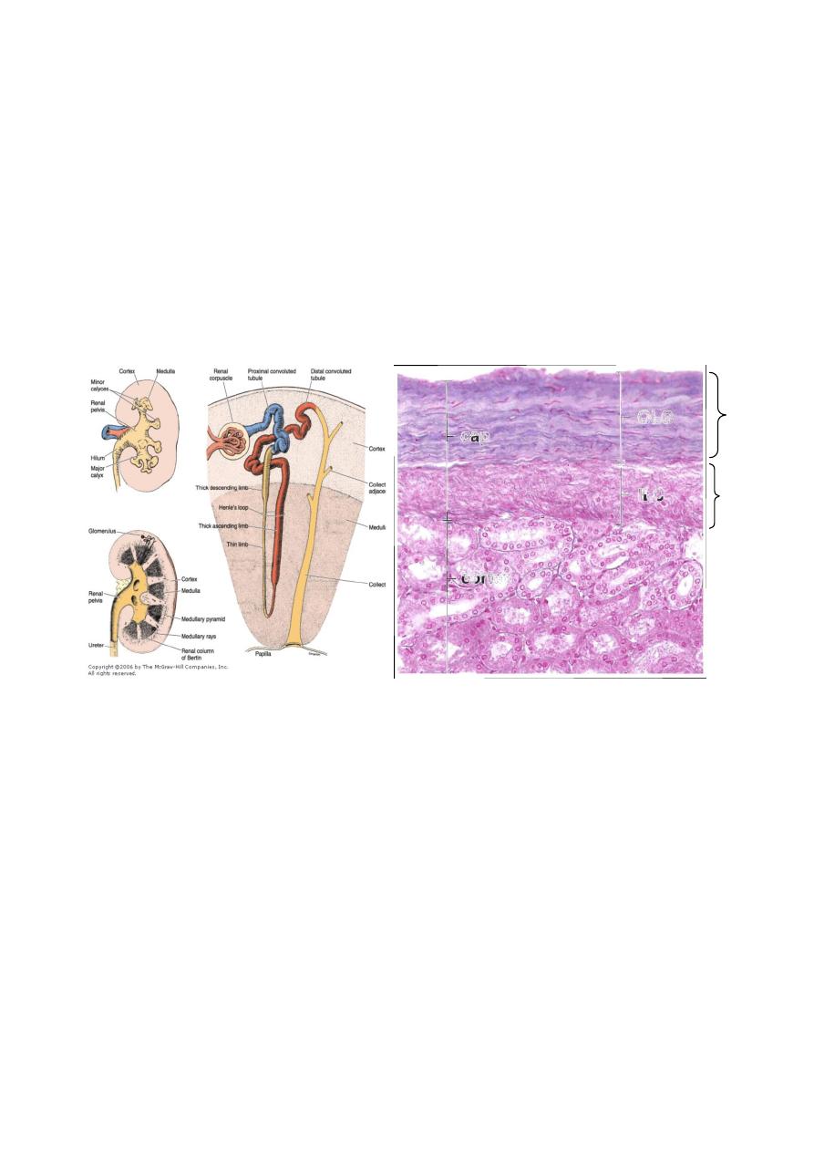
2
The kidney is divided into an outer cortex and an inner medulla. Renal medulla
consists of 10-18 conical or pyramidal structures; the medullary pyramids, whose
bases form the cortico-medullary junction and vertices protrude into the minor
calyces. The tips of pyramids are called renal papillae, where the collecting ducts
open into. From the base of each medullary pyramids parallel tubules; the
medullary rays, penetrate the cortex.
Kidney lobe consists of medullary pyramid and associated cortical tissue at its
base and sides, and they are about 8-18 in each kidney.
Renal pelvis is the dilated upper portion of the ureter which is divided into 2-3
major calyces that branch into several smaller minor calyces.
Structure of the kidney
structure of kidney capsule( two layers)
3
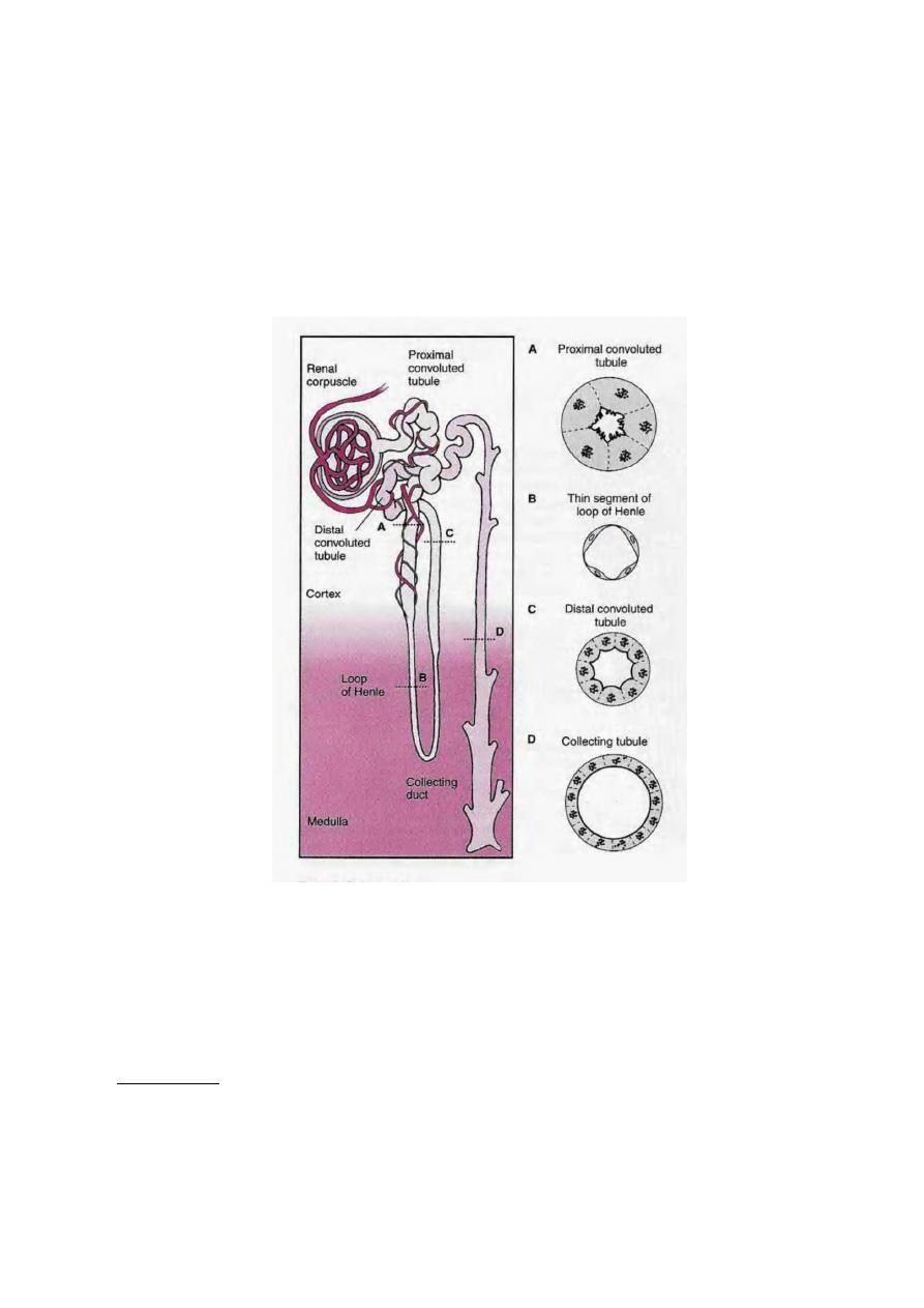
NEPHRON
It is the functional unit of the kidney. There are about 1-4 millions in each
kidney. Each nephron consists of a dilated portion; the renal corpuscle, proximal
convoluted tubule (PCT), thin and thick limbs of Henle's loop, and distal
convoluted tubule (DCT).
Some investigators consider the collecting tubules and
ducts to be part of the nephron.
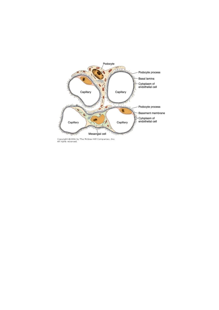
RENAL CORPUSCLE
It is about 200 µ in diameter, and consists of a tuft of capillaries; the glomerulus,
and a double-walled epithelial capsule; glomerular(Bowman's capsule).
Renal corpuscle has a vascular pole, where afferent arteriole enter, and efferent
arteriole leave, and a urinary pole where the PCT begins.
Glomerulus:
When afferent arteriole enter the renal corpuscle, it will divide into 2-5 primary
branches which will further divide into capillaries forming the glomerulus. The
endothelial cells have thin cytoplasm except around the nucleus. It is of the
fenestrated type with 70-90 nm in diameter, with no diaphragm.
4
Bowman's capsule
Composed of two layers; parietal and visceral, with a space in between called
urinary space which receives the fluid filtered through capillary wall.
Parietal layer consists of simple sequamus epithelium supported by a basal
lamina and a thin reticular fibers. At the urinary pole, this epithelium will change
into simple cuboidal epithelium of the PCT.
Visceral layer is modified during embryonic life and the cells are called
Podocytes. The cell body gives rise to several primary processes from which arise
numerous secondary processes that called pedicels. These pedicels embrace the
capillary of glomerulus and come in contact with the basal lamina at a periodic
distance of 25nm. The pedicels interdigitate with each other defining a space about
25nm wide; the filtration slit which is bridged by a diaphragm of 6nm thick.
Podocytes have bundles of actin microfilaments to give them the contractile
capacity.
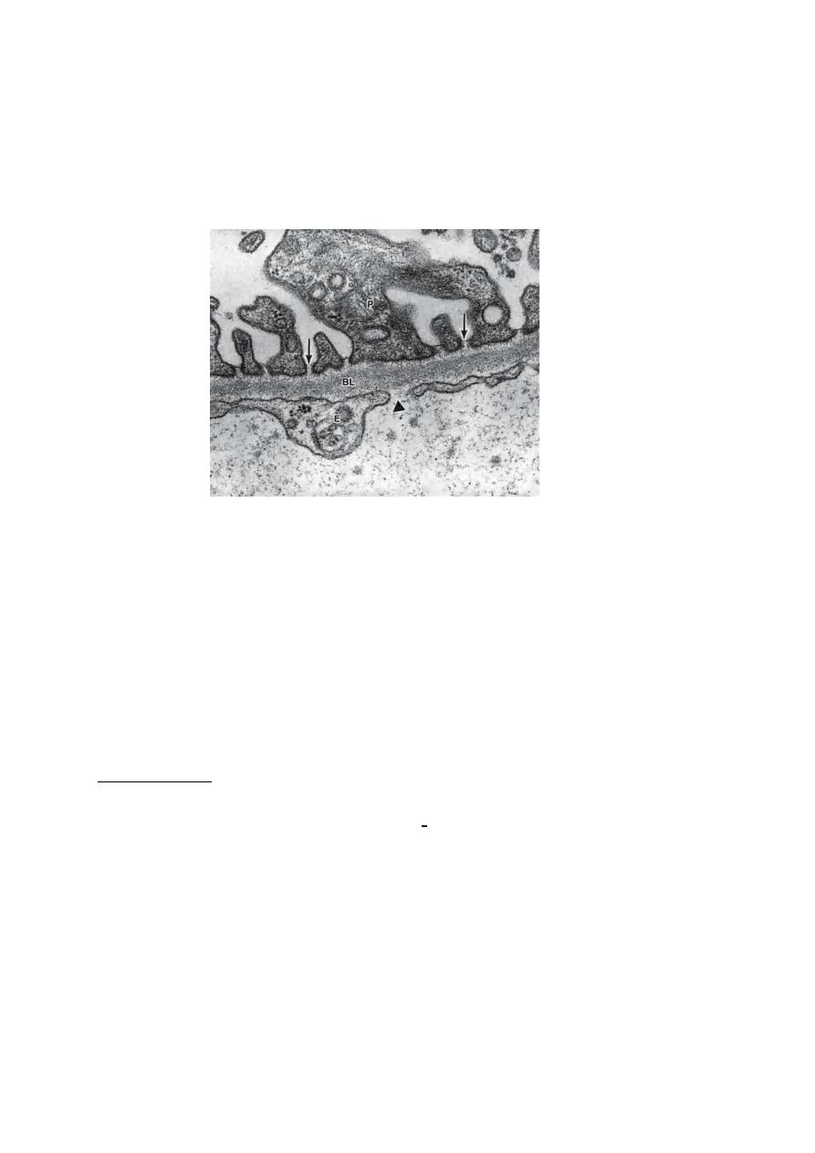
5
A thick basement membrane (0.1µm) is formed by fusion of the basal laminae
of both capillary and podocytes, forming a filtration barrier that separates urinary
space and capillary blood. By E.M,the basement membrane consist of a central
electron-dense layer called lamina densa, and, on each sides, a more lucent layer
called lamina rara which contains fibronectin to bind them to the cells. Lamina
densa contains typeIV collagen.
The filtration apparatus(barrier) consists of:
1- Endothelium of glomerular capillaries: they are fenestrated, with no diaphragm,
and posse a large number of aquaporin-1(AQP-1) water channels that allow
fast movement of water through epithelium.
2- Glomerular basement membrane.
3- Podocytes.
In diseases such as diabetes mellitus and glomerulonephritis, the glomerular
filter is altered and becomes much more permeable to proteins, with the subsequent
release of protein into the urine (proteinuria).
Mesangial cell: This cell is found adherent to the wall of glomerular capillaries
where the basal lamina is shared by two or more capillaries. It has cytoplasmic
processes that extend to the endothelial cells. They have receptors for angiotensin
II. When these receptors are activated, the glomerular flow is reduced. Mesangial
cells also have receptors for the natriuretic factor produced by cardiac atria cells.
This factor is a vasodilator that relaxes the mesangial cells, probably increasing the
blood flow and the effective surface area available for filtration.
Mesangial cells also have several other functions: they give structural support to
the glomerulus, synthesize extracellular matrix, endocytose and dispose of normal
and pathological (immune complex) molecules trapped in the glomerular basement
membrane,
and
probably
produce
chemical
mediators
such
as
cytokines,prostaglandins, interleukin-1, and platelet-derived growth factor(PDGF)
6
In certain diseases of the kidney it has been noticed that mesngeal cells
proliferate, in which abnormal amount of proteins trapped in the basement
membrane.
Although mesangeal cells act as macrophages, embryologically they originate
from smooth muscle cell precursors, rather than the precursors of mononuclear
phagocytic system.
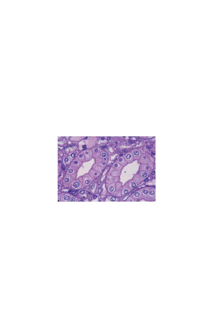
PROXIMAL CONVOLUTED TUBULE(PCT)
Begins at the urinary pole of the renal corpuscle, where the simple sequamus
epithelium change into simple cuboidal or simple columnar epithelium.
This tube is long and tortuous so it is more frequently seen in renal cortex than
DCT. The cytoplasm is acidophilic due to the presence of numerous mitochondria.
The cell apex has abundant microvilli, of 1µm in length, and they form the brush
border. Each cell has a spherical, centrally located nucleus. In living animal PCT
has wide lumen, but in histological section the lumen is collapsed and the brush
border is disorganized. The apical cytoplasm has numerous canaliculi between the
bases of microvilli. They are important in absorbing macromolecules. The basal
portion of PCT has membrane invaginations and the mitochondria are concentrated
at the bases of these cells parallel to the long axis of the cell. These are the
characteristics of ion transporting cells. There is an extensive interdigitations of the
lateral membrane of adjacent cells, so there is no discrete cell margins when
observed by L.M. Cells of PCT are large, so they appear only 3-5 nuclei in
transverse section.
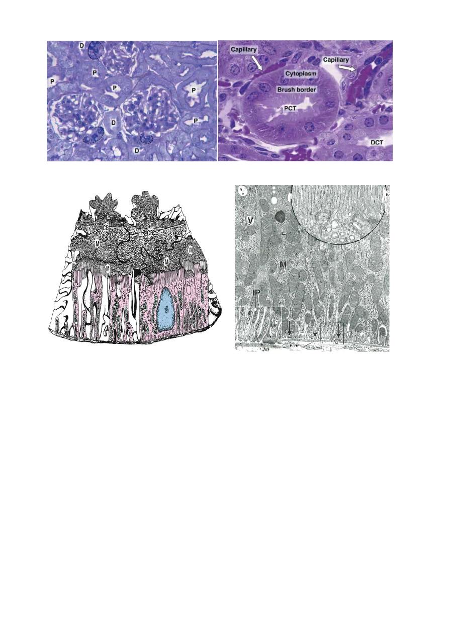
7
E.M. photo of PCT showing apical microvilli, large no.of mitochondria, and basal membrane
folding.
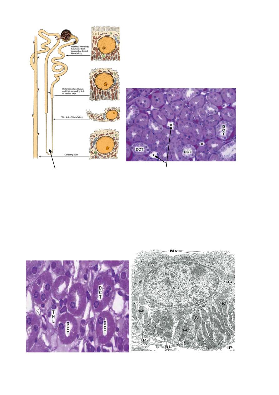
HENLE'S LOOP
It is a U shaped structure consist of thick and thin descending limbs, thin and
thick ascending limbs. It starts as a thick limb in the outer medulla with a diameter
of 60µm, lined by simple cuboidal epith. , then suddenly narrows to about 12µm in
diameter as the thin descending limb. This segment has a wide lumen, and lined by
simple seq.epith., whose nuclei protrude slightly into the lumen. This segment runs
deep into the medulla, then continues upwards as the ascending thin limb, which
will suddenly change into simple cuboidal epith. of the thick ascending limb.
Most of nephrons are cortical, and they have very short thin descending limb
with no thin ascending limb. Few number of nephrons are located near the cortico-
medullary junction and called juxtamedullary nephrons. They have very long
Henle's loop. Juxtamedullary nephrons are important in producing concentrated
urine. Intermediate or midcortical nephrons have their corpuscles in the mid
region of cortex, and their loop of Henle is of the intermediate length.
8
Loop of Henle Loop of Henle (star)
DISTAL CONVOLUTED TUBULE(dct)
It is the continuation of the thick ascending limb of Henle's loop, when it
penetrates the cortex. It is tortuous and lined by simple cuboidal epith. The cells of
DCT are small and flat, so more nuclei are seen in histological section. The cells
lack brush border or apical canaliculi and they appear less acidophilic than the cells
of PCT. Their lumen is wide. Lateral boundaries between adjacent cells are not
seen by L.M due to the interdigitations between these cells. Like cells of PCT, the
cells of DCT have numerous basal membrane invaginations with large number of
mitochondria.
9
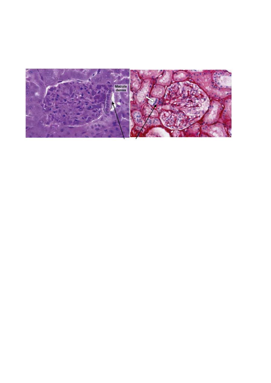
When DCT come in contact with the afferent arteriole of its parent nephron, its
cells will be modified. They will become columnar and their nuclei are closely
packed together, with the Golgi complex at the basal region. This modified area of
the DCT appears darker in L.M. and called macula densa, which is sensitive to
ionic contents and volume of tubular fluid.
Macula densa
Juxtaglomerular apparatus:
Tunica media of afferent arteriole of the renal corpuscle will be modified and
called juxtaglomerular cells(J.G.C.) . The nucleus is spherical, the internal elastic
membrane will disappear, and the cytoplasm is full of secretary granules. By E.M.,
J.G.C. show characteristics of protein-secreting cells; abundant RER, highly
developed Golgi complex, and the presence of secretary granules of 10-40 nm in
diameter.
Macula densa of the DCT is usually located close to the J.G.C. Together,they
form the J.G.apparatus. J.G.C. produce renin which act on a plasma protein;
angiotensinogen, to produce an inactive angiotensin Ι, which will be converted
into angiotensinΙΙ by a converting enzyme.
Part of the J.G.apparatus are some light-staining cells whose function is not well
understood, called extraglomerular mesangeal cells or Lacis cells.
After a significant hemorrhage (decreased blood volume promotes a decreased
blood pressure), there is an increase in renin secretion. Angiotensin II is produced,
enhancing blood pressure by both constricting arterioles and stimulating the
secretion of the adrenocortical hormone aldosterone. Aldosterone acts on cells of
the renal tubules (mostly the distal tubules) to increase the absorption of sodium
and chloride ions from the glomerular filtrate. This increase in sodium and chloride
ions, in turn, expands the fluid volume (particularly blood plasma volume), leading
to an increase in blood pressure due to increased blood volume. Decreased blood
pressure caused by other factors (eg, sodium depletion, dehydration) that decrease
blood volume also activates the renin “ angiotensin II“ aldosterone mechanism that
contributes to the maintenance of blood pressure. Aldosterone deficiency in
adrenalectomized animals and in humans with Addison disease results in an
excessive loss of sodium in the urine.
11
COLLECTING TUBULES AND DUCTS
DCT will open into a straight collecting tubules in the medullary rays. They lie
side by side and make up most of the pyramids. Collecting tubules are lined by
simple cuboidal epith., with a diameter of 40µm. these tubules join each other
forming a larger straight collecting ducts, which widen gradually as they approach
the tips of pyramids. The lining epith. of collecting ducts is simple columnar epith.,
with a diameter of 200µm. The cells of both tubules and ducts are lightly stained,
with few organellls in their cytoplasm. There is no basal invagination, or lateral
membrane interdigitations, so the intercellular limits are clearly seen. The epith. of
collecting duct is under the control of the anti-diuretic hormone(ADH)
There are two types of cells in collecting tubules and ducts:
1- Light cells: pale staining cells with basal infoldings, single apical cilium and
few microvilli. The cytoplasm contains small spherical mitochondria.
2- Dark cells( intercalated cells): They are fewer in number, and appear denser,
and their cytoplasm contains many mitochondria. Their apical cytoplasm
contains microvilli and vesicles. They don’t show basal infoldings, but have
basal interdigitations with neighboring cells. Dark cells gradually decrease in
number as the collecting duct approach the papilla.
Blood circulation of the kidney
Renal artery will enter the kidney through the hilum, and divide into two
branches; one supplies the anterior part, the other supplies the posterior part of the
kidney. An inter -lobar artery will run between the medullary pyramids. At the
cortico-medullary junction, inter lobar artery forms the arcuate artery, from which
arise the interlobular arteries at right angle to the renal capsule. From inter lobular
arteries, arise the afferent arterioles which supply the glomerular capillaries, then
blood goes to the efferent arterioles which branch to give the peritubular capillary
net work, to supply PCT and DCT.
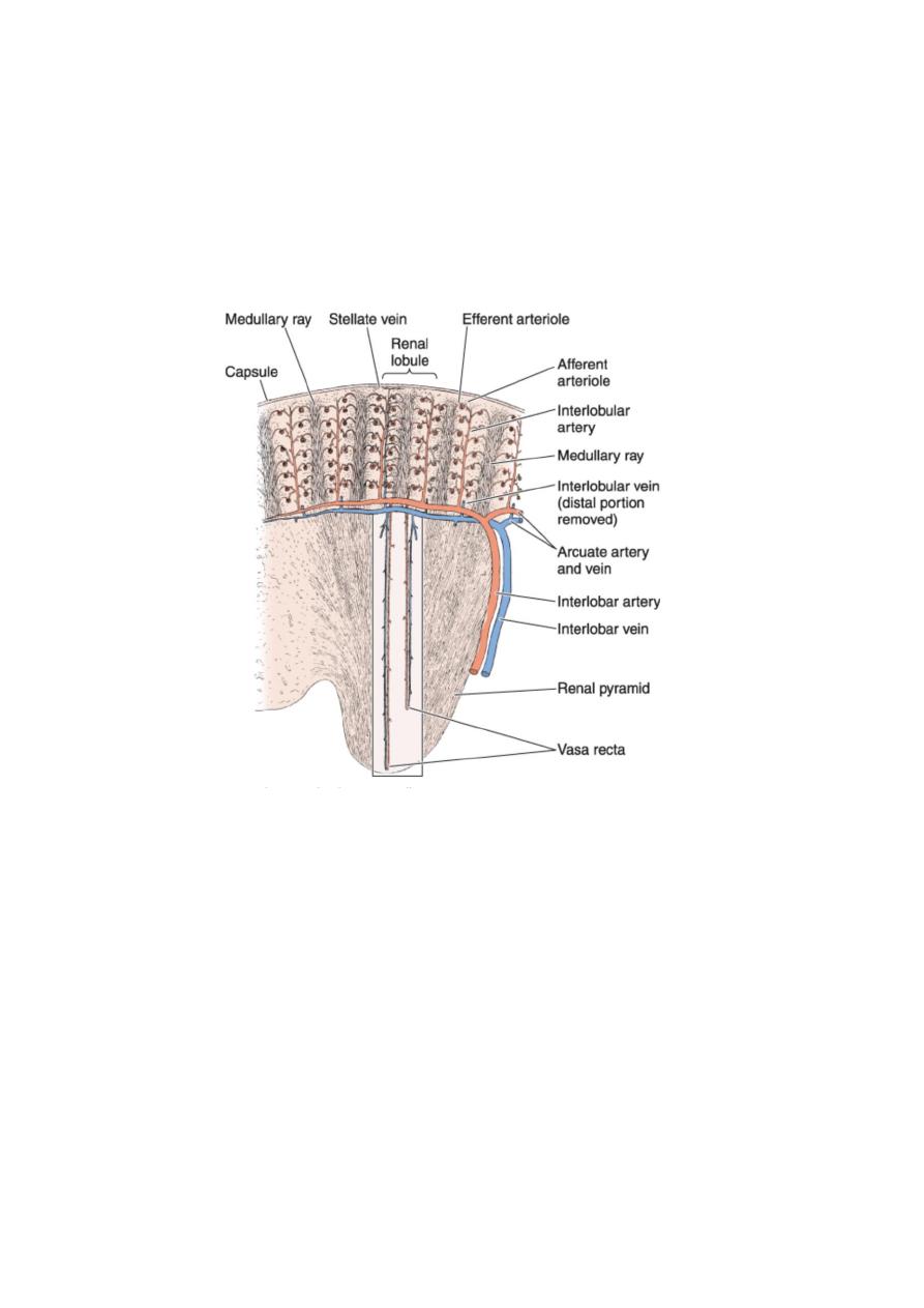
11
The efferent arterioles which are associated with juxta-medullary nephrons are
long and thin capillaries and they follow a straight path into the medulla, then loop
back towards the cortico-medullary boundaries and called vasa recta. The
descending vessel is continuous, while the assending one is fenestrated. These
capillaries are important in the nourishment of medulla.
Capillaries from outer cortex and capsule converge to form the stellate veins that
empty into the interlobular vein. Veins follow the same course of arteries in a
reverse way.
Renal interstitium
It represents the space between tubules, blood, and lymph vessels. It is little in
the cortex, but increase in the medulla. It consists of small amount of connective
tissue, collagen fibers, ground substance, and secreting cells; the interstitial cells.
There are two types of cortical interstitial cells; fibroblasts and macrophages.
Fibroblasts are found in between tubules and capillaries, and they synthesize and
secrete collagen and glycosaminoglycans of the extracellular matrix of the
interstitium.
In the medulla, the principle interstitial cells resemble myofibroblasts. Their
cytoplasm contains abundant actin filaments, abundant RER, well developed Golgi
complex, and lysosomes. Some evidence suggests that these cells may secrete a
hormone-like material that reduces blood pressure. Prostaglandins and prostacyclin
may also been synthesized in the interstitium.
12
URINARY BLADER
It stores the urine temporarily, before its exsecretion to the outside. The wall of
the bladder consists of three layers:
1-Mucosa: It is the inner layer, composed of transitional epith. and lamina
propria.
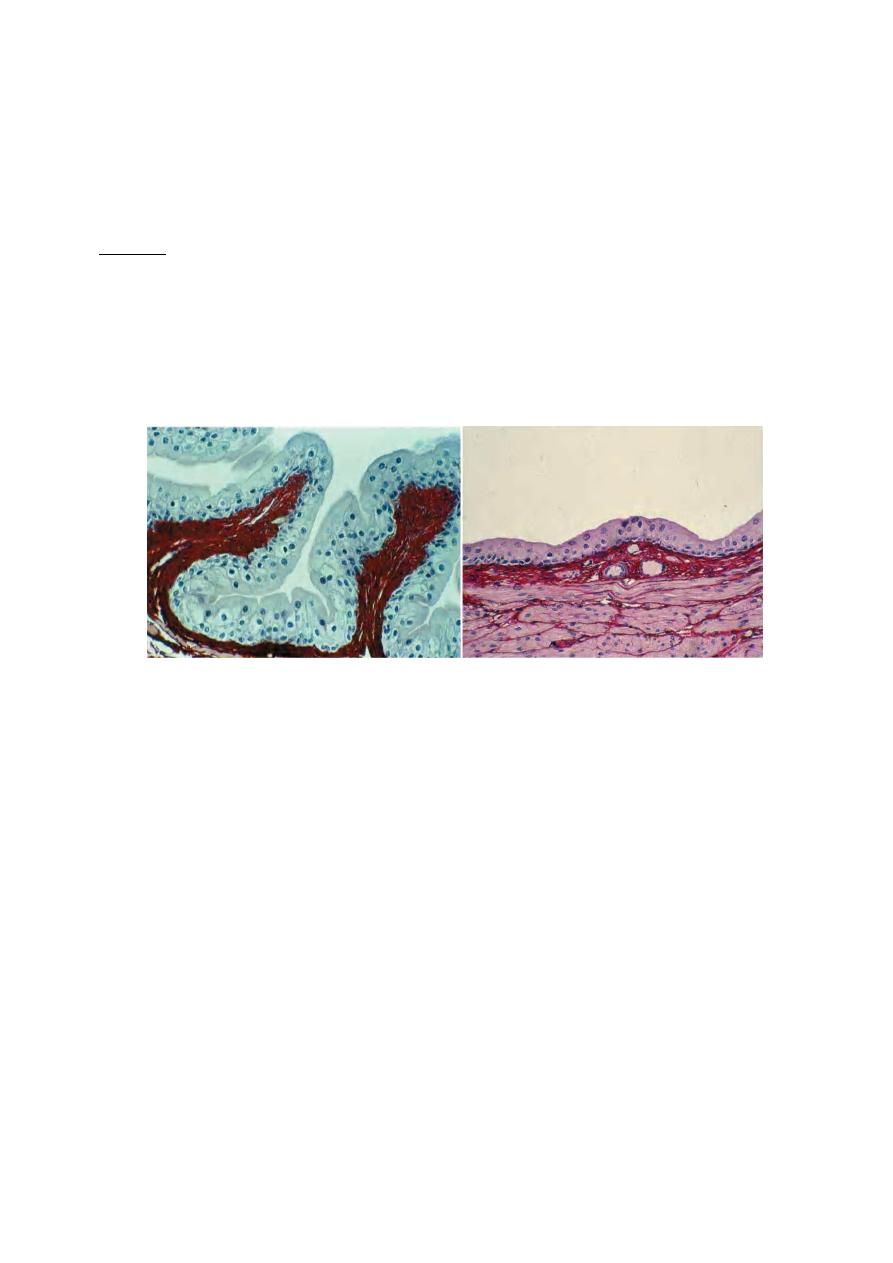
In an empty bladder, the transitional epith. is 5-6 cells in thickness, the
superficial cells are rounded and bulge into the lumen, and they are usually
polypoid or binucleated. When bladder is full of urine, the epith. will stretch and
cells will become 3-4 only in thickness and the superficial cells become
sequamous.
Empty bladder Full bladder
The bladder contains three orifices; two upper ureteric orifices, and one lower
internal urethral orifice. The triangular region defined by these three orifices is
called trigone, is smooth and has constant thickness, while the rest of the wall of
bladder has variable texture and thickness in relation to the fullness of bladder.
The trigone originates embryologically from mesonephric ducts, while the rest of
bladder wall originates from cloaca.
The superficial cells of the transitional epithelium have a special membrane of
thick plates separated by narrow bands of thinner membrane that are responsible
for the osmotic barrier between urine and tissue fluids.
When bladder contracts, the membrane folds along the thinner regions, and the
thicker plates invaginate to form cytoplasmic vesicles. These vesicles represent a
reservoir of these thick plates that can be stored in the cytoplasm of the cells of the
empty bladder and used to cover the increased cell surface in the full bladder.
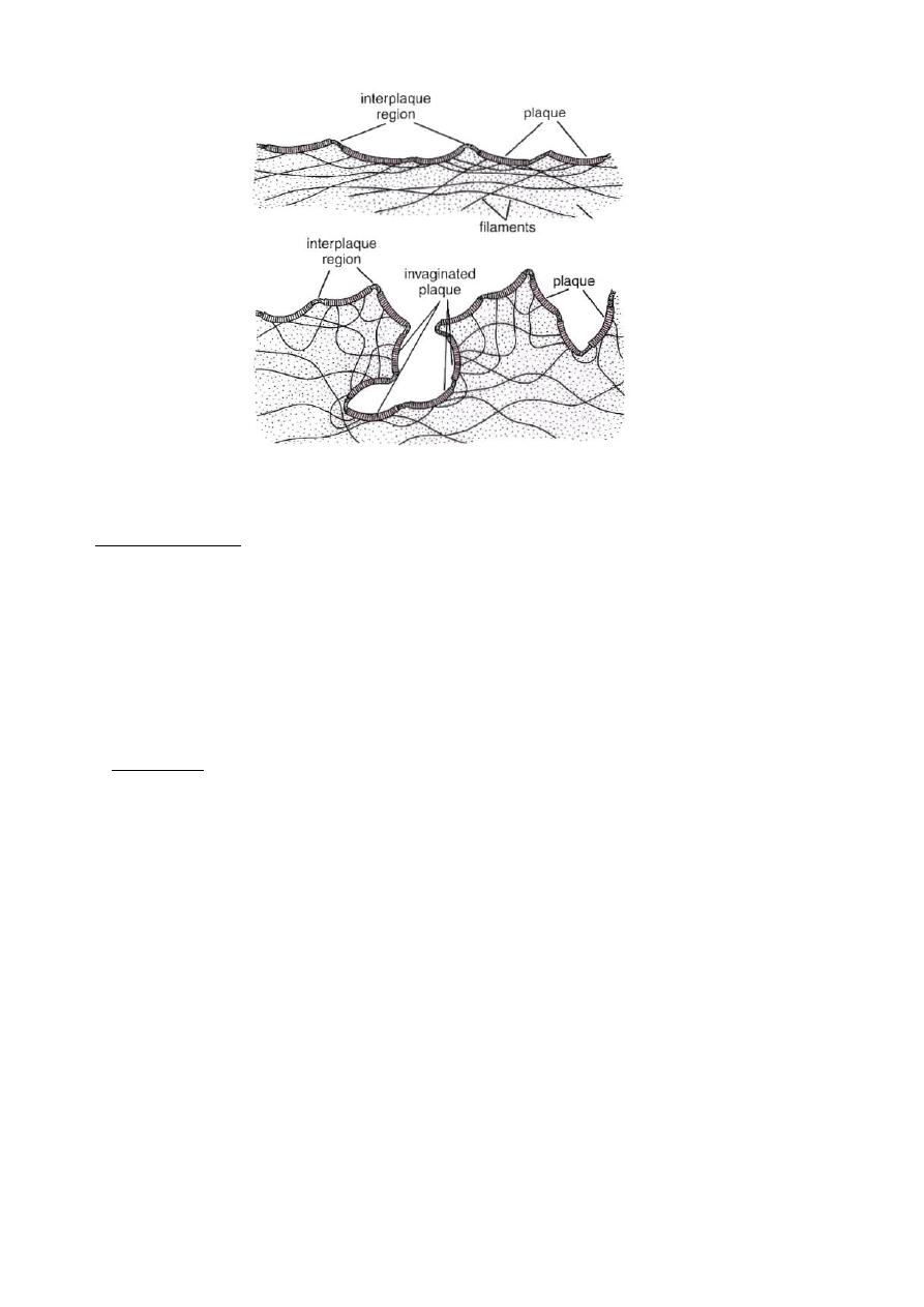
13
Plates of the membrane of superficial transitional epithelial cells
Lamina propria consists of loose connective tissue and diffuse lymphoid tissue.
2-Muscular layer: Thick layer that runs in every direction, without distinct
orientation. At the bladder neck(origin of urethra), the muscular layer will arrange
in three layers:
a- Inner longitudinal layer: it will become circular distal to the bladder neck and
surrounds the prostatic urethra in the male, and external meatus of the female,
forming the true involuntary urethral sphincter.
b- Middle circular layer: it ends at the bladder neck.
c- Outer longitudinal layer: continues to the end of the prostate in male, and the
external urethral meatus in female.
3- Adventitia: Loose connective tissue rich in blood vessels, except the upper part
of the bladder which is covered by serosa(because it is an intra peritoneal part).
14
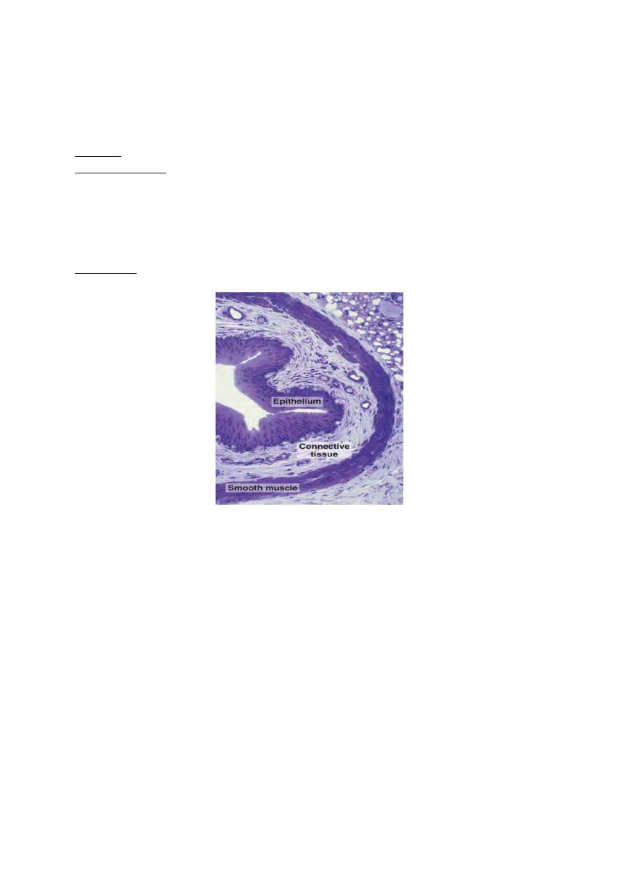
URETER
Renal calyces, renal pelvis, and ureters have the same histological structure. The
wall consists of three layers:
1- Mucosa: similar to that of the urinary bladder.
2- Muscular layer: it has a helical arrangement, then near the bladder, it will
become two layers; an inner longitudinal and outer circular. When the ureter
pass through the wall of the bladder, the muscles will become longitudinal only.
The ureter pass obliquely through the bladder forming a valve to prevent the
back-flow of urine. In addition, there is a flap of bladder mucosal membrane act
as a valve.
3- Adventitia: loose connective tissue.
URETHRA
It is a tube that carries urine to the outside.
Male urethra
It is divided into three parts:
1- Prostatic urethra: represents the initial part, surrounded by the prostate gland,
very close to the bladder. In the dorsal and distal part, there is an elevation;
verumontanum, that protrudes into the interior. The two ejaculatory ducts open
on the sides of the verumontanum, in addition to the prostatic glands. Seminal
fluid enter through these ducts to be stored before ejaculation. Prostatic urethra
is lined by transitional epith.
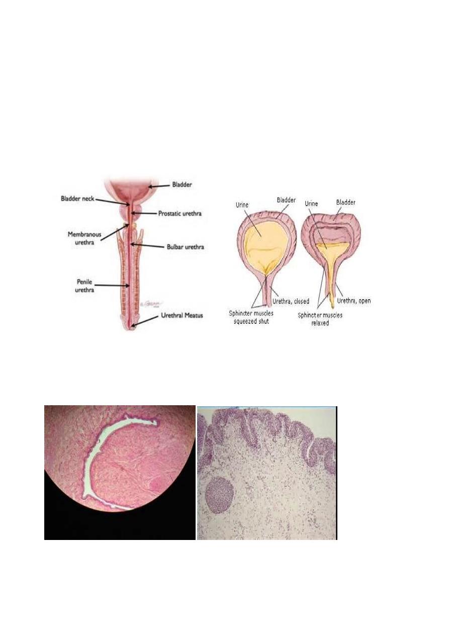
15
2- Membranous urethra: about 1cm long, extends from prostatic urethra to the
bulb of corpora cavernosa. It is lined by stratified or pseudostratified columnar
epith., and surrounded by striated muscle; the voluntary urethral sphincter.
3- Penile urethra: include both bulbous and pendulous urethra. Both extend
through the corpus spongiosum of the penis. They are lined by pseudostratified
columnar epith., or columnar epith. At the distal part, the lumen of urethra
dilates forming fossa navicularis, which is lined by stratified sequamous epith.
The lamina propria of the urethra is composed of loose connective tissue
rich in blood vessels and mucous glands; Littre's gland(mostly at the pendulous
part).
Female urethra
Short tube; about 4-5cm long, lined by pseudostratified epith., and near the
external orifice, it becomes stratified sequamous epith. The middle portion is
surrounded by the external voluntary sphincter.
Female urethra external orifice