
Dr. Thanaa Al-Khishali
THE EAR
VESTIBULOAUDITORY SYSTEM
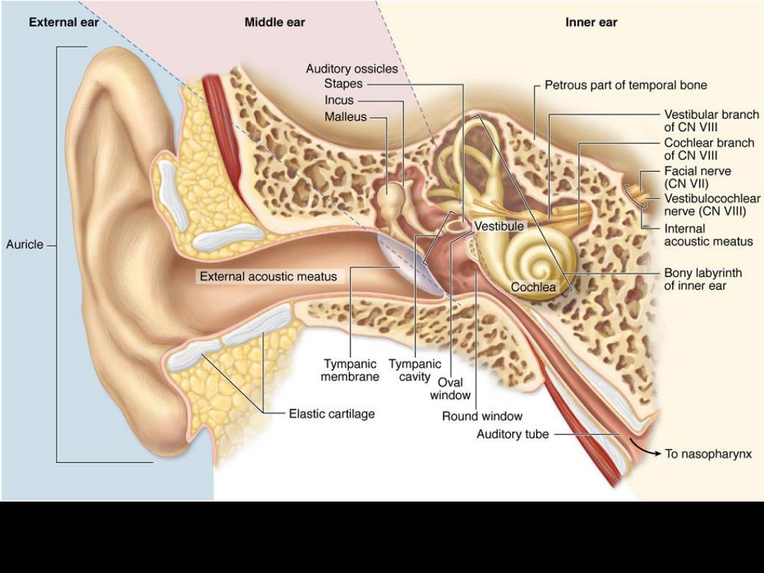
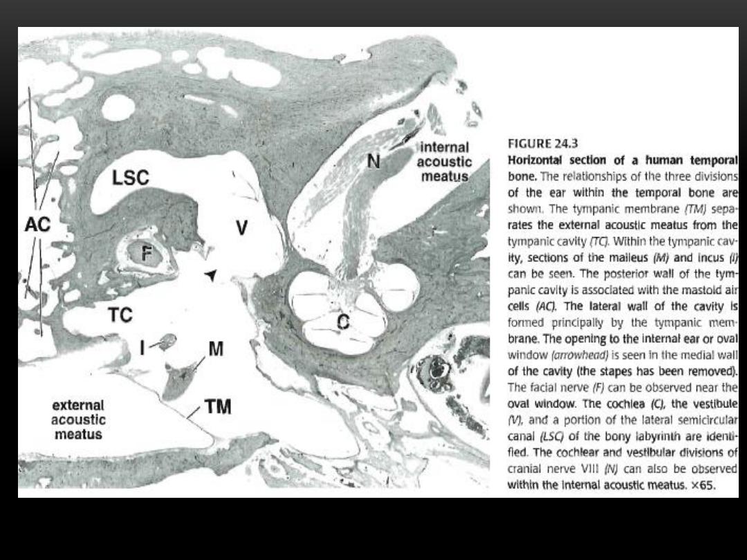
Major divisions of the ear, The external, middle, and internal region of the right ear
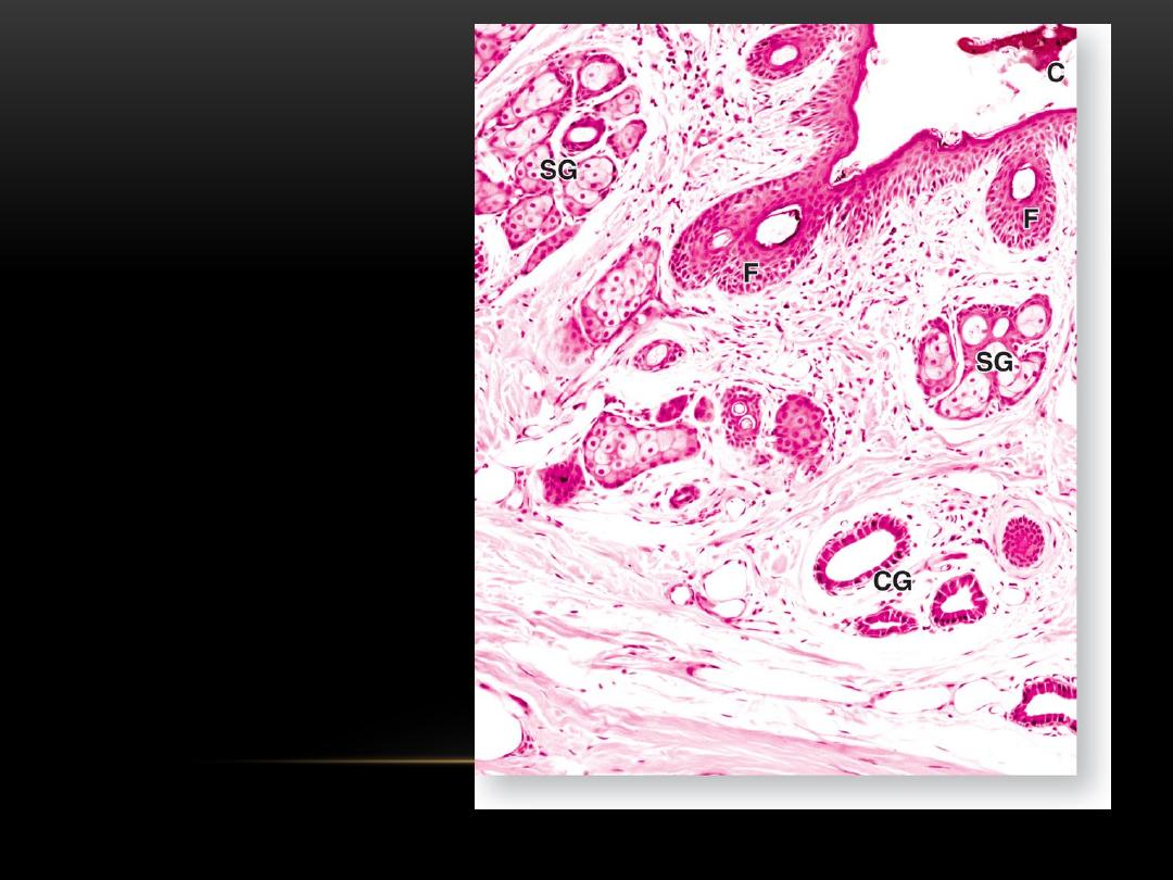
A section through the
external auditory meatus,
(SG) sebaceous gland,
(CG)ceruminous gland, (F)
hair follicle,
(C) cerumen
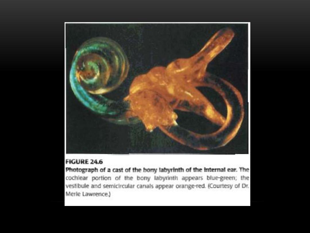
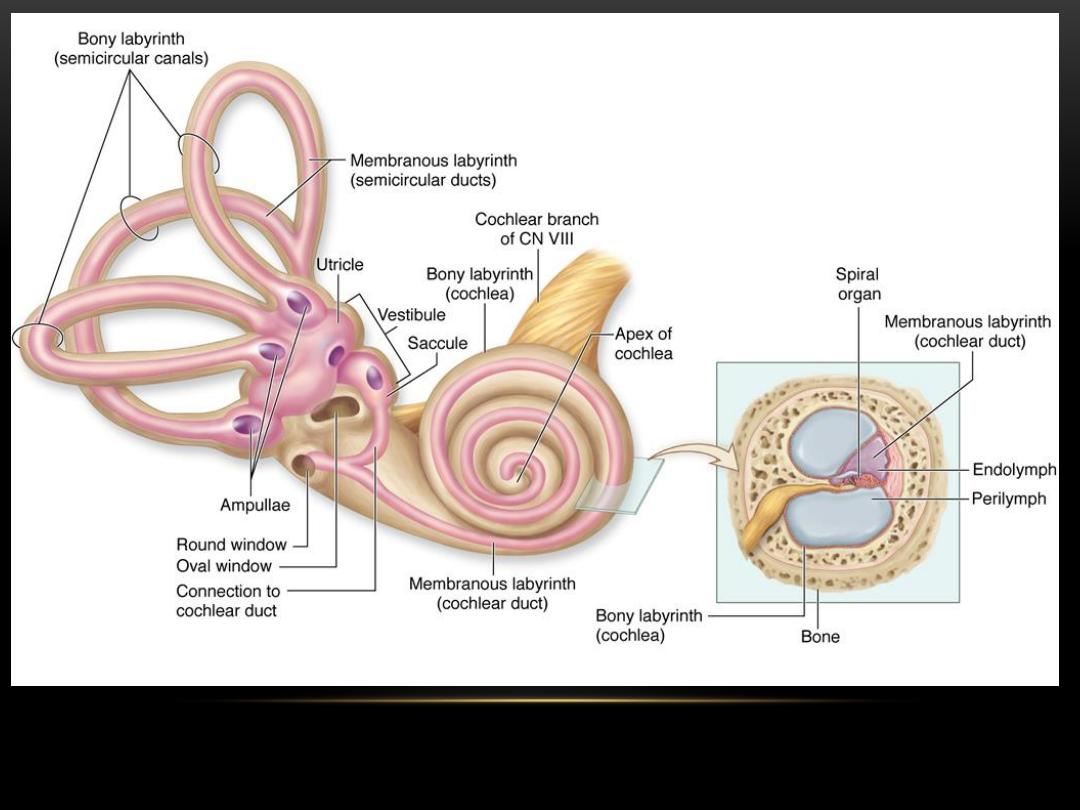
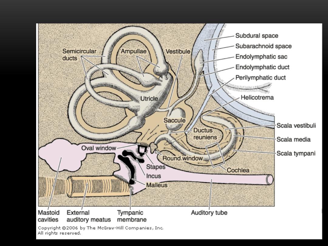
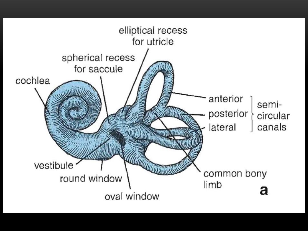
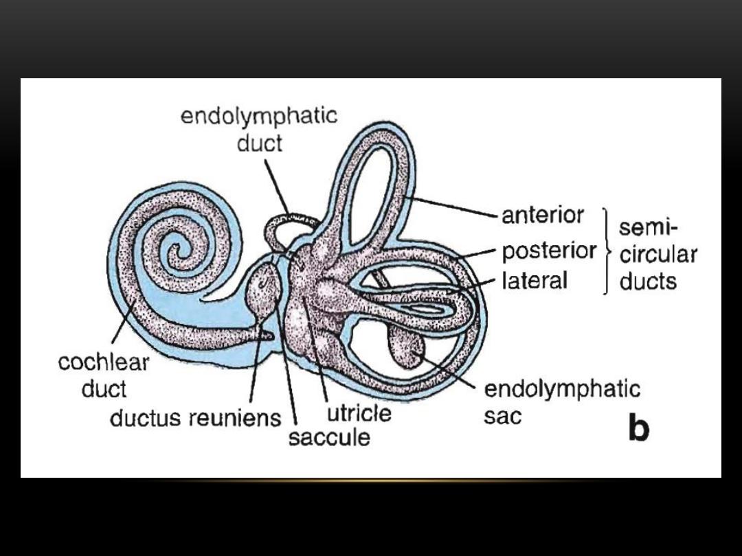
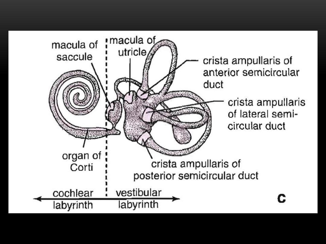
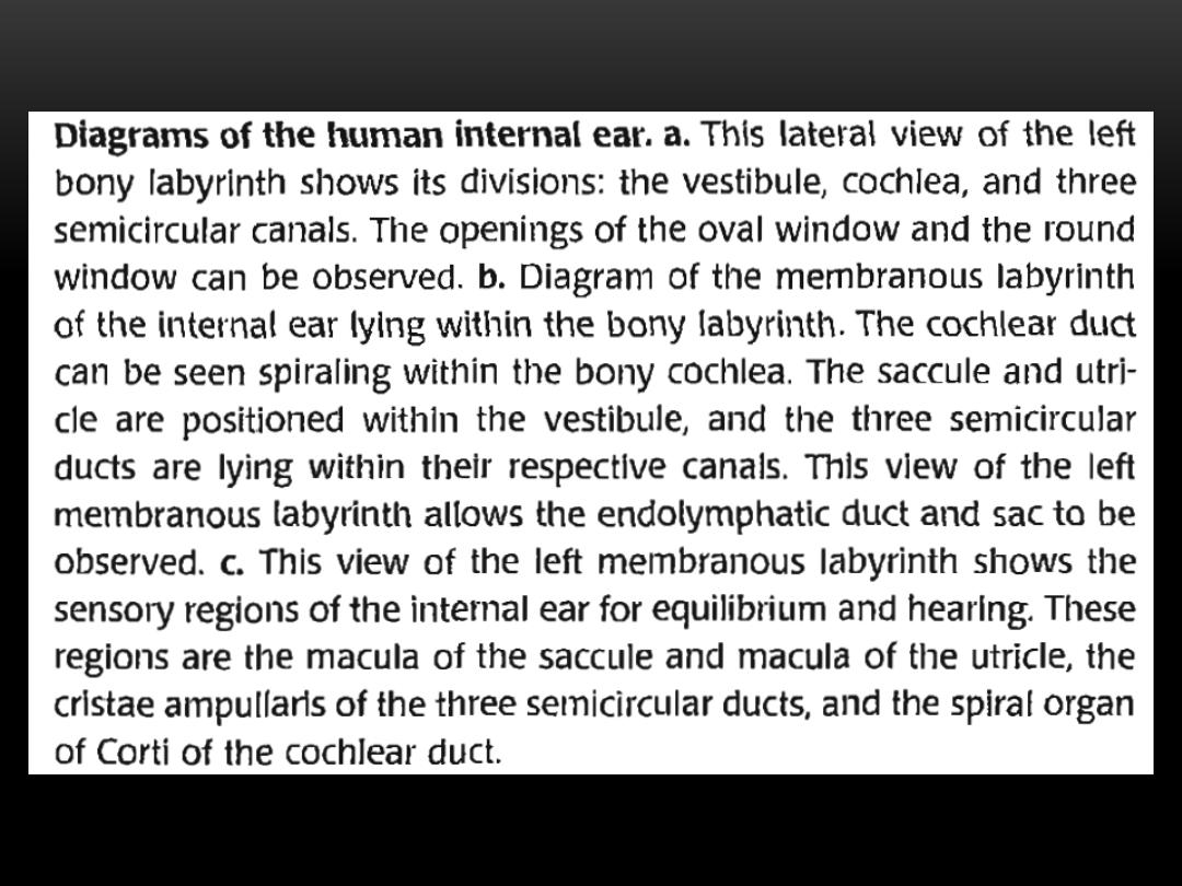
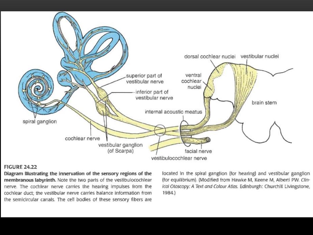
Internal ear
, the bony labyrinth, housing the membranous labyrinth which includes the saccule,
utricle, and semicircular ducts(equilibrium) and the cochlea for the sense of (hearing)
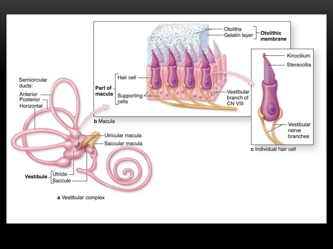
Structure of the macula
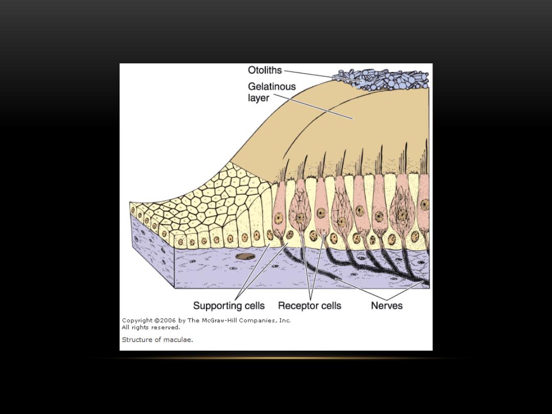
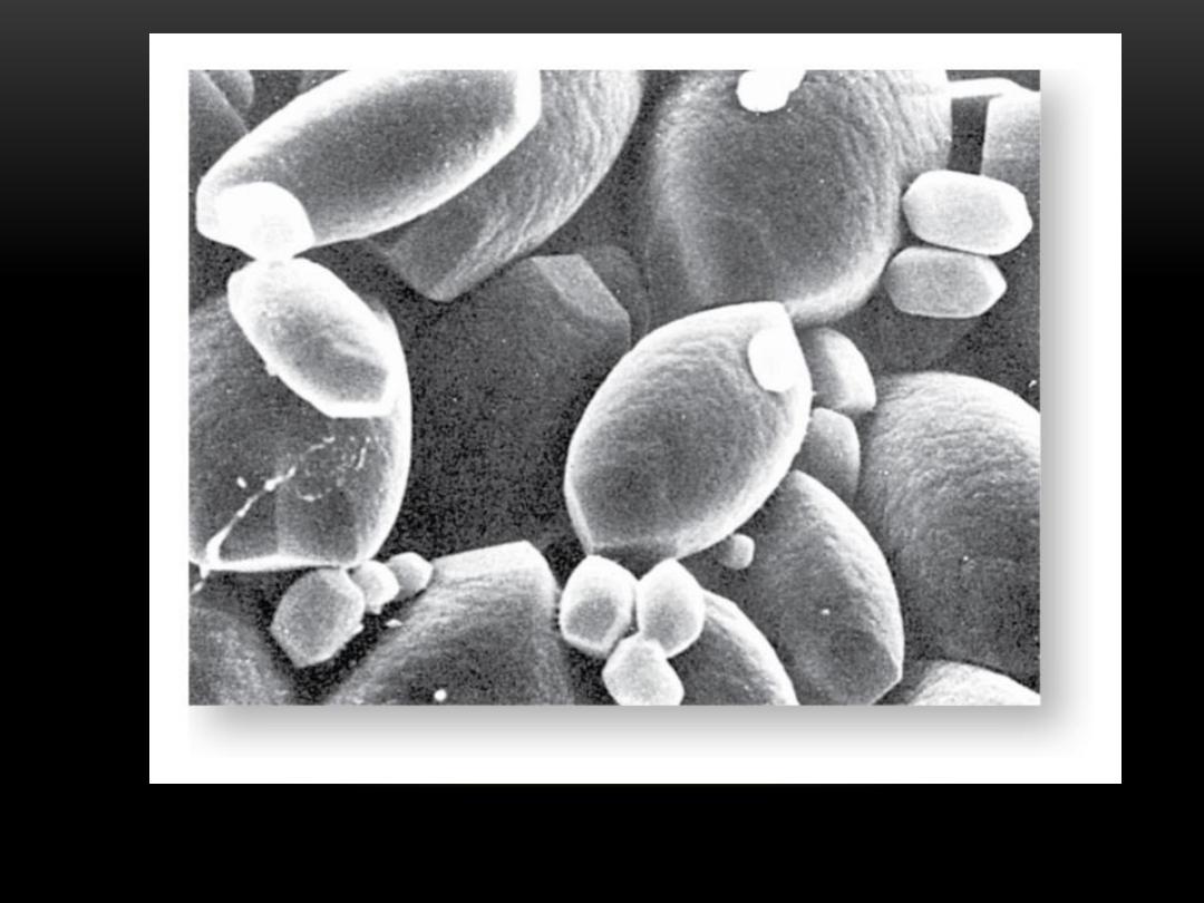
Scanning electron micrograph of the macula showing the otoliths
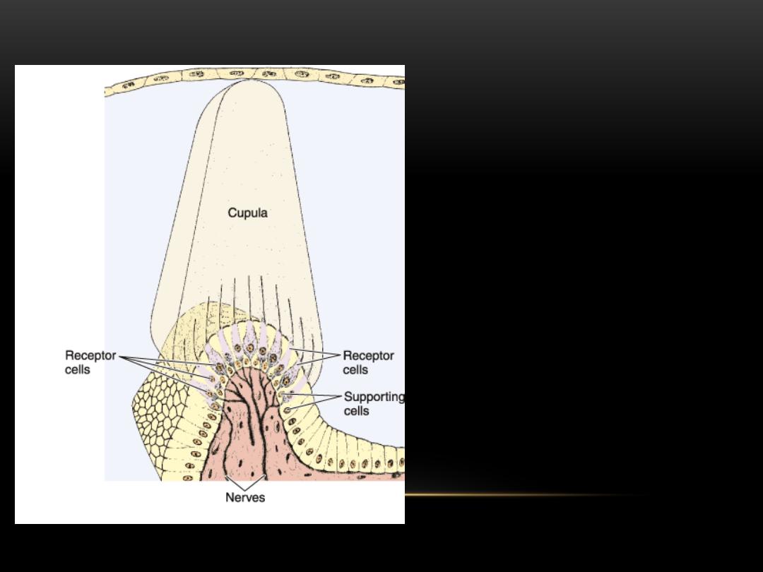
A diagram showing
the structure of
crista ampullaris
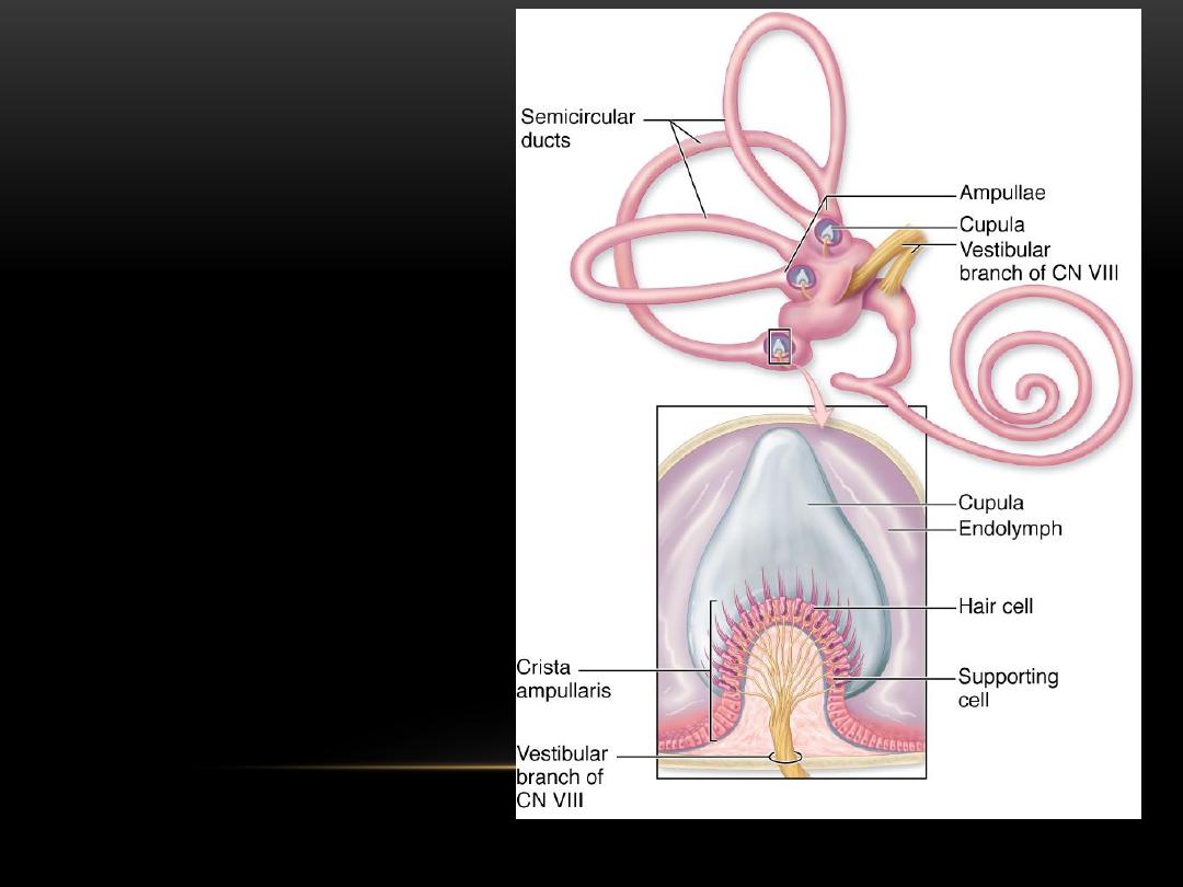
Diagram
showing the
structure of
crista ampullaris
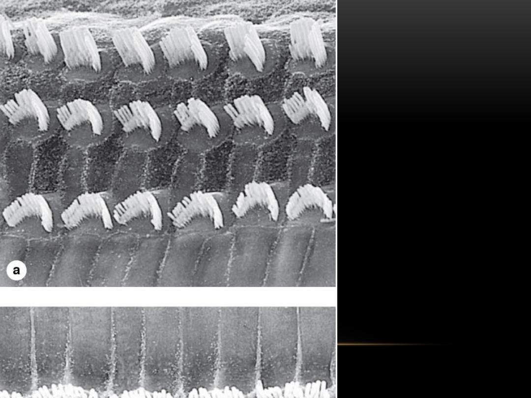
A scanning electron
micrograph showing
the hair cells
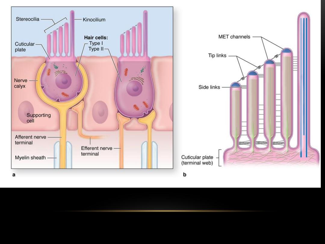
Hair cells
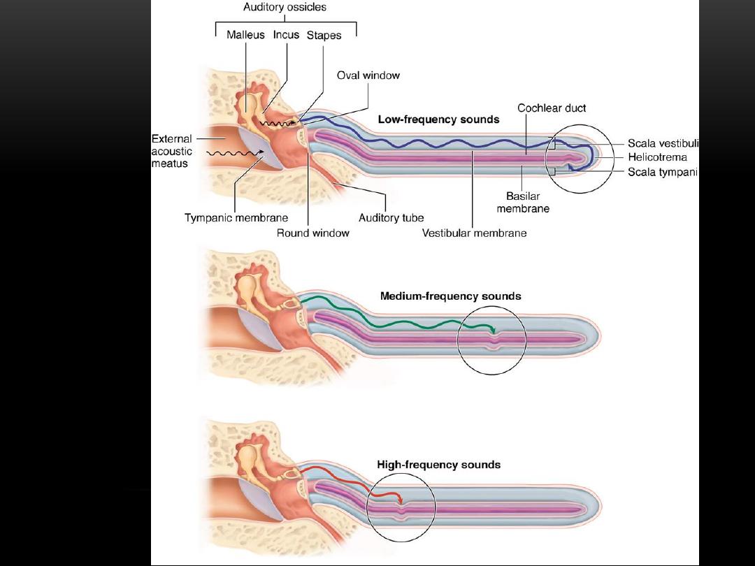
Cochlea
CoCchleaC
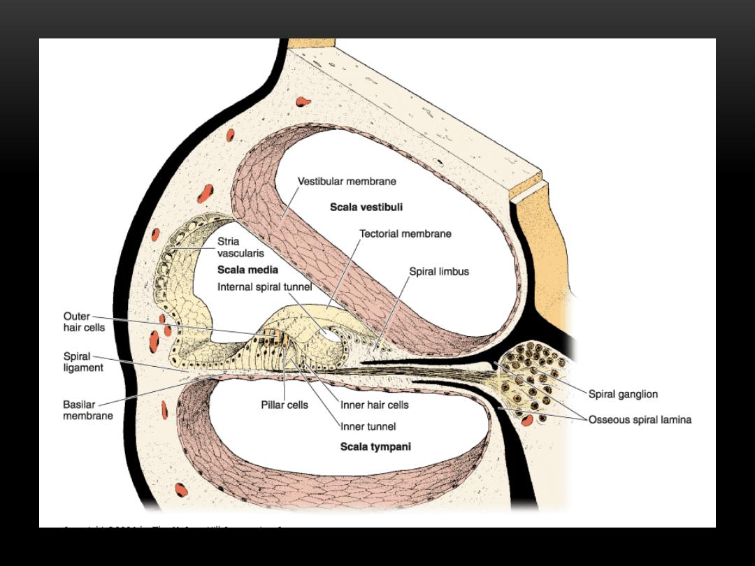
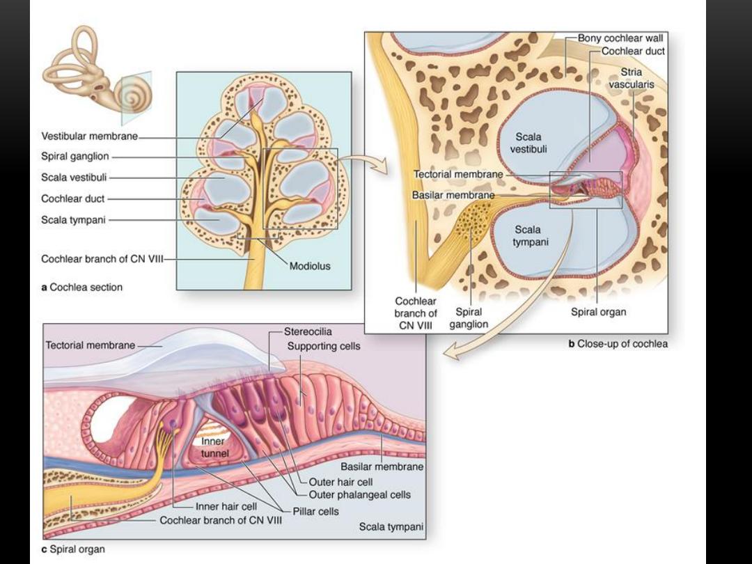
Structure of the cochlea
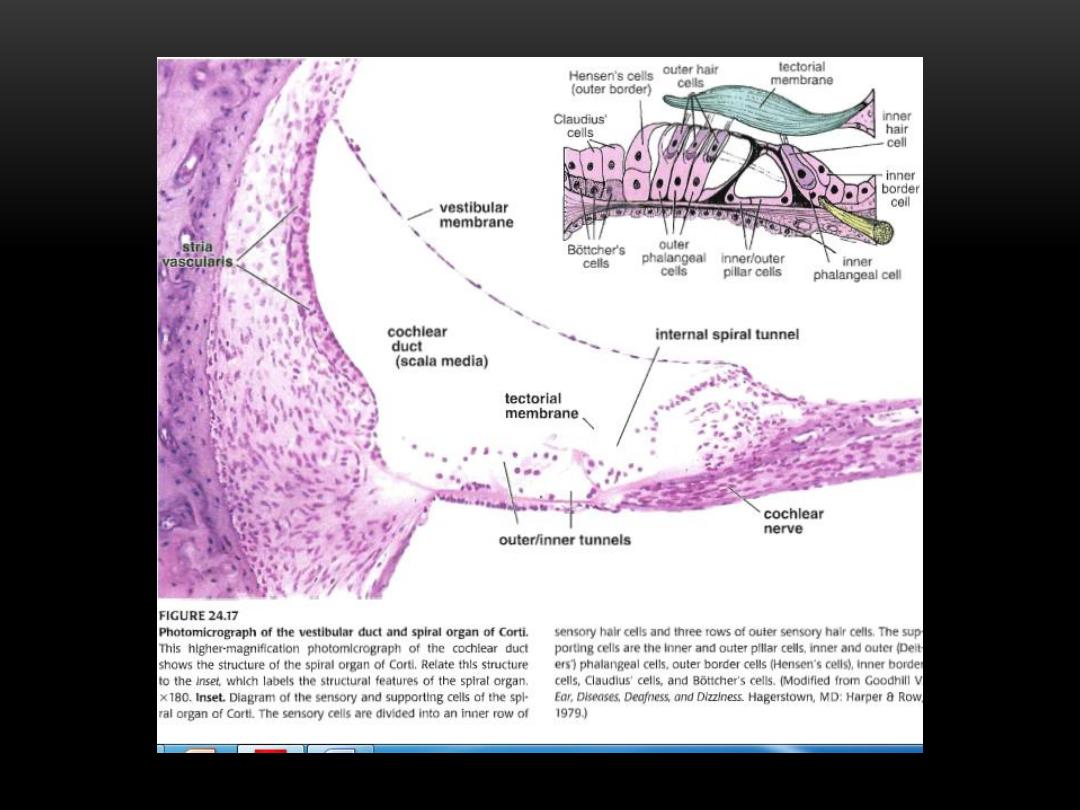
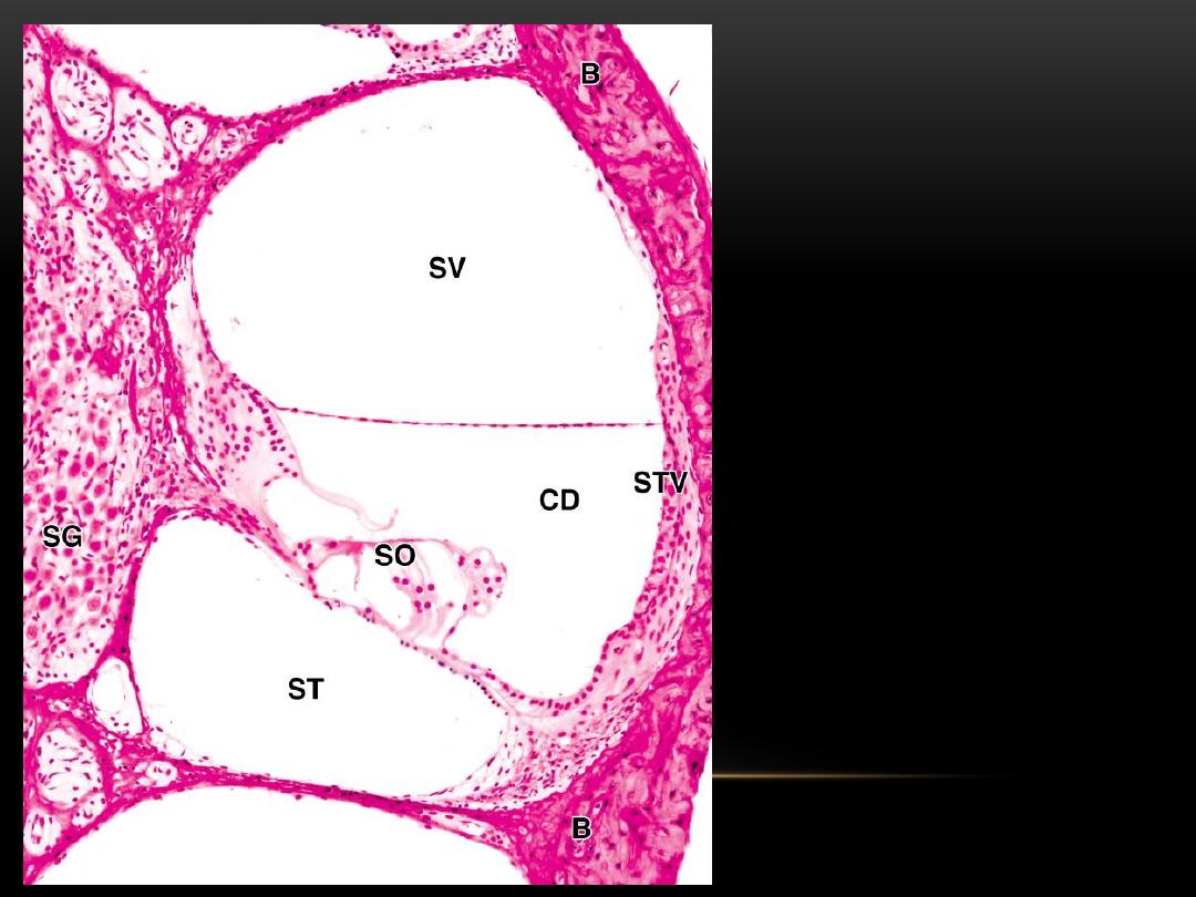
Cochlear duct and
spiral ganglion. (SO)
spiral organ, (CD)
cochlear duct filled with
endolymph produced by
stria vascularis (STV), B
bone. (SV) scala
vestibuli, and(ST) scala
tympani are filled with
perilymph. (SG) spiral
ganglion
THANK YOU