The Reproductive system
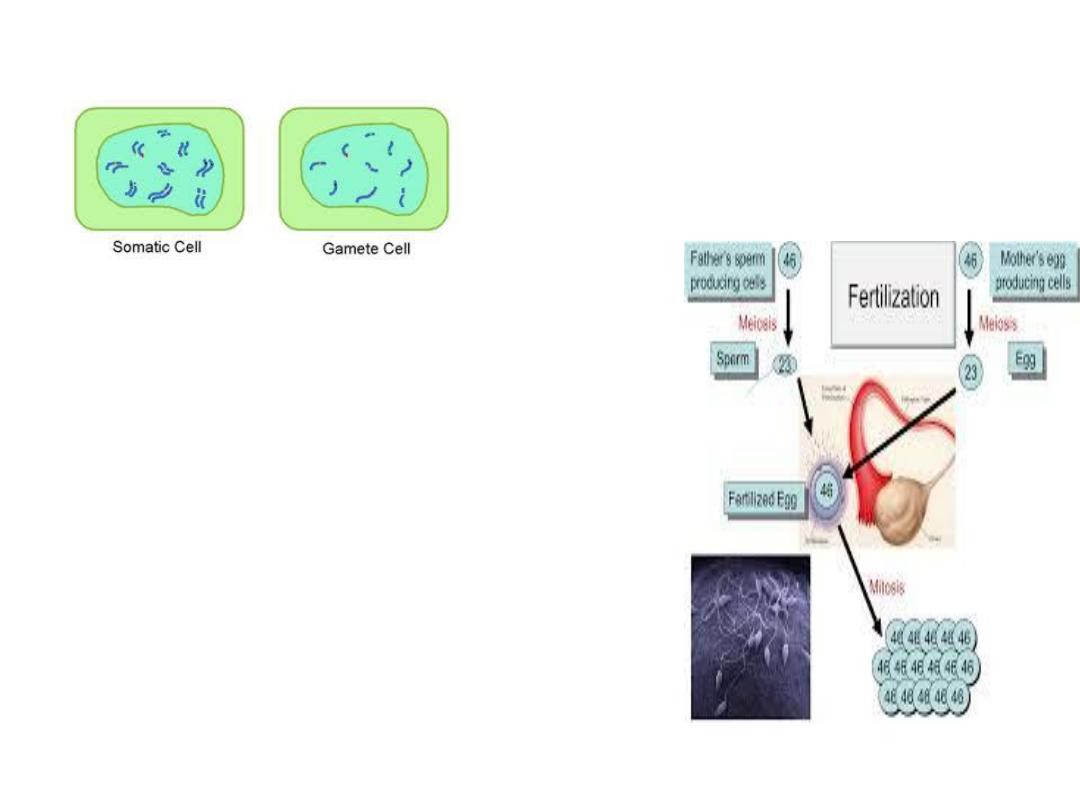
in males, 22 pairs of autosomes
plus an X chromosome and a Y
chromosome;
in females, 22 pairs of autosomes
plus two X chromosomes.
As a consequence of meiosis
during gametogenesis, each
normal ovum contains a single X
chromosome, but half the
normal sperms contain an X
chromosome and half contain a
Y chromosome .

• The differentiation of the primitive gonads in
utero is genetically determined in humans,
• but the formation of male genitalia depends
upon the presence of a
functional testis.
There is evidence that male sexual behavior
and, the male pattern of gonadotropin
secretion are due to the action of male
hormones on the brain in early development.
Development
of
the
Gonads
The gonad develops a
cortex and a medulla.
Until the
sixth
week of
development,
called
bipotential
gonads
which contains primitive
germ cells and has both
male
and
female
primordial genital ducts.
After the 6
th
week the
differentiation started.
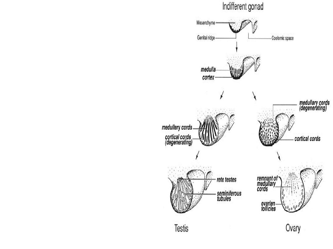
In genetic males, the
medulla develops into a
testis, and the cortex
regresses. Leydig and
Sertoli cells appear, and
testosterone
and
mullerian
inhibiting
substance are secreted.
In genetic females, the
cortex develops into an
ovary and the medulla
egresses. It will not
secret estrogen inutero,
so the secondary sex
organs
will
develop
without any hormonal
influence.
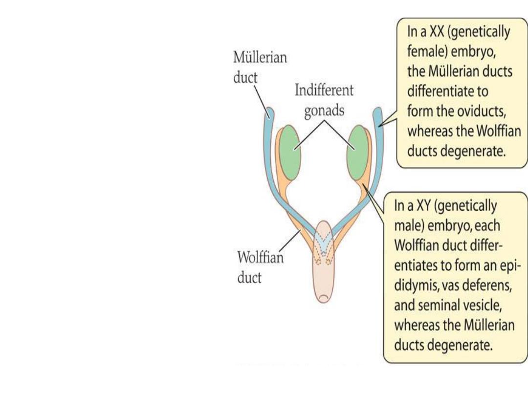
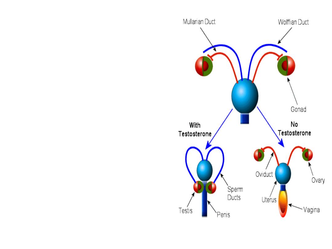
In a normal female
fetus, the mullerian
duct system then
develops into uterine
tubes (oviducts) and
a uterus. In the
normal male fetus,
the wolffian duct
system on each side
develops into the
epididymis and vas
deferens.
The
external genitalia are
similarly bipotential
until
the
eighth
week
.

When there are functional testes in the
embryo, male internal and external genitalia
develop.
The Leydig cells of the fetal testis secrete
testosterone (androgens), and the Sertoli
cells secrete mullerian inhibiting substance
(MIS) .
MIS causes regression of the mullerian ducts,
and testosterone fosters the development of
the vas deferens and related structures from
the wolffian ducts. The testosterone
metabolite induces the formation of male
external genitalia and male secondary sex
characteristics.

Note
that the determination of the
internal
sex organs
is
not under the influence of
androgens,
that’s why if we introduce
androgen into a genetically female fetus,
this does not influence the female pattern
of the internal sex organ,
but it will affect
the
external sex organ
and it will show a
male pattern.
ABERRANT SEXUAL
DIFFERENTIATION
Chromosomal Abnormalities
Abnormalities of sexual development
could be caused by genetic or
hormonal abnormalities or other
teratogenic causes.
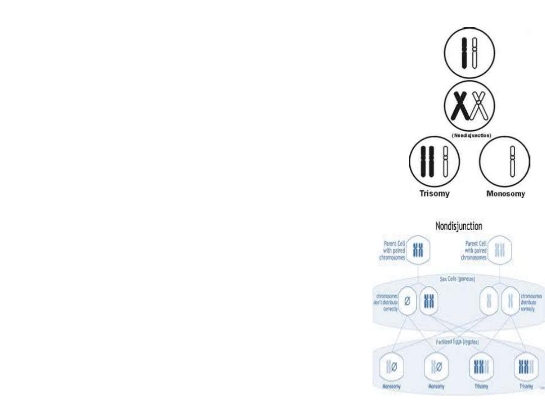
An established defect in
gametogenesis is nondisjunction,
a phenomenon in which a pair of
chromosomes fails to separate, so
that both go to one of the
daughter cells during meiosis.
Examples of these abnormalities
: 1- a syndrome called
gonadal dysgenesis
(or agenesis) or,
alternatively,
ovarian agenesis or
Turner's syndrome.
In these individuals
there is an
XO
chromosomal
pattern,

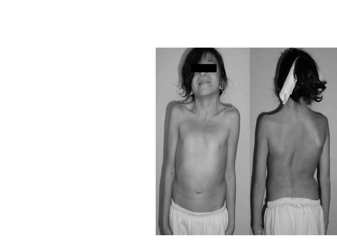
TURNER’S
• the gonads are rudimentary or absent, there is
female external genitalia. Stature is short,
webbing of the neck, other congenital
abnormalities are often present, and no
maturation occurs at puberty.

2- A syndrome known as seminiferous tubule
dysgenesis or
Klinefelter's syndrome
.
Individuals with the
XXY
pattern, the most
common sex chromosome disorder, have the
genitalia of a normal male. Testosterone
secretion at puberty is often great enough for
the development of male characteristics.
However, the seminiferous tubules are
abnormal, and there is a higher than normal
incidence of mental retardation, and usually
these patients are long.
.

3- The
XXX
("superfemale") pattern, it is more
common in general population, since it does
not seem to be associated with any
characteristic abnormalities.
4- The
YO
combination is probably lethal and
incompatible with life.

Nondisjunction or simple loss of a sex
chromosome can occur during the early
mitotic divisions after fertilization. The
result of faulty mitoses in the early zygote
is the production of a
mosaic
, an individual
with two or more populations of cells with
different chromosome complements.
.

True hermaphroditism
,
the condition in which
the individual has both ovaries and testes, is
probably due to XX/XY mosaicism and related
mosaic patterns, although other genetic
aberrations are possible.

• A pseudohermaphrodite
is an individual with
the genetic constitution and gonads of one sex
and the genitalia of the other. After the
thirteenth week, the genitalia are fully
formed, but exposure to androgens can cause
hypertrophy of the clitoris. Its causes either
due to congenital adrenal hyperplasia ,or it
may be caused by androgens administered to
the mother.

nondisjunction of several different autosomal
chromosomes is known to occur. For example,
nondisjunction of chromosome 21 produces
trisomy 21, the chromosomal abnormality
associated
with
Down's
syndrome
(mongolism).
In most instances, nondisjunction occurs in the
ovary rather than the testis and the incidence
of Down's syndrome increases with advancing
age
of
the
mother
.

Hormonal Abnormalities
Development of the male external genitalia
occurs normally in genetic males in response to
androgen secreted by the embryonic testes.
Genetic females exposed to androgens from
some other source during the eighth to the
thirteenth weeks of gestation, may develop
male genetalia. The syndrome that results is
female pseudohermaphroditism.
Male pseudohermaphroditism: development of
female external genitalia in genetic males. It
occurs when the embryonic testes are
defective. Because the testes also secrete MIS
(mullerian inhibiting substance or mullerian
regression factor), genetic males with
defective testes have female internal genitalia.

Another
cause
of
male
pseudohermaphroditism
is
androgen
resistance, in which, male hormones
cannot exert their full effects on the tissues.
One form of androgen resistance is a 5α-
reductase deficiency, in which the enzyme
responsible for the formation of the active
form of testosterone is decreased.
This androgen resistance may also due to
mutations in the androgen receptor gene.

When the loss of receptor function is
complete,
the
testicular
feminizing
syndrome, now known as complete
androgen resistance syndrome, results. In
this condition, MIS is present and
testosterone is secreted at normal or even
elevated rates.
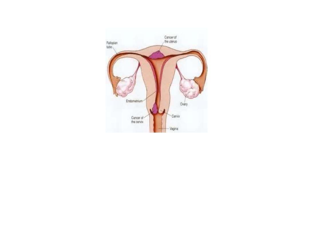
Female reproductive system
Female reproductive functions can be divided into two major
phases: (1) preparation of the female body for conception and
pregnancy, and (2) the period of pregnancy itself.
Ovaries:
From the sixth week of intrauterine life the embryonic ovaries
develop which contain the primordial follicles
the female needs only one ovum in each menstrual cycle.

Functions of the ovarian hormones—the ovary
secrets 3 hormones:
Estrogens
Progestins
Relaxin
ESTROGENS:
It is a steroid synthesized in the ovaries mainly
from cholesterol and secreted by the ovarian
theca
interna
cells,
corpus
luteum,
fetoplacental units and small amounts by the
adrenal cortices and the testes
.

There are three estrogens present in
significant quantities in the plasma of the
human female: b-estradiol, estrone, and
estriol, but The estrogenic potency of b-
estradiol is more than the others, it is
considered the major estrogen.
They are transported in the blood bound
mainly with plasma albumin and with
specific estrogen binding globulins.
Functions of the Estrogens
Growth of the tissues of the sex
organs
1-Effect of Estrogens on the Uterus
and External Female Sex Organs.
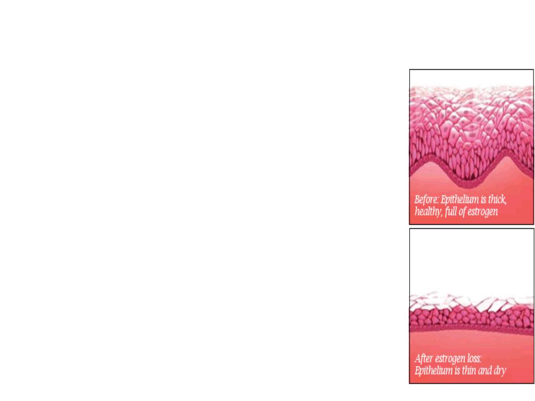
Estrogens change the type of cervical
mucosa and change the vaginal
epithelium from a cuboidal into a
stratified type (more resistant to
infection),
.
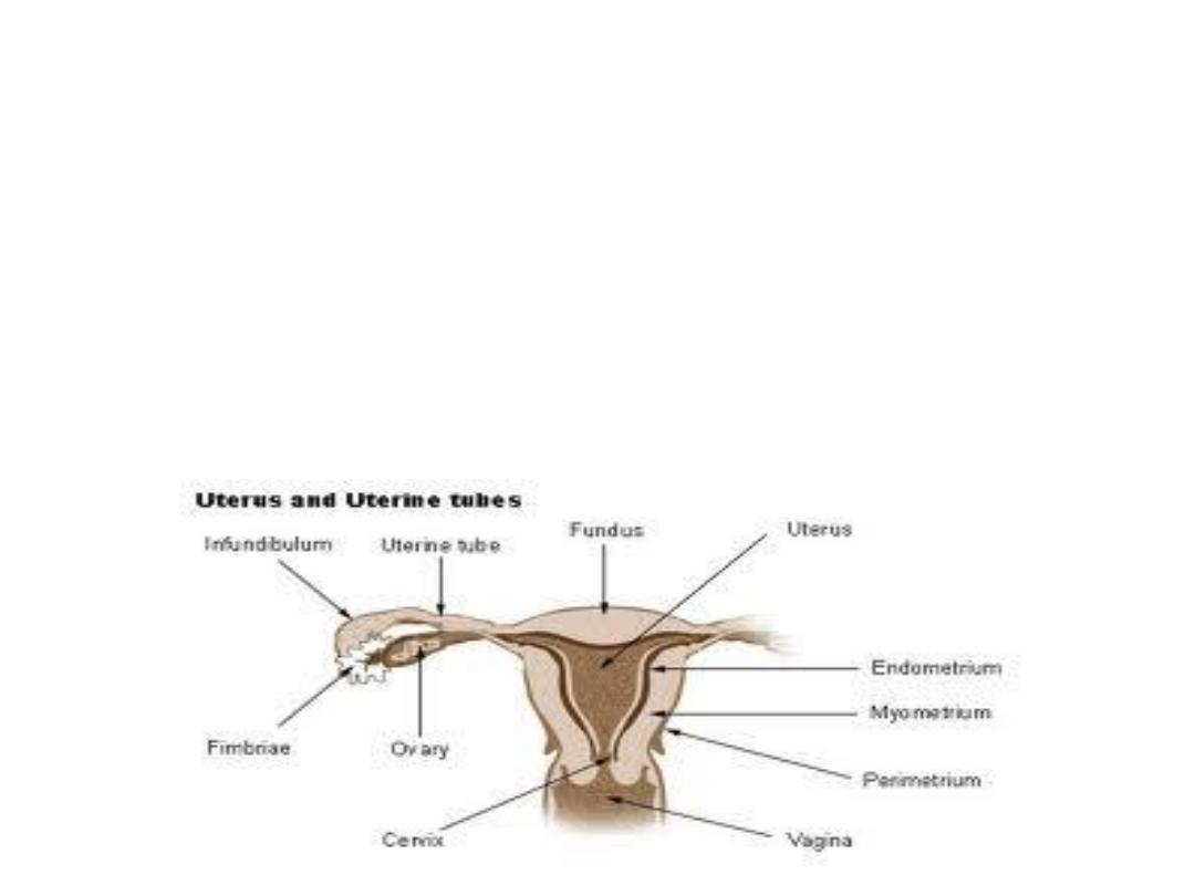
The external genitalia enlarge. The size of the
uterus will increase and the excitability of its
muscle will increase also. It also cause marked
proliferation of the endometrial stroma
(especially in the first half of the menstrual
cycle).
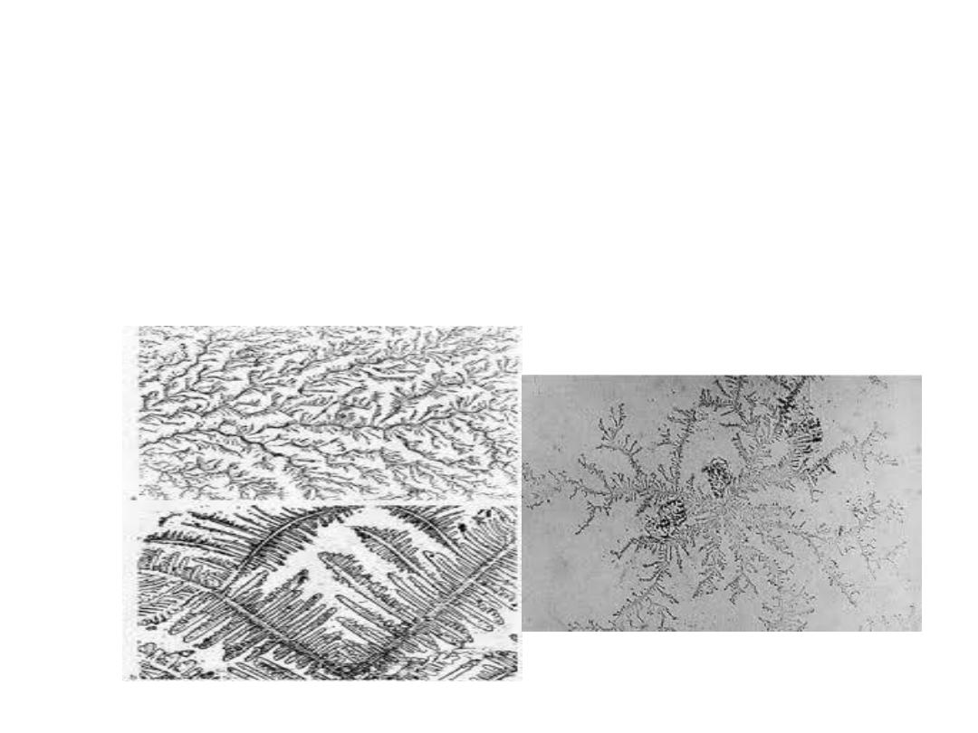
Estrogen makes the cervical mucosa
thinner and more alkaline, these
changes promotes survival and
transport of sperms

2-Effect of Estrogens on the Fallopian Tubes:
it causes increase in the mobility of the
fallopian tubes .
3-Effect of Estrogens on the Breasts.
It causes development of the stroma and
the ductile tissue of the breast in addition
to that it causes breast growth

4-Effect of Estrogens on the Skeleton.
It stimulates bone growth (and causes a slight
increase in total body protein). Also it
causes uniting of the epiphyses with the
shafts of the long bones. This effect of
estrogen in the female is much stronger
than the similar effect of testosterone in the
male.
After menopause and because of estrogen
deficiency there will be decrease of the
bone matrix (increase in osteoclastic
activity) and osteoporosis usually occurs.

5-Female sexual characteristics: estrogen
causes deposition of fat in the breasts and
subcutaneous tissues, also in the buttocks
and thighs (broad hip), which is
characteristic of the feminine figure.
6-Effect of Estrogens on the Skin. Estrogens
cause the skin to develop a texture that is
soft and usually smooth, also it causes high
ratio of scalp hair to the body hair.

7-Effect of Estrogens on Electrolyte Balance.
It Causes sodium and water retention by
the kidney tubules.
8-Estrogen also causes decrease in the
plasma cholesterol level, that’s why the
incidence of ischemic heart disease in
female is higher after menopause.

Mental health
Estrogen is considered to play a significant role
in women’s mental health. Sudden estrogen
withdrawal, fluctuating estrogen, and periods
of sustained estrogen low levels correlate with
significant mood lowering..