Hearing: and Equilibrium
Hearing
Organ of hearing: organ of Corti
Sensory receptor :cochlea duct
Nerve: cochlear nerve
Equilibrium:
Dancing with music
o
dynamic equilibrium
Organ of equilibrium
o
dynamic: crista ampularis (hair cells + cupula) of the semicircular
canals
vestibular nerve
o
static: maculae
Common characteristics – receptor for hearing & equilibrium
Hair cells bathed in endolymph
o
Hairs (cilia) of the cells are embedded in a dense mass
Movement of the mass of hair cells relative to one another stretches/bends the
cilia
o
Stretching/ bending of cilia in 1 direction increases impulse generation
Hearing (Audition)
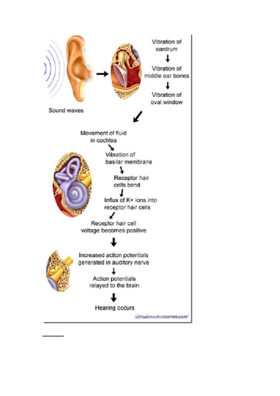
Outline of hearing (3 major steps)
1.Airborne sound consists of vibrations: alternate phases of condensation &
rarefaction; the auditory apparatus convert these vibrations in air to vibrations in the
inner ear fluid
2.Vibrations lead to bending of cilia of hair cells (of the Organ of Corti); Generate
nerve impulse: transmit along auditory nerve to higher centers of hearing
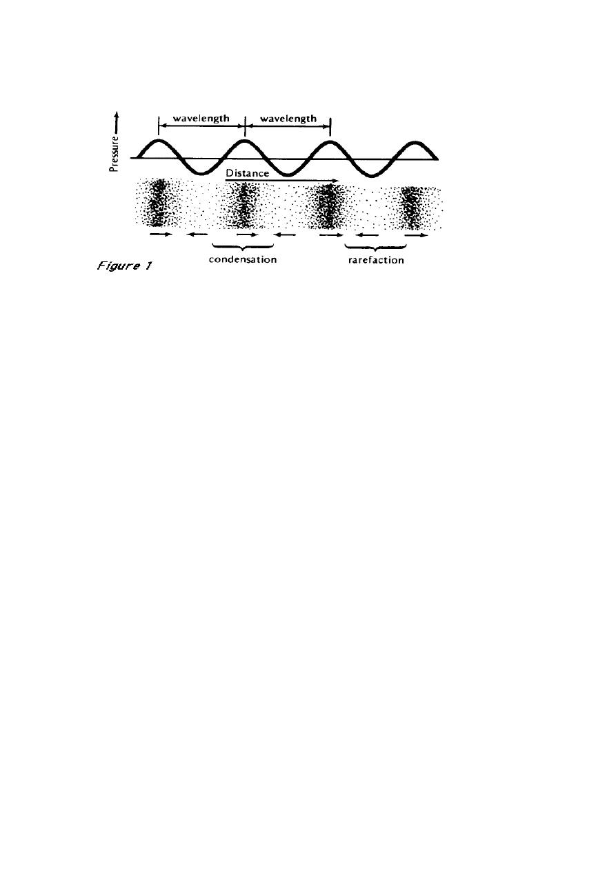
Sound
pressure wave produced by air by a vibrating body
o
results from the back & forth vibration of the particles of the medium
(air)
o
resulting in alternate phases of condensations (compressions) and
rarefactions
Properties
o
Greater amplitude (intensity of sound wave)
louder the sound
expressed as decibel (dB)
1 dB = 0.1 bel
1 bel
the logarithm of the ratio of the intensity of the
sound and a standard sound
0 dB
not absence of sound
sound level of an intensity equal to that of the
standard
threshold pressure
140 dB
potentially damaging to auditory receptor
organ of Corti
o
Greater frequency
higher the pitch
o
Soundwaves that have repeating patterns
perceived as musical sounds
o
Aperiodic nonrepeating vibrations
noisiness
Threshold for human hearing
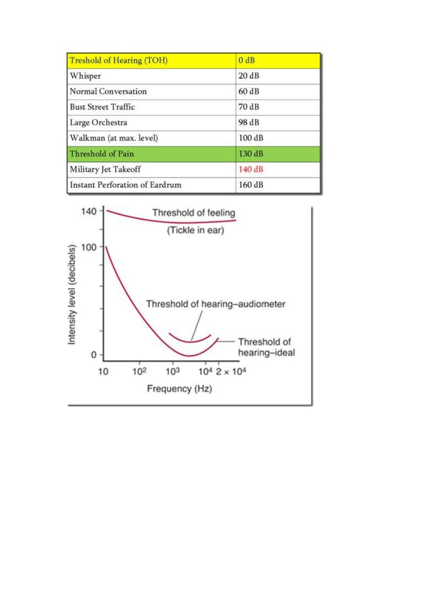
Threshold varies with the pitch of sound
Greatest sensitivity range
o
1000-4000 Hz
Audible frequency range
o
20 – 20,000 Hz
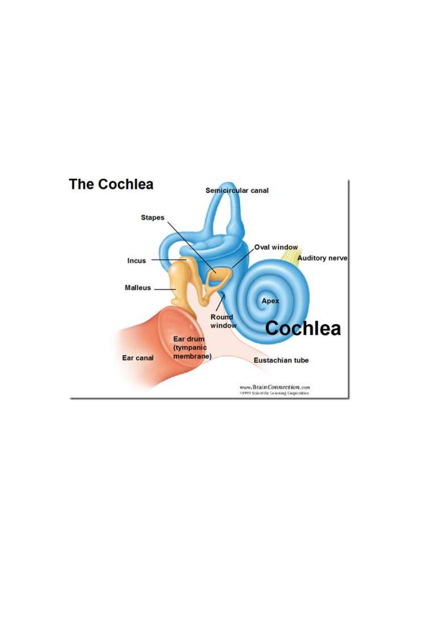
Hearing mechanism
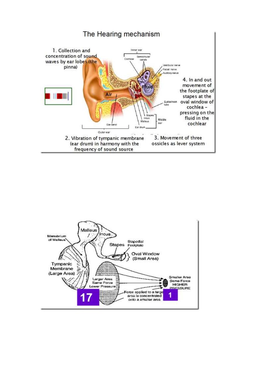
Collection & concentration of sound waves
o
by ear lobes (pinna)
Vibration of tympanic membrane
o
in harmony with the frequency of sound source
Movements of 3 ossicles
In & out movement of the footplate of stapes at the oval window of cochlea
o
pressing on the fluid in the cochlear
Impedance matching – function of the middle ear
Auditory receptors of the inner ear
o
operate in a fluid environment
o
underwater sound receiver
Effective transfer of sound energy from air (lower acoustic
resistance/impedance) to fluid (higher acoustic resistance/impedance)
o
is due to amplification of the pressure by:
large ratio btwn the areas of tympanic membrane & stapes
footplate-oval window (17:1)
amplified 17 times
pressure = force/area
mechanical advantage of the ossicular lever system
1:3
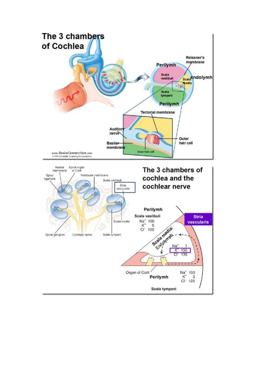
3 chambers of cochlea & cochlea nerve
Round window allows for fluid displacement in the cochlea
o
because fluid of the inner ear is not compressible
inward movement of the stapes footplate is allowed because of
the yielding of the thin membrane which covers the round
window
o
this is essential to the transmission process
since it provides elastic relief for the fluid of the inner ear
thus permitting movement of the stapes & the structure of the
inner ear
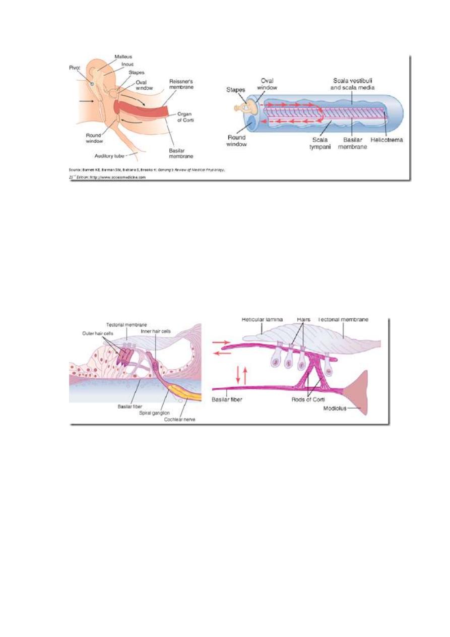
Mechano-electrical event
Hair cells & their stereocilia are ‘wedged’ between tectorial membrane &
reticular lamina
Vibrations transmitted by stapes
o
produce displacement of basilar membrane
o
up & down movement
Shearing movement between the tectorial membrane & reticular lamina
o
bends the hairs (stereocilia) of the hair cells of organ of Corti
o
Opening of mechanically-gated K+ channes in the stereocilia
K+ influx from endolymph into the stereocilia
depolarization of the hair cell membrane
o
Opening of voltage-gated Ca2+ channels
Ca2+ influx
o
Release excitatory neurotransmitter
glutamate
When basilar membrane bends towards scala vestibuli (medially)
o
hair cells depolarized
o
Opposite direction
hyperpolarize
o
generating alternating hair cell receptor potential
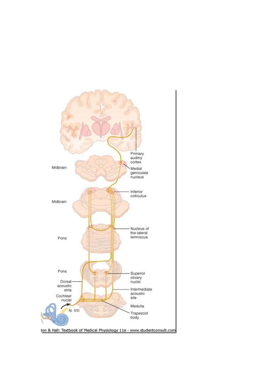
Auditory Pathways
Receptors
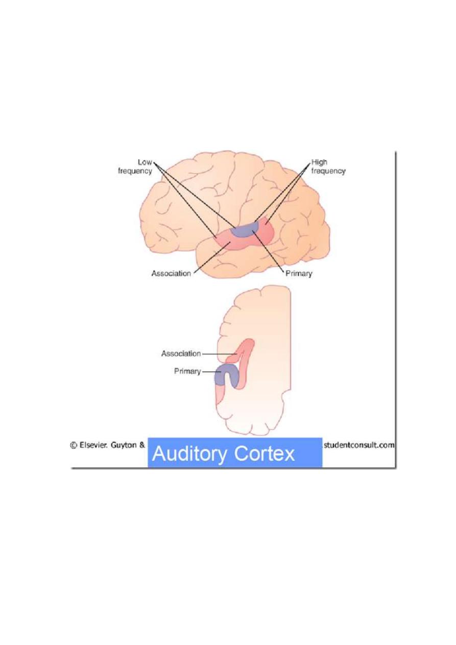
o
cochlear division of 8th CN
Cochlear nuclei in the medulla oblongata
Inferior colliculi
Medial geniculate bodies of thalamus
Thalamic radiation
Cortical auditory centres in temporal lobe
Auditory cortex
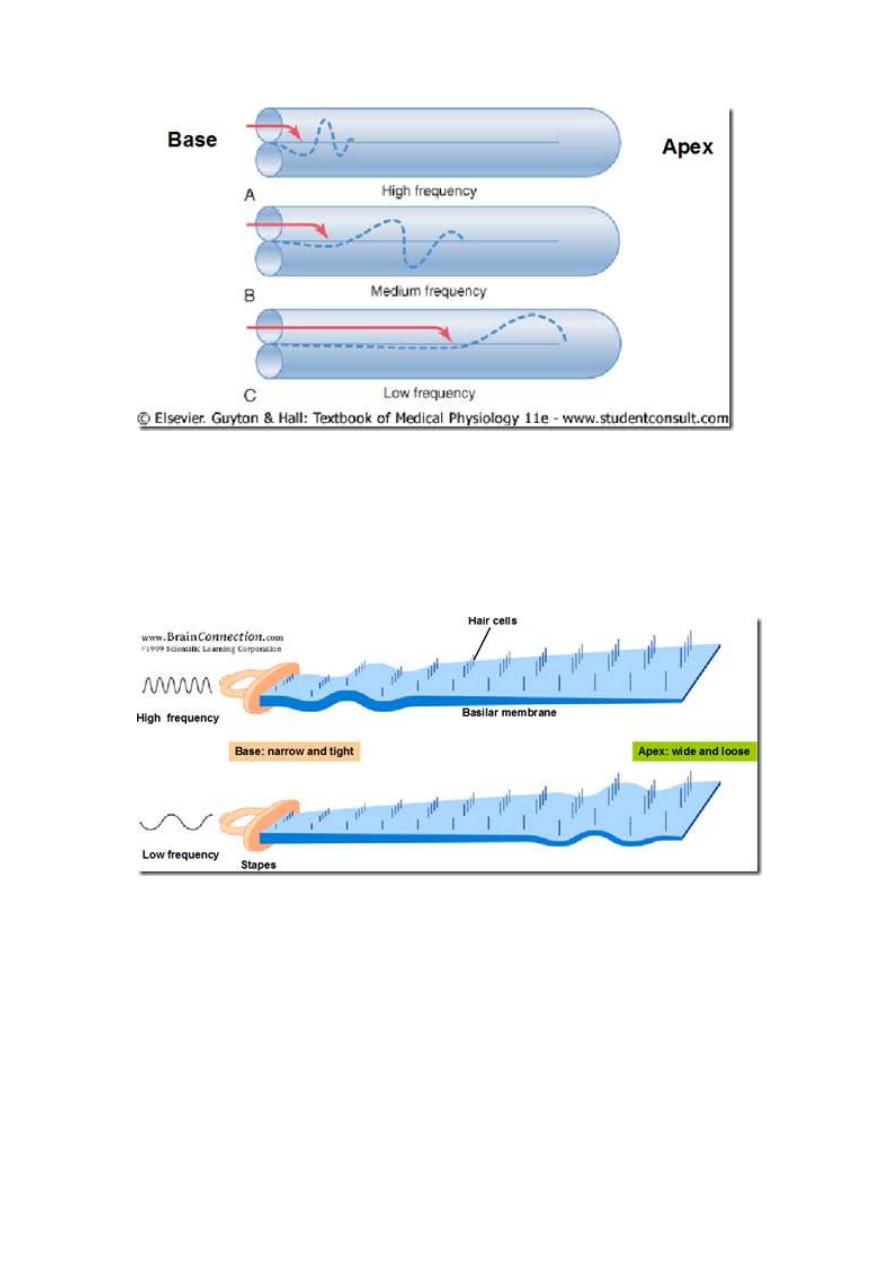
Pitch discrimination
Sounds of different frequencies travel different distances down the basilar
membrane
o
low pitched – apex
o
high pitched – base
Why?
o
the width & stiffness of the basilar membrane vary from the apex to
the base
Intensity discrimination
Greater the degree of displacement of the hair cells
o
more hair cells displaced
o
hence more nerve fibres are stimulated
Summary of hearing physiology
Deafness
Types of deafness

Conductive deafness
o
pathology in external/middle ear
o
impaired sound conduction
o
common causes
plugging of the external auditory canals with wax (cerumen) or
foreign bodies
otitis externa
inflammation of the outer ear
swimmer’s ear
otitis media
inflammation of the middle ear
causing fluid accumulation
hyperemic swelling & increased mucus
production associated with an upper respiratory
infection leads to temporary closing of the
Eustachian tube
-ve pressure develops within the middle
ear
distention of the tympanic membrane
perforation of the eardrum
osteosclerosis
bone is resorbed and replaced with sclerotic bone
grows over oval window
fixation of the stapes in the oval window
Sensorineural deafness/nerve deafness
o
pathology in cochlear/auditory neural pathways
o
common causes
presbycusis
hearing loss with aging
loss of hair cells & neurons
ototoxicity
aminoglycoside antibiotics (streptomycin, gentamycin)
obstruct the mechanosensitive channels in the
stereocilia of the hair cells
cause cells to degerate
damage to hair cells by prolonged exposure to noise
tumours in
8th cranial nerve
cerebellopontine angle
vascular damage in the medulla
Mixed deafness
Tinnitus
What is tinnitus?
o
conscious experience of sound that originates within the head
not originating from external source
o
may take many forms
roaring noise
tones and clicks
intermittent/continuous
Bone conduction
What is bone conduction?
o
direct conduction of sound to the inner ear through the bones of the
skull
bypassing the external auditory canal & middle ear
Differentiate between conduction and sensorineural deafness
Some hearing aids employ bone conduction
o
effect equivalent to hearing directly from ear
Recreational use
o
Audio Bone 1.0
o
safer for eardrums
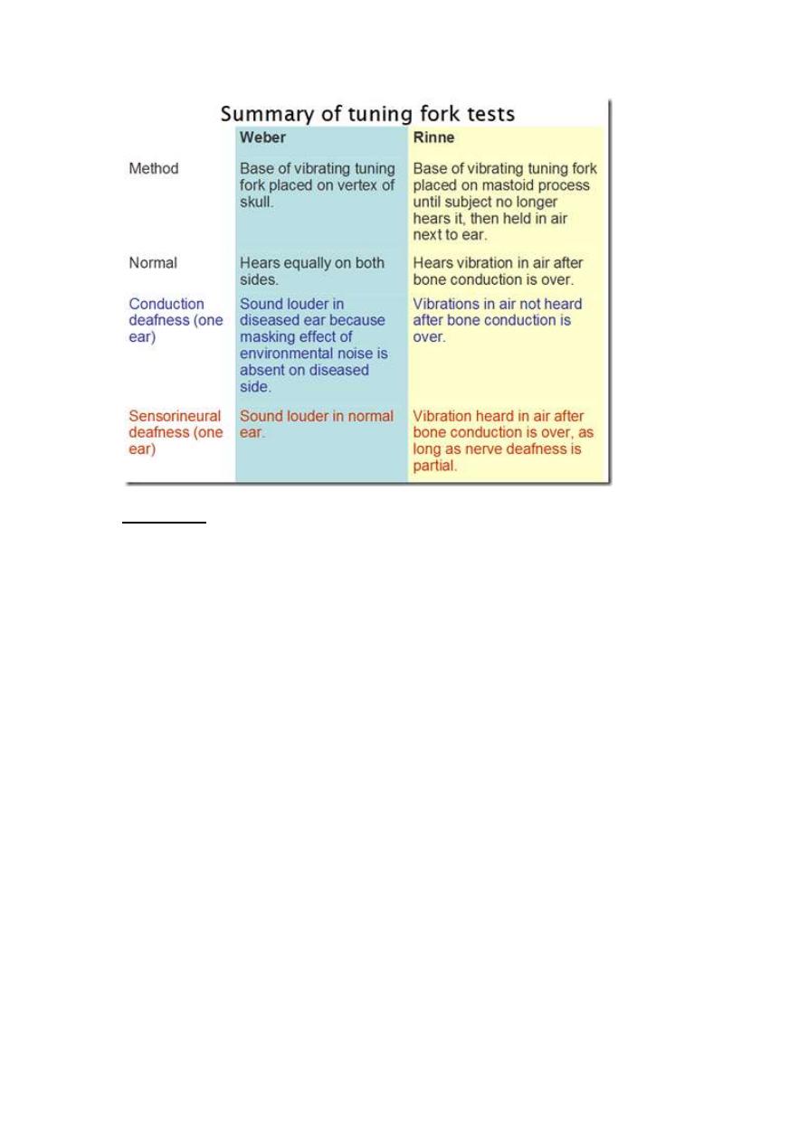
Tuning fork tests
Differentiate between conduction and sensorineural deafness
Based on principle
o
air conduction (AC) is better than bone conduction (BC)
o
AC is subjected to the ‘masking efect’ of environmental noise
Rinne’s test
Vibrating tuning fork placed on mastoid process
o
then place beside the ear when the sound stops
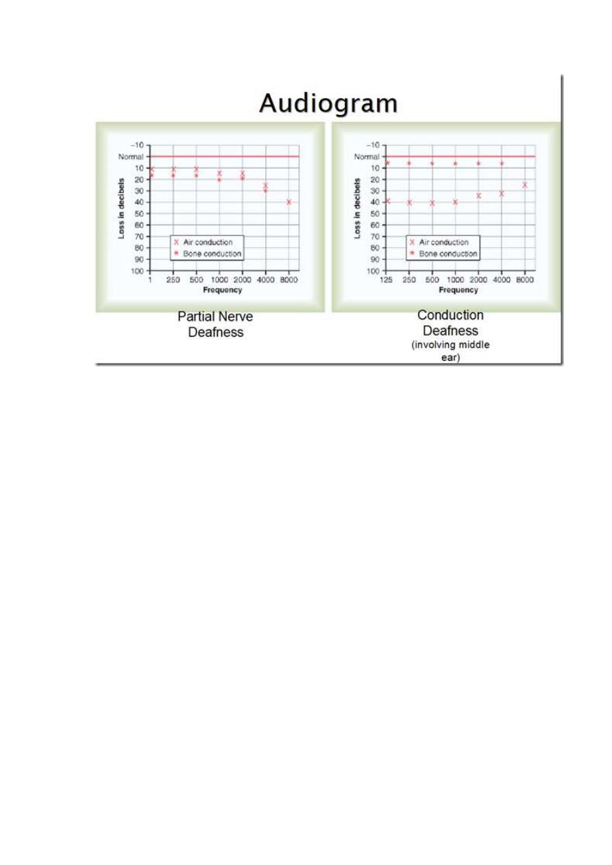
Results:
o
sound heard better when held infront of ear (AC>BC)
+ve test
normal hearing/ partial nerve deafness
o
sound heard better over the bone (BC>AC)
-ve test
conduction deafness*
Can’t really tell if there’s sensorineural deafness with this test
Weber’s test
Vibrating tuning fork placed centrally on the forehead
Results:
o
Sound is heard equally on both sides
normal
o
Sound is localized on 1 side
side of sound: conduction deafness
there is masking of sound from environment
no nerve damage, just bone conduction is defective
no sound at all on 1 side: nerve deafness
Useful in differentiating the type of hearing loss
Audiometry
Audiometer – measure auditory acuity
o
pure tones of various frequencies through earphones
At each frequency
o
threshold intensity is determined & plotted on a graph as a % of
normal hearing
Provides objective measurement of the degree of deafness
o
and the tonal range that is most affected
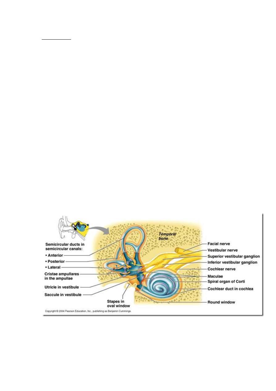
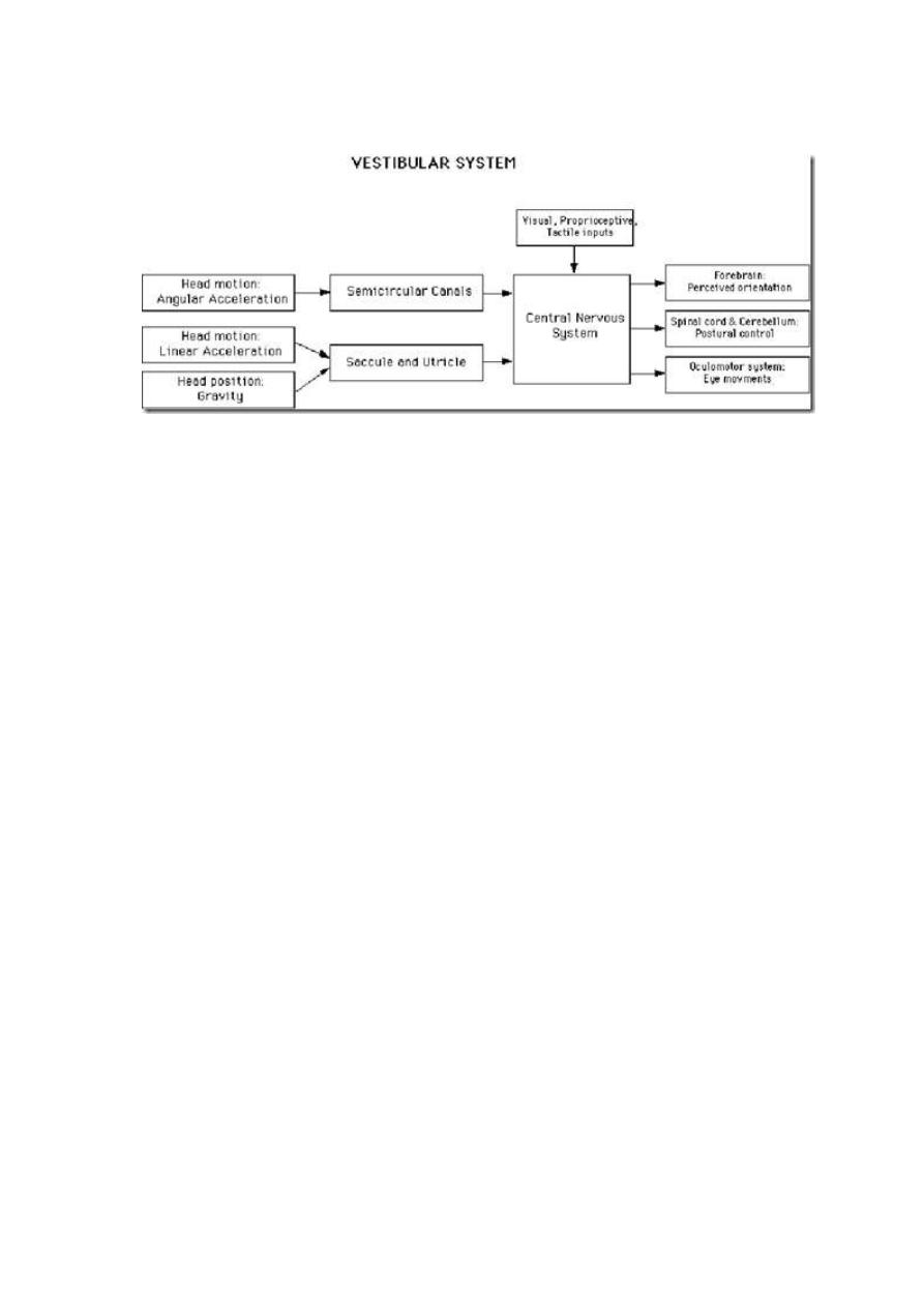
Equilibrium
Sensory organ
o
vestibular apparatus
encased in a system of bony tubes & chambers located in the
petrous portion of the temporal bone
bony labyrinth
within this system are membranous tubes & chamgers
membranous labyrinth
functional part of the vestibular apparatus
Vestibular functions
o
Responding to gravity & acceleration
maintains:
body posture
labyrinthine reflexes
equilibrium
vestibulocerebellar connections
o
Gives subjective sensation to motion & spatial orientation
along with visual, proprioceptive and cutaneous (exteroceptive)
inputs
o
Vestibular input to regions of the nervous system controlling eye
movements
helps stabilize the eye in space during head movements
reduced movement of the image of a fixed object on the retina
vestibulo-ocular reflex (VOR)
Vestibular apparatus
Vestibular portion of the labyrinth (filled with endolymph) consists of:
o
vestibule
utricle
saccule
o
3 semicircular canals
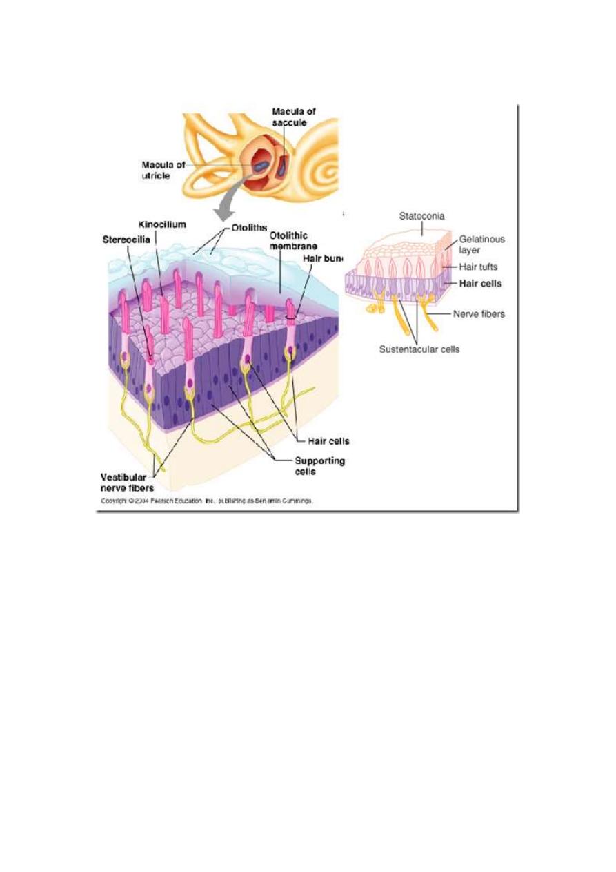
Vestibular receptors
Macula (otolith organ) in
o
Utricle
oriented in horizontal plane
Responds to:
changes in head position
fore & aft lift
linear acceleration in horizontal plane
running
o
Saccule
oriented in vertical plane
Responds to:
changes in head position
lateral lift
linear acceleration in vertical plane
jumping down
o
Crista ampularis
in each of the expanded ends (Ampulla) of the 3 semicircular
canal
detect angular/rotational acceleration
Detection of linear & rotational acceleration
Linear acceleration
When the body is suddenly thrust forward-that is, when the body accelerates-
o
the statoconia, which have greater mass inertia than the surrounding
fluid, fall backward on the hair cell cilia, and information of
dysequilibrium is sent into the nervous centers, causing the person to
feel as though he or she were falling backward.
o
This automatically causes the person to lean forward until the resulting
anterior shift of the statoconia exactly equals the tendency for the
statoconia to fall backward because of the acceleration.
o
At this point, the nervous system senses a state of proper equilibrium
and leans the body forward no farther.
o
Thus, the maculae operate to maintain equilibrium during linear
acceleration in exactly the same manner as they operate during static
equilibrium
Rotational acceleration
-when stereocilia bend towards kinocilium –> stimulation
-when sterocilia bend away from kinocilium –> inhibition
Rotational acceleration in the plane of a given semicircular canal stimulates its
crista.
The endolymph, because of its inertia, is displaced in a direction opposite to
the direction of rotation. The fluid pushes on the cupula, deforming it. This
bends the processes of the hair cells.
When a constant speed of rotation is reached, the fluid spins at the same rate
as the body and the cupula swings back into the upright position.
When rotation is stopped, deceleration produces displacement of the
endolymph in the direction of the rotation, and the cupula is deformed in a
direction opposite to that during acceleration.
It returns to mid position in 25 to 30 s.
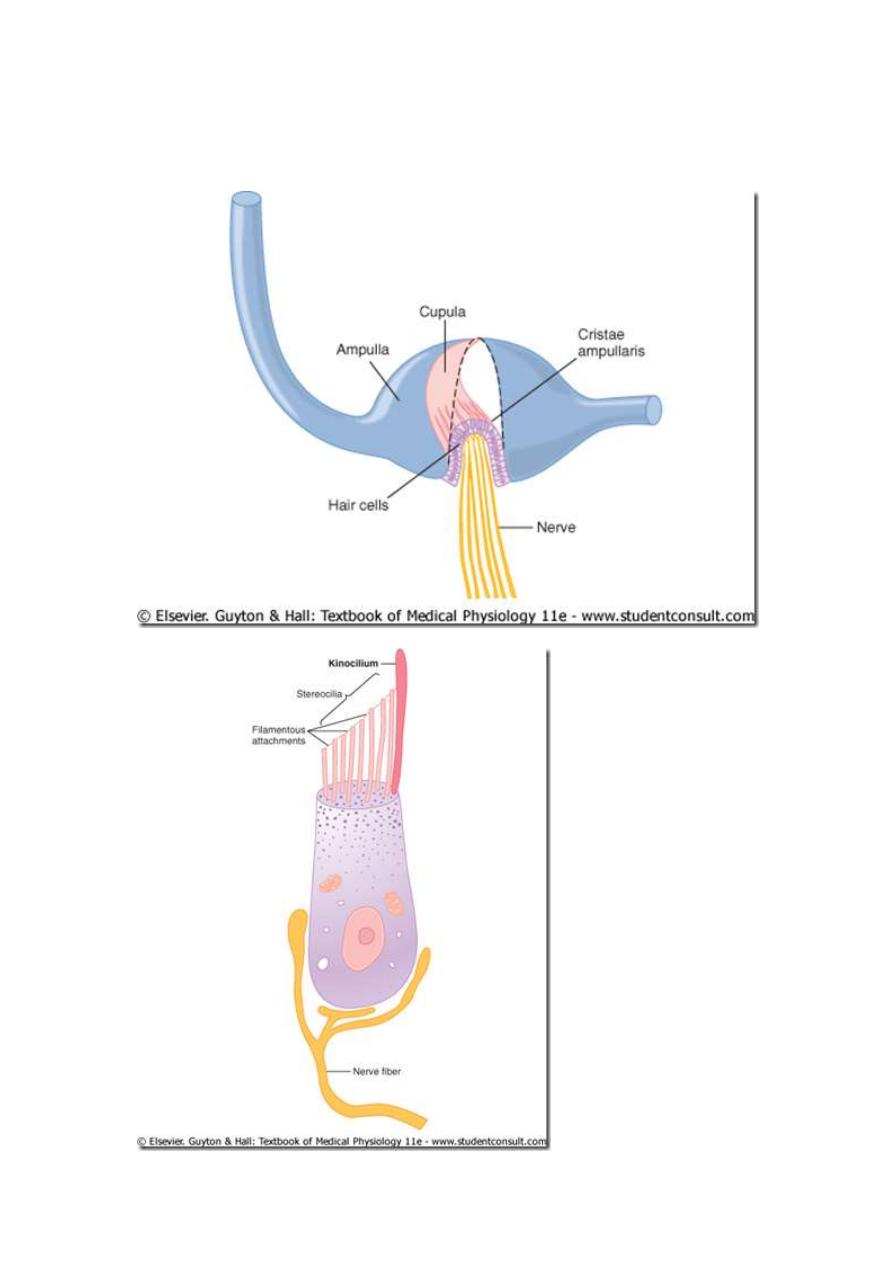
Movement of the cupula in one direction commonly causes an increase in the
firing rate of single nerve fibers from the crista, whereas movement in the
opposite direction commonly inhibits neural activity
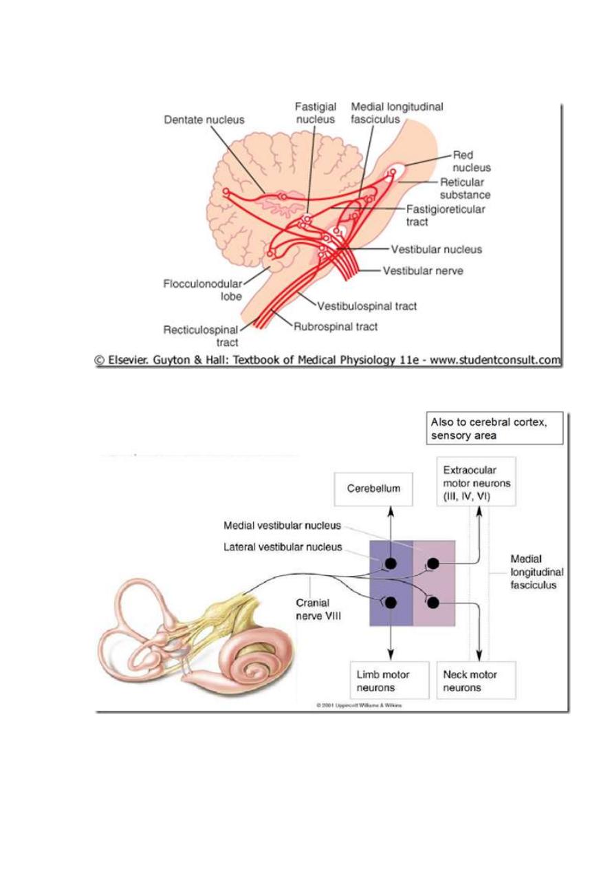
Vestibular connections
Vestibular pathways
Vestibular dysfunction
Impairment
o
loss of equilibrium & postural adjustments

o
Absence of nystagmus on vestibular stimulation
caloric test:
setting up convection currents in endolymph of the
lateral semicircular canal (made vertical) by instilling
water hotter/cooler than body temperature into external
auditory canal
Overstimulation
o
motion sicknss
giddiness
nausea
vomiting
o
irritative lesions in vestibular pathways
vestibular neuronitis