
Dr. Nabeel Al-Dawoodi
Lec. 1
ULCERS, SINUSES &
FISTULAE
Tues. 24 / 3 / 2015
DONE BY : Ali Kareem
مكتب اشور لالستنساخ
2014 – 2015
Ulcers, Sinuses & Fistulae Dr. Nabeel Al-Dawoodi
24-3-2015
2
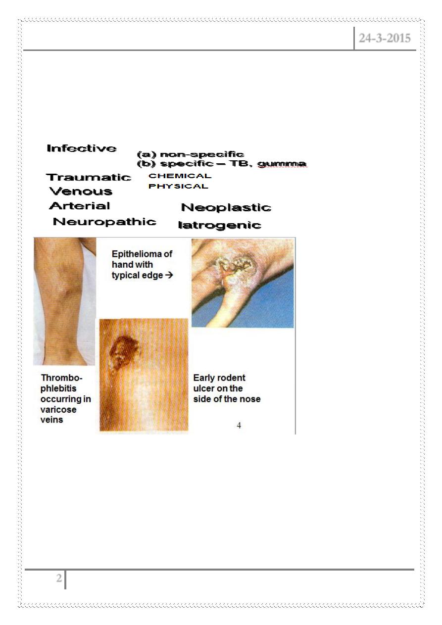
ULCER
Breach or discontinuity of an epithelium or
mucosal surface.
Classification
Diagnosis
–
1.
History
:
–
age
(young – infective, >40 Ca, 60 Rodent)
–
sex
(female – varicose, male – squamous cell Ca)
–
Race
(Chinese – Varicose ulcer)

Ulcers, Sinuses & Fistulae Dr. Nabeel Al-Dawoodi
24-3-2015
3
–
Religion
(Muslim/Jews - squamous cell Ca)
–
Occupation
- prolong standing (baker/hairdresser – varicose veins)
2. Mode of onset:
–
acute
inflammation/trauma sudden
-chronic
chronic inflammation very slowly
malignant rapid
3. Duration:
– very short (days) – acute infection;
– short (month) – malignant;
– long - chronic inflammation
4. Progress:
– very rapid – acute inflammation,
– rapid – malignant,
– slow – chronic inflammation
5. Pain:
– acute inflammation, arterial ulceration– painful (venous – not usually
very painful)
– Neuropathic – less pain
– malignant – early – usually painless.
6. Fever:
infective/TB
7. Loss of weight: malignant/TB
8. Smoking:
Buerger's disease, artherosclerosis
Past History – TB, Syphilis, Diabetes Mellitus, Hypertension
Ulcers, Sinuses & Fistulae Dr. Nabeel Al-Dawoodi
24-3-2015
4
Responses to Antibiotics: (+) in infection (-) in malignancy
PHYSICAL EXAMINATION
General
– cachexia, anaemia,weight loss (Ca, TB)
Local
:
1.
Number
(single or multiple –TB)
2.
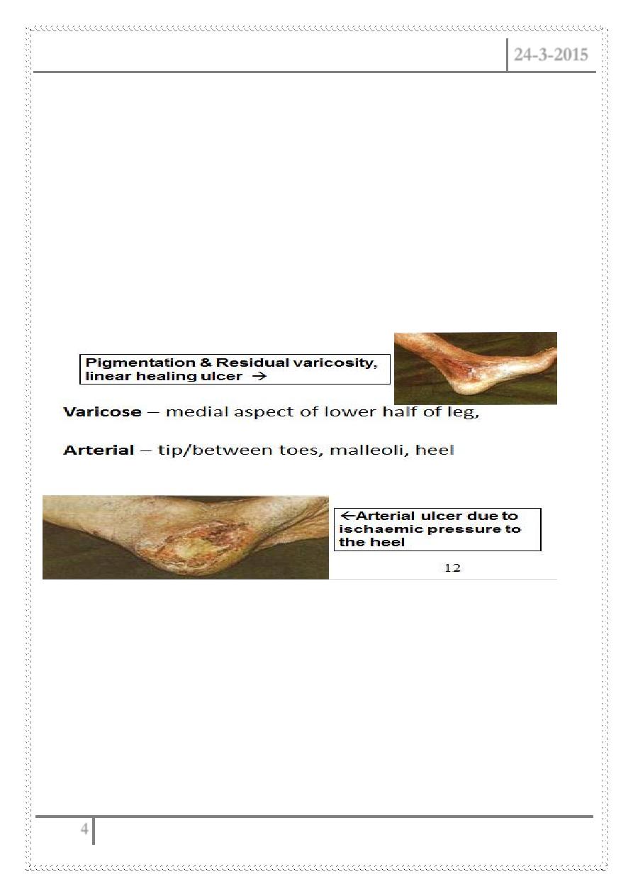
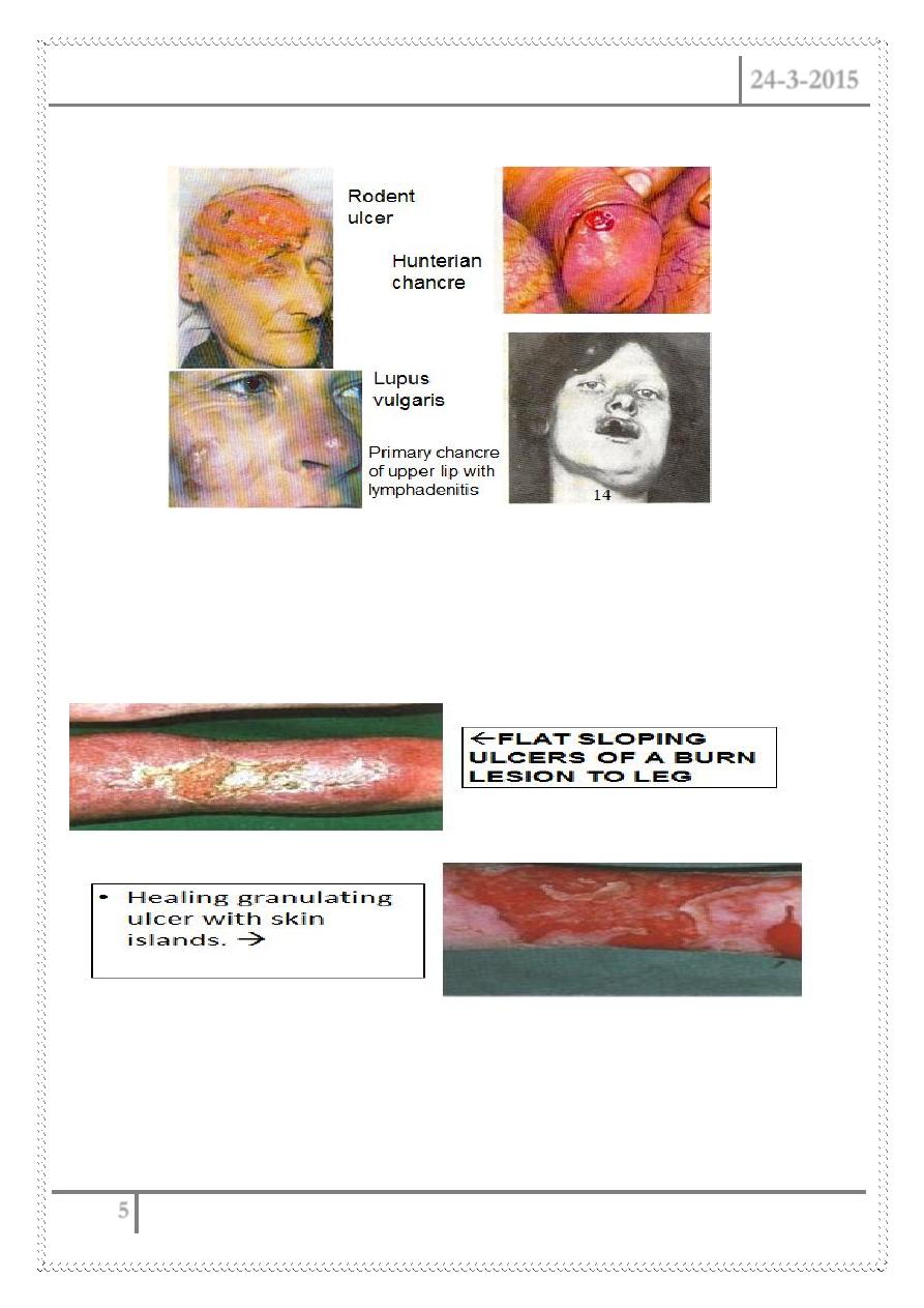
Site
Rodent –lobule of ear & angle of the mouth;
SCC – lower lip, Gumma – s/c
bone ~ tibia/sternum/skull
Diabetic/Perforating/trophic –
heel/ball of the foot (head of 1
st
/2
nd
metatarsals
TB – neck, axilla, groin
Lupus – face, fingers, hands,
Chancre/soft sore – ext.genitalia
Ulcers, Sinuses & Fistulae Dr. Nabeel Al-Dawoodi
24-3-2015
5
• 3.
Size
(depends on duration
& rate of growth)
• 4. Shape (oval – varicose,
circular – rodent,
irregular – malignant)
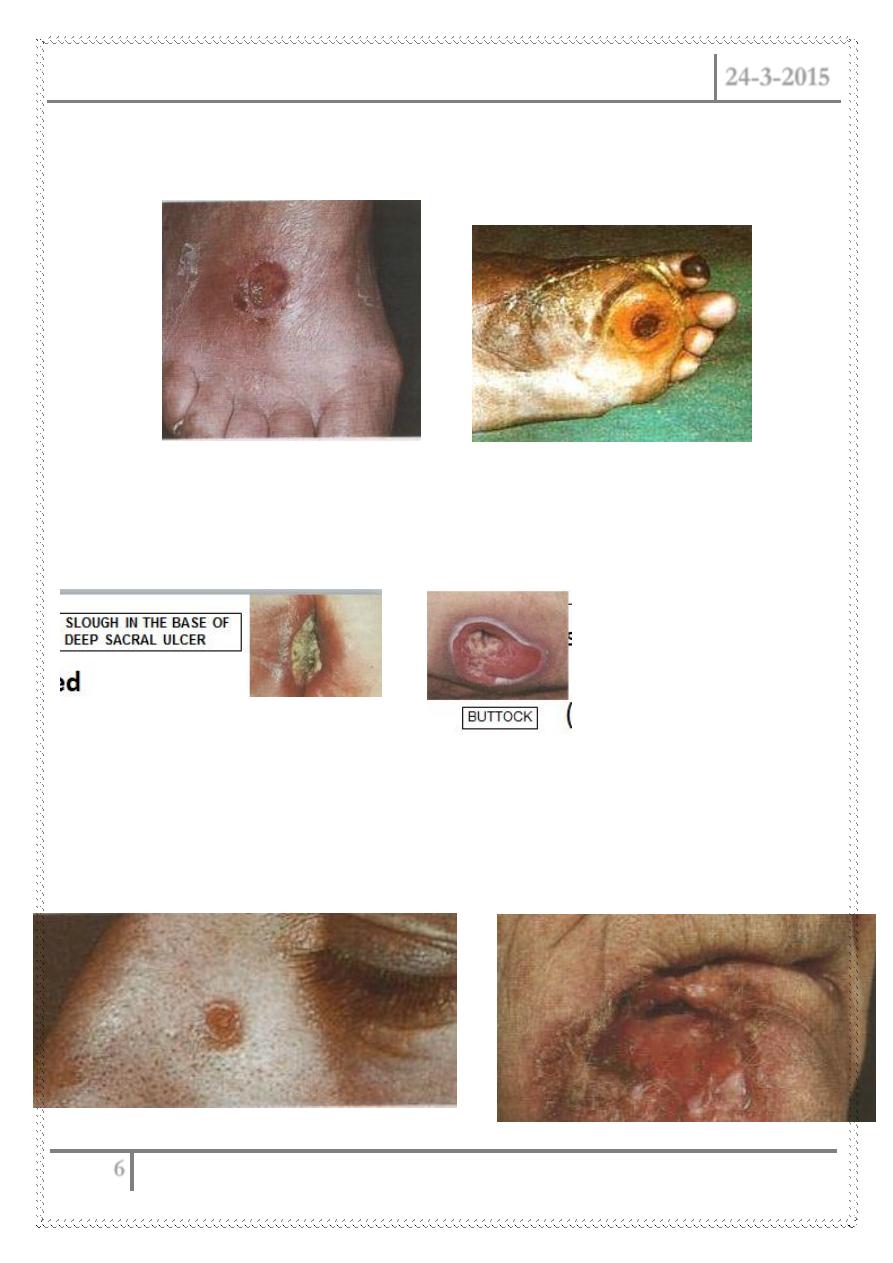
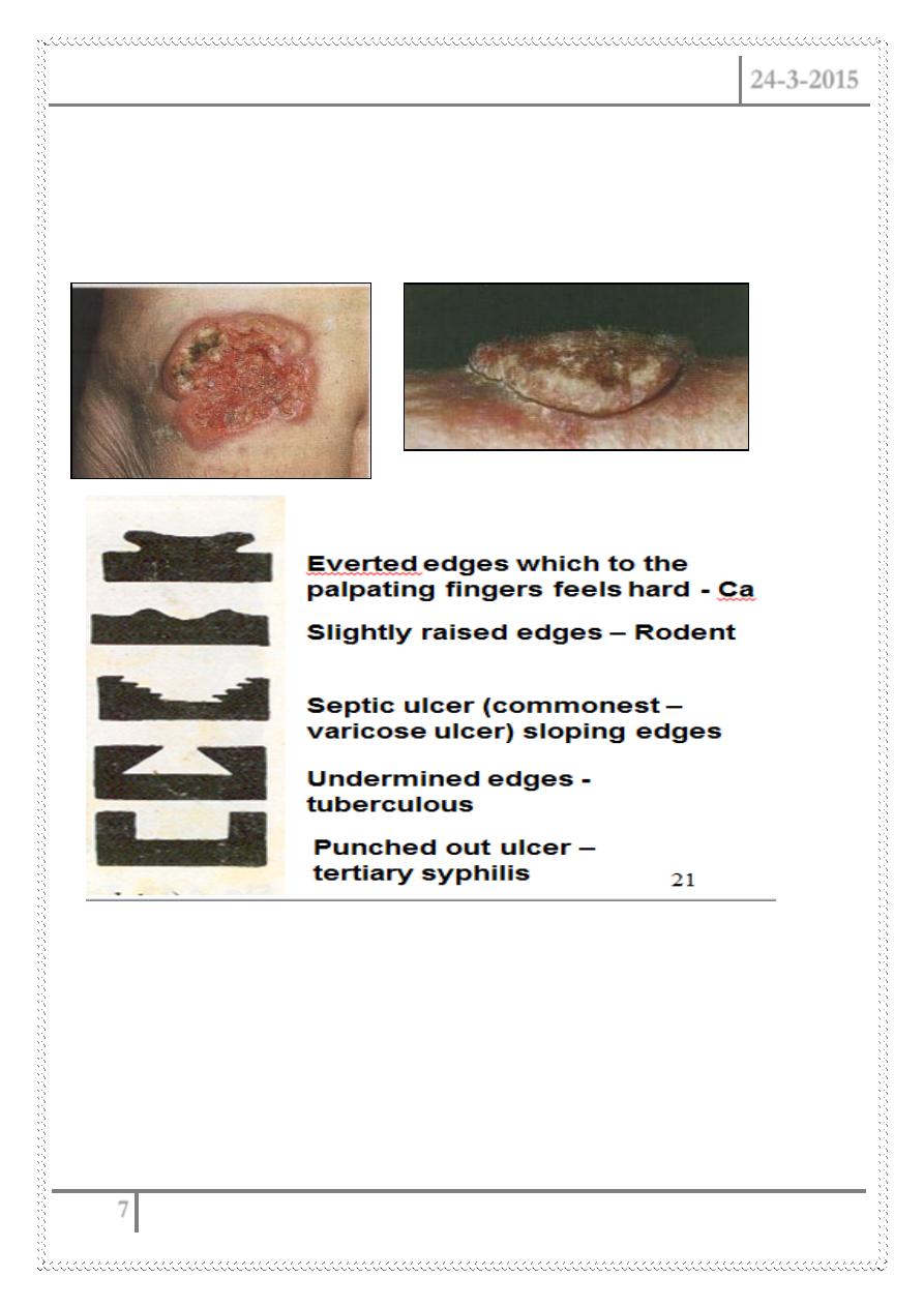
• 5. Edge: (a) flat sloping ~
simple or healing ulcer,
venous ulcer (edge red,
blue, transparent zone)
(b) square cut or punched
out
– gumma, trophic, diabetic
- Chronic GU/DU, leprosy
Ulcers, Sinuses & Fistulae Dr. Nabeel Al-Dawoodi
24-3-2015
6
(rapid death & loss of whole
thickness of skin without much
attempt by the body
to repair the defect)
(c) undermined
– TB, amoebic, bed sore, carbuncle
(infection affects underneath tissue more than epithelial
surface)
(d) raised & rolled up
– rodent/BCC
(slow growth of tissue in the edge of ulcer, edge pale pink or white
with clumps or cluster of cells visible through paper thin superficial coverings of
squamous cell)
Ulcers, Sinuses & Fistulae Dr. Nabeel Al-Dawoodi
24-3-2015
7
(e) raised & everted
–
malignant ulcer, epithelioma
(tissue in edge growing quickly and spilling out of the ulcer to overlap
normal skin or mucosa)
Ulcers, Sinuses & Fistulae Dr. Nabeel Al-Dawoodi
24-3-2015
8
Colour of the Edge
Red inflammation
Pale or cyanosed ischaemia
Late blue, purple, black
Pigmentation venous ulcer, malignant melanoma
Pearly edge BCC
Keratinization Neuropathic ulcer
6. Floor:
Haemorrhage & necrotic slough–malignant
purulent - acute infection
washed leather- gumma
bluish unhealthy granulation tissue TB (whitish in brownish space/
apple jelly)
solid brown or gray – dead tissue full thickness skin death

Ulcers, Sinuses & Fistulae Dr. Nabeel Al-Dawoodi
24-3-2015
9
7. Discharge:
On dressing gauze – serous,
sero- sanguinous, purulent,
offensive, copious, or so
slight – dries up into a
scab.
8.
Surrounding skin, state of local tissue, blood supply,
innervation
ss of infl
n
– infective, scar – TB
PALPATION
Temperature difference/tenderness - in acute infected ulcer
Base – induration +/-,
Mobility of ulcer over underlying structures –
– fixed – malignant; bleed on touch +/-
regional lymphatics –
enlarged -> inflammation
hard – malignant
Systemic Examination:
Infection – constitutional symptoms - TB
cachexia, anaemia, loss of weight-- malignant
hypertension, artherosclerosis –- ischaemic ulcer
Hensens’, tabes dorsalis, peripheral numbness – neuropathic
Done by
Ali Kareem