
Structure of Bacteria
Prof.Dr. Mohammad Alfaham
Microbiology

Chapter 4
Size of Bacteria
• Average bacteria 0.5 - 2.0 um in diam.
– RBC is 7.5 um in diam.
• Surface Area ~12 um^2
• Volume is ~4 um
• Surface Area to Volume is 3:1
• Typical Eukaryote Cell SA/Vol is 0.3:1
• Food enters through SA, quickly reaches all
parts of bacteria
• Eukaroytes need structures & organelles
Chapter 4
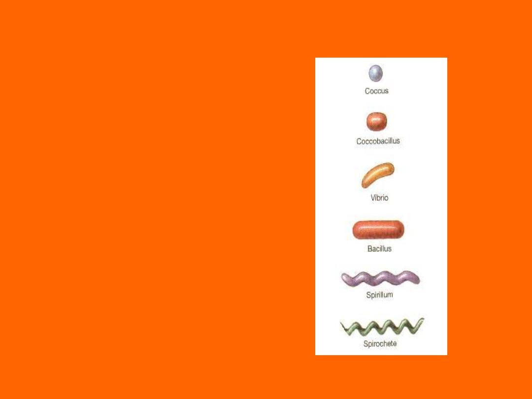
Shapes of Bacteria
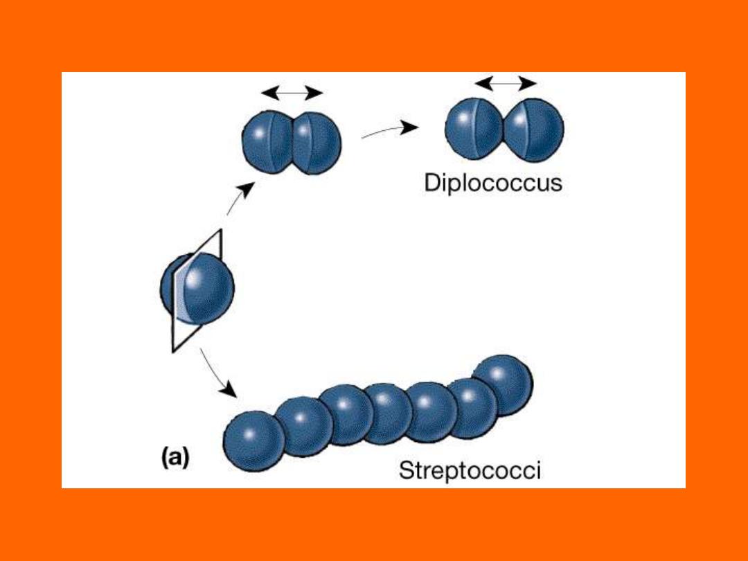
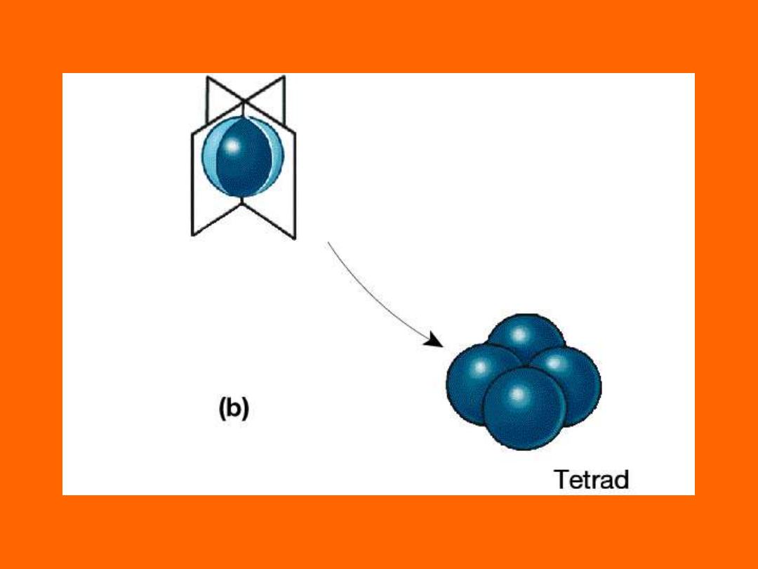
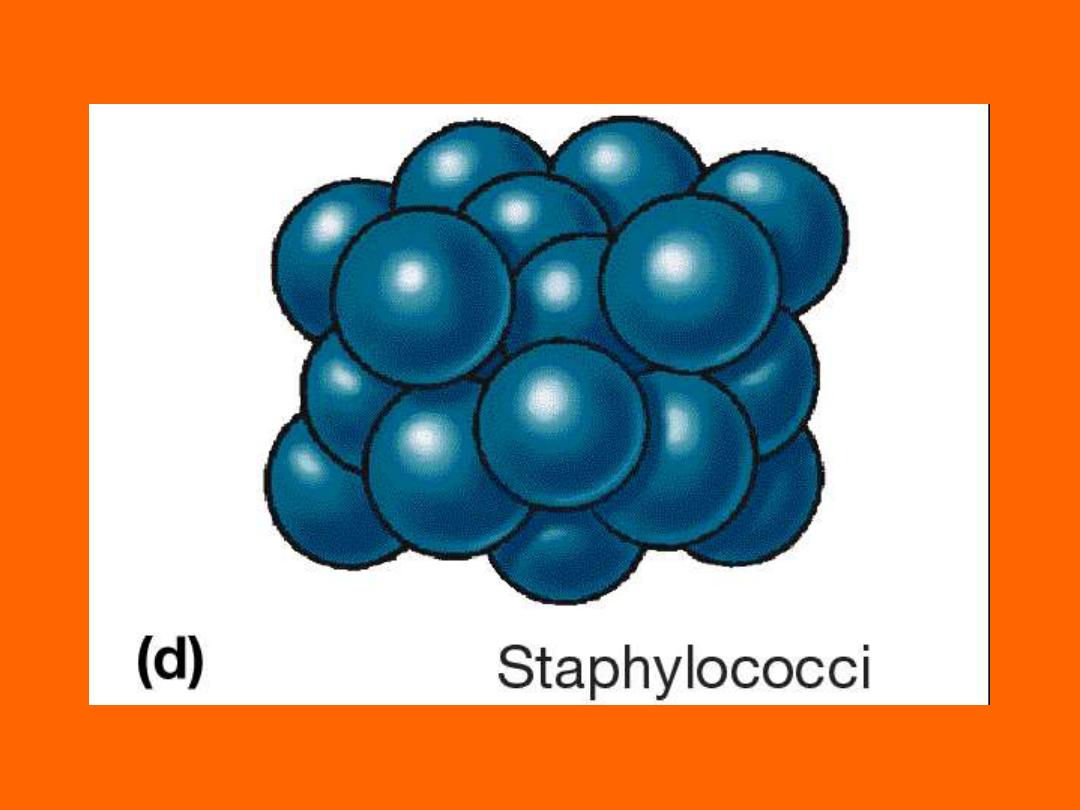
• Coccus
– Chain = Streptoccus
– Cluster = Staphylococcus
• Bacillus
– Chain = Streptobacillus
• Coccobacillus
• Vibrio = curved
• Spirillum
• Spirochete
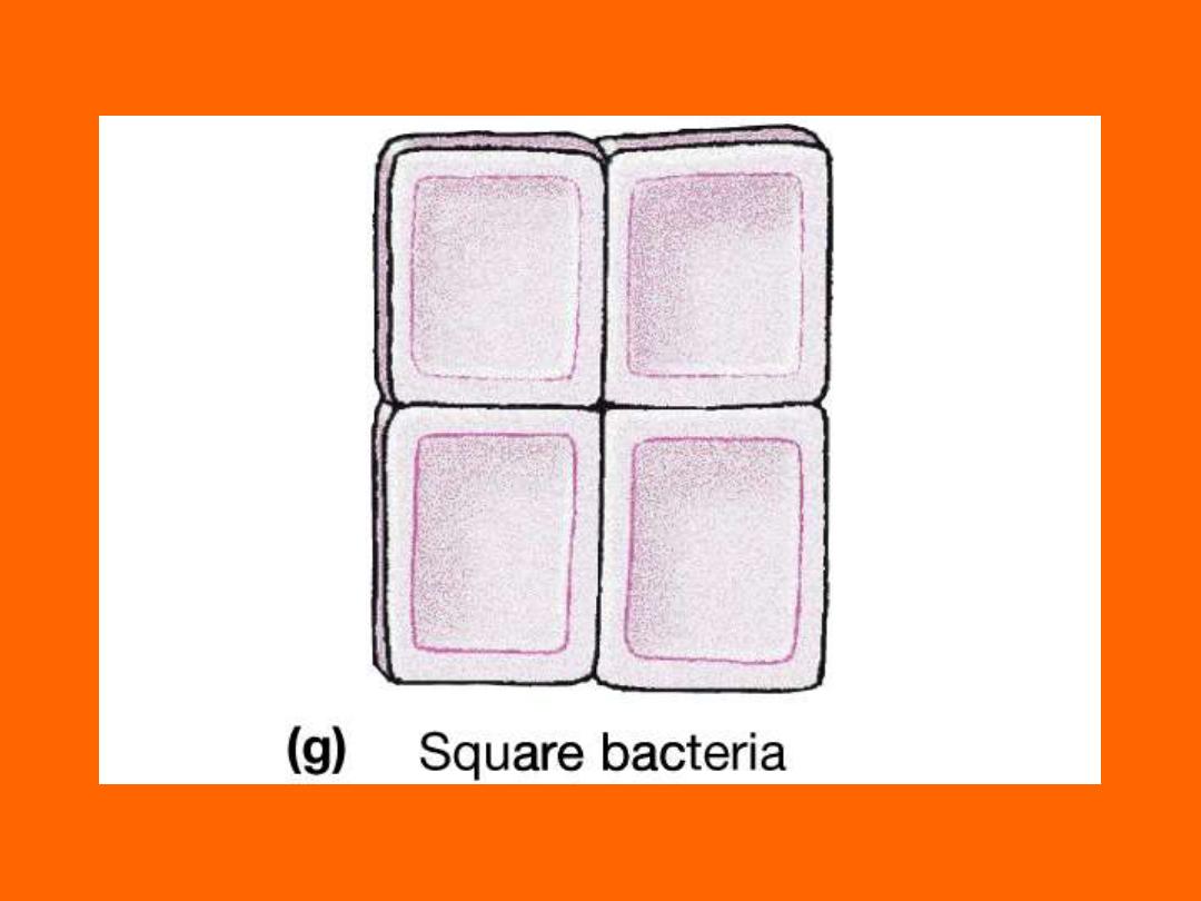
• Square
• Star
Chapter 4
Chapter 4
Chapter 4
Chapter 4
Chapter 4
Chapter 4
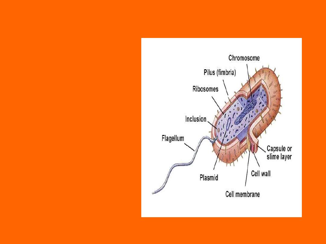
Bacterial Structures
• Flagella
• Pili
• Capsule
• Plasma Membrane
• Cytoplasm
• Cell Wall
• Lipopolysaccharides
• Teichoic Acids
• Inclusions
• Spores

Chapter 4
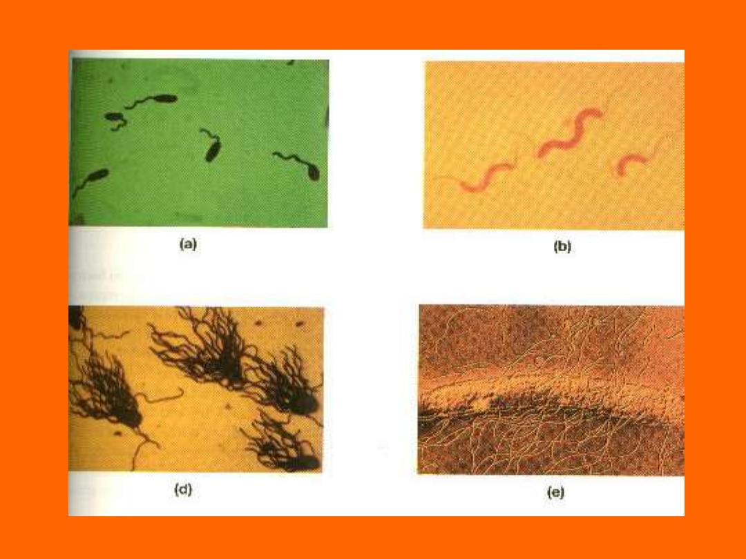
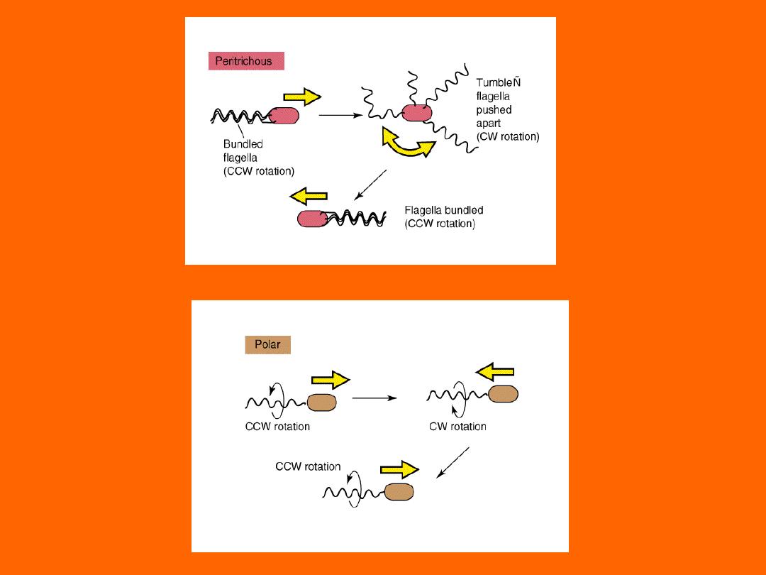
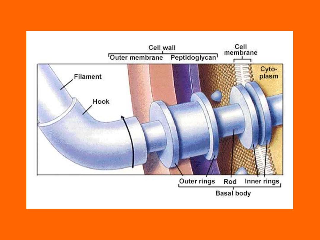
Flagella
• Motility - movement
• Swarming occurs with some bacteria
– Spread across Petri Dish
– Proteus species most evident
• Arrangement basis for classification
– Monotrichous; 1 flagella
– Lophotrichous; tuft at one end
– Amphitrichous; both ends
– Peritrichous; all around bacteria
• Observe Picture in Micro Lab.
Chapter 4
Chapter 4
Mono- or Lophotrichorus
Chapter 4

Chapter 4
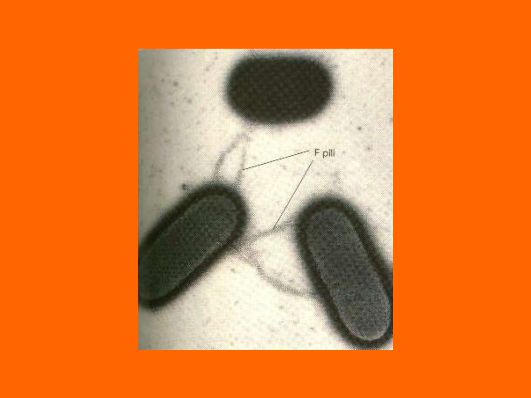
Pili
• Short protein appendages
– smaller than flagella
• Adhere bacteria to surfaces
– E. coli has numerous types
• K88, K99, F41, etc.
– Antibodies to will block adherance
• F-pilus; used in conjugation
– Exchange of genetic information
• Flotation; increase boyancy
– Pellicle (scum on water)
– More oxygen on surface
Chapter 4
F-Pilus for Conjugation

Chapter 4
Capsule or Slime Layer
• Glycocalyx - Polysaccharide on external
surface
• Adhere bacteria to surface
– S. mutans and enamel of teeth
• Prevents Phagocytosis
– Complement can’t penetrate sugars

Chapter 4
Cytoplasm
• 80% Water {20% Salts-Proteins)
– Osmotic Shock important
• DNA is circular, Haploid
– Advantages of 1N DNA over 2N DNA
– More efficient; grows quicker
– Mutations allow adaptation to environment
quicker
• Plasmids; extra circular DNA
– Antibiotic Resistance
• No organelles (Mitochondria, Golgi, etc.)

Chapter 4
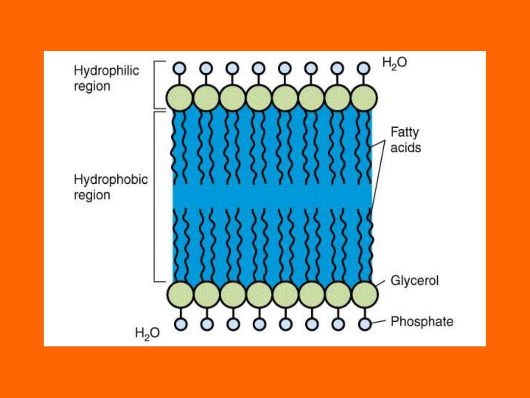
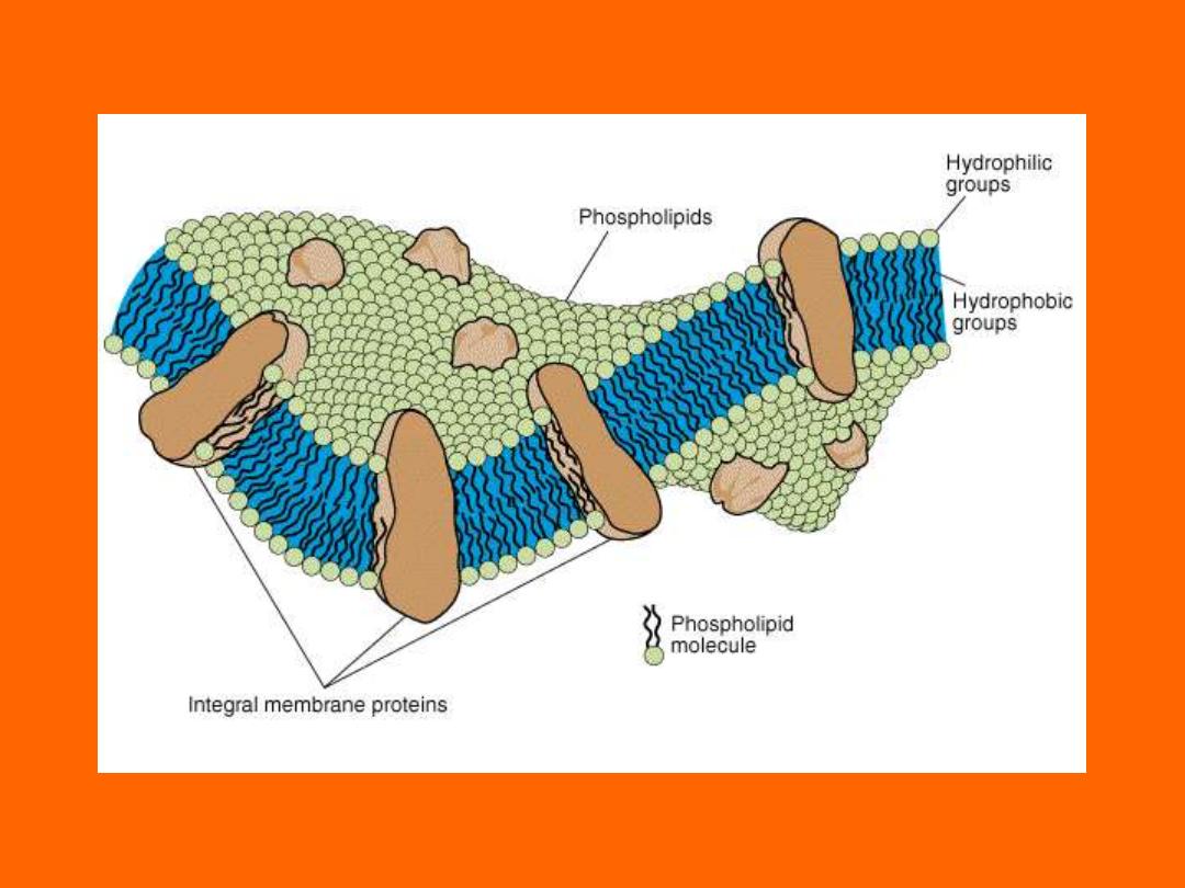
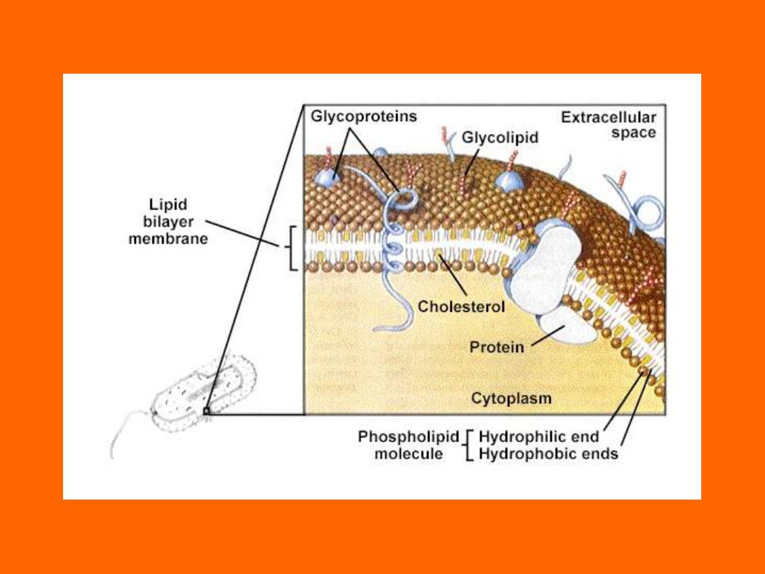
Cell Membrane
• Bilayer Phospholipid
• Water can penetrate
• Flexible
• Not strong, ruptures easily
– Osmotic Pressure created by cytoplasm
Chapter 4
Chapter 4
Chapter 4

Chapter 4
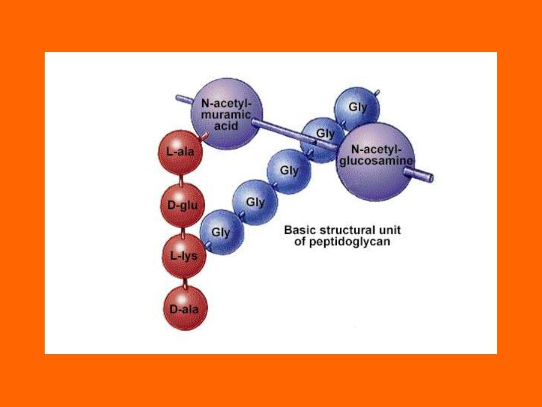
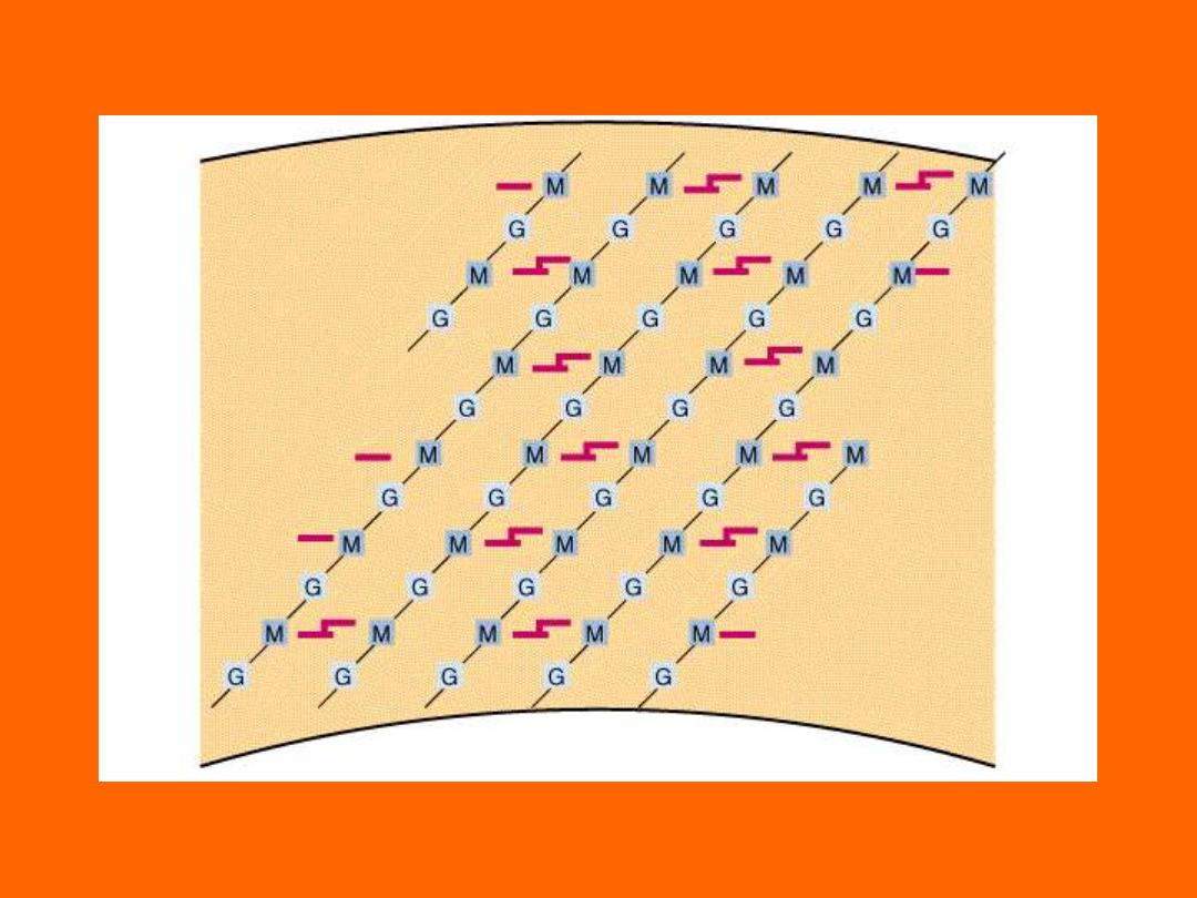
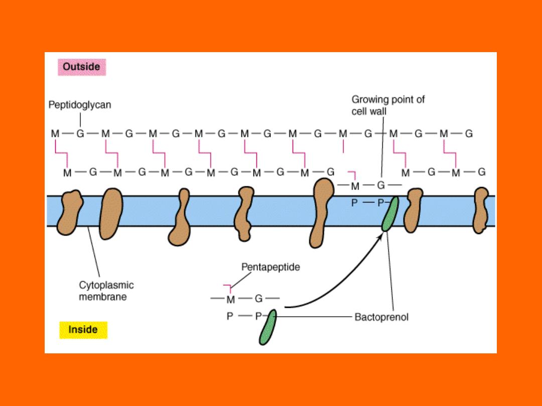
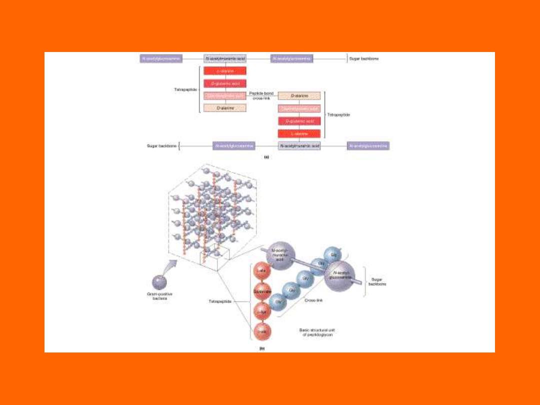
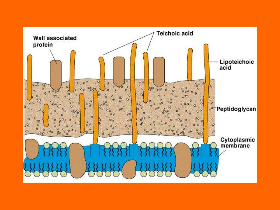
Cell Wall
• Peptido-glycan Polymer (amino acids +
sugars)
• Unique to bacteria
• Sugars; NAG & NAM
– N-acetylglucosamine
– N-acetymuramic acid
• D form of Amino acids used not L form
– Hard to break down D form
• Amino acids cross link NAG & NAM
Chapter 4

Chapter 4
Chapter 4
Chapter 4
Chapter 4
Chapter 4

Chapter 4
Cell Wall Summary
• Determine shape of bacteria
• Strength prevents osmotic rupture
• 20-40% of bacteria
• Unique to bacteria
• Some antibiotics effect directly
– Penicillin
Chapter 4
Video Clip on Cell Wall

Chapter 4
Teichoic Acids
• Gram + only
• Glycerol, Phosphates, & Ribitol
• Attachment for Phages
Chapter 4

Chapter 4
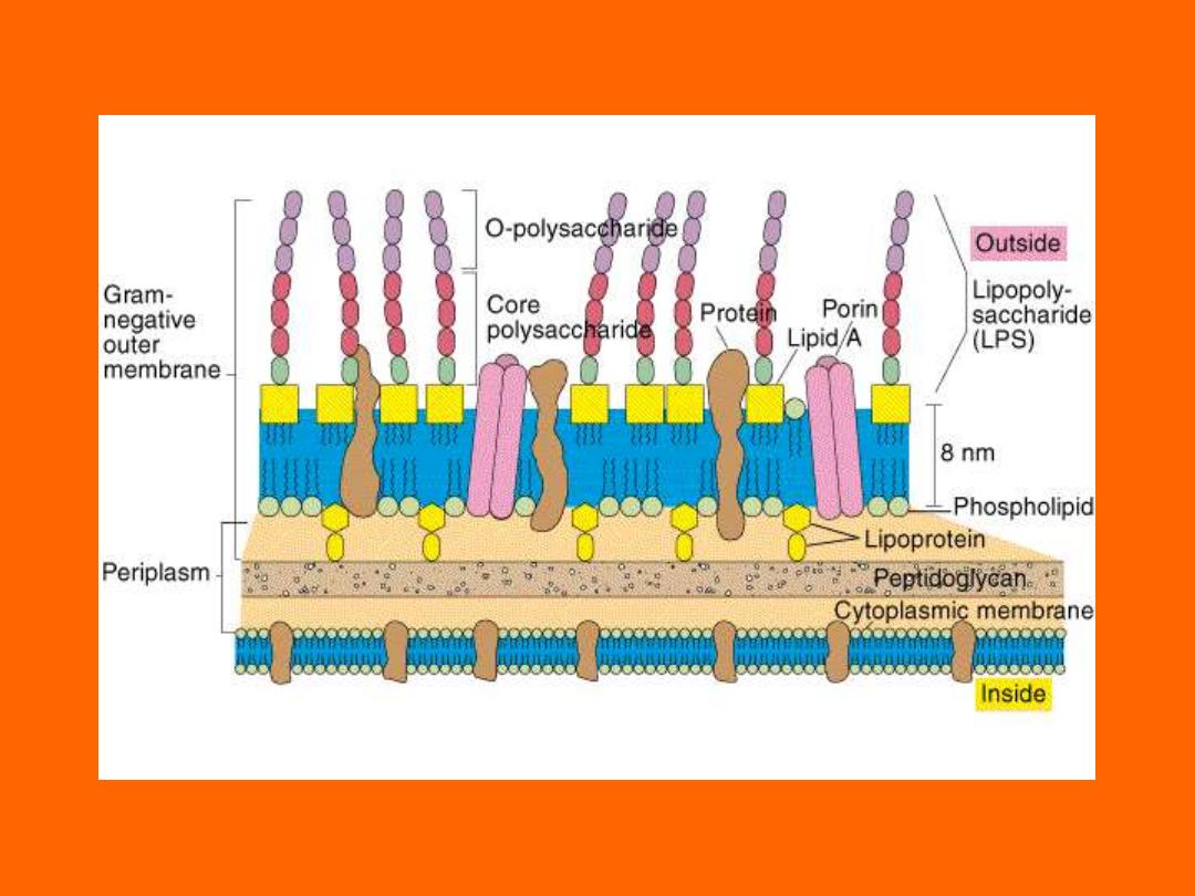
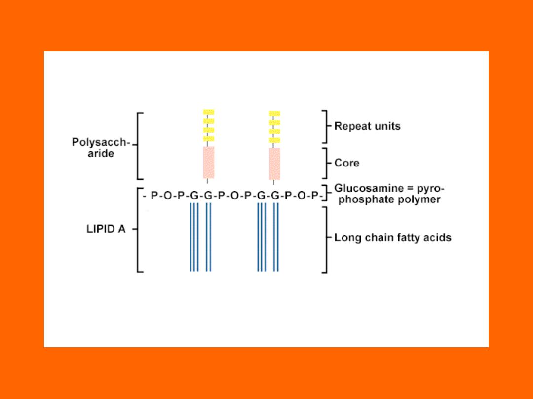
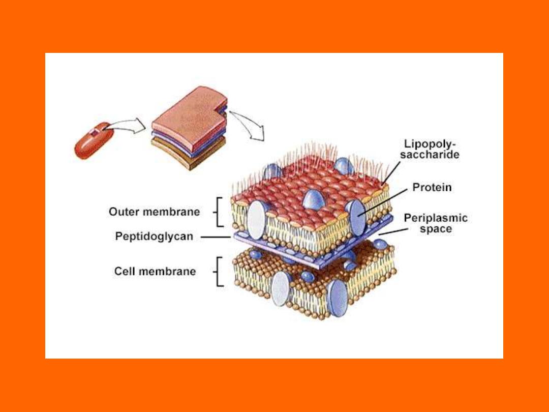
Lipopolysaccharide (LPS)
• Endotoxin or Pyrogen
– Fever causing
– Toxin nomenclature
• Endo- part of bacteria
• Exo- excreted into environment
• Structure
– Lipid A
– Polysaccharide
• O Antigen of E. coli, Salmonella
• G- bacteria only
– Alcohol/Acetone removes
Chapter 4
Chapter 4

Chapter 4
LPS (cont’d)
• Functions
– Toxic; kills mice, pigs, humans
• G- septicemia; death due to LPS
– Pyrogen; causes fever
• DPT vaccination always causes fevers
– Adjuvant; stimulates immunity
• Heat Resistant; hard to remove
• Detection (all topical & IV products)
– Rabbits (measure fever)
– Horse shoe crab (Amoebocytes Lyse in
presence of LPS)

Chapter 4
LPS (cont’d.)
• Appearance of Colonies
– Mucoid = Smooth (lots of LPS or capsule)
– Dry = Rough (little LPS or capsule)
• O Antigen of Salmonella and E. coli
– 2,000 different O Ags of Salmonella
– 100’s different O Ags of E. coli
• E. coli O157
• O Ags differ in Sugars, not Lipid A

Chapter 4
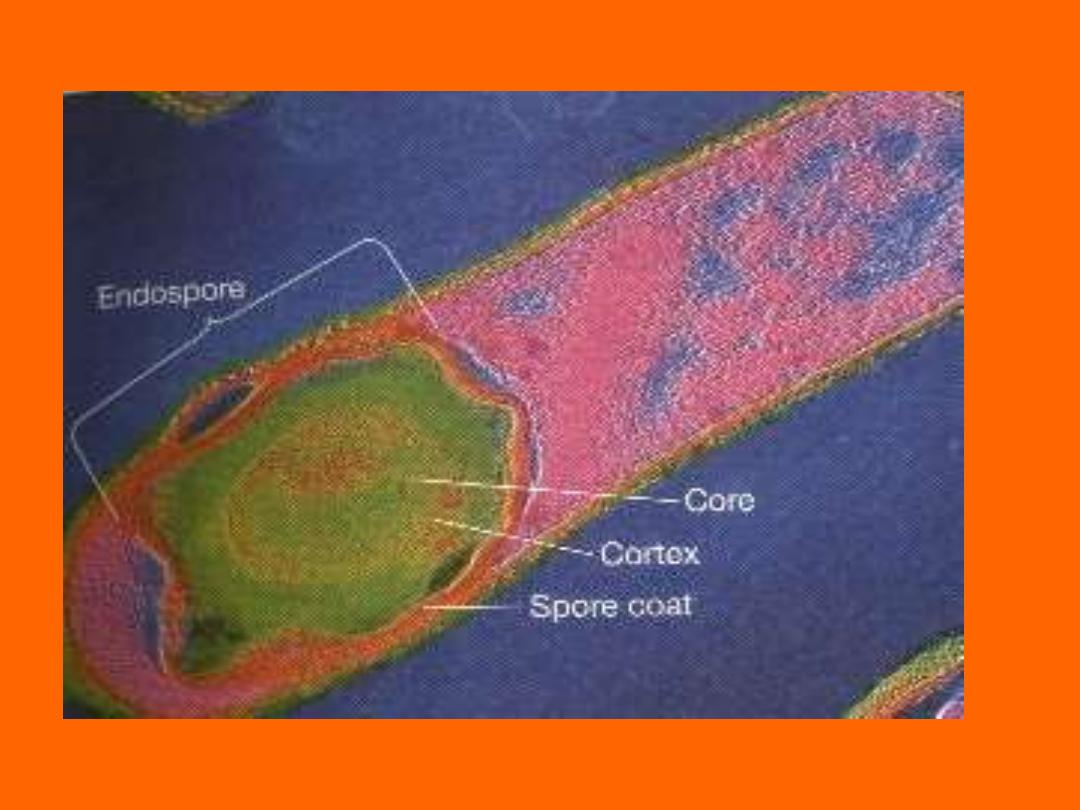
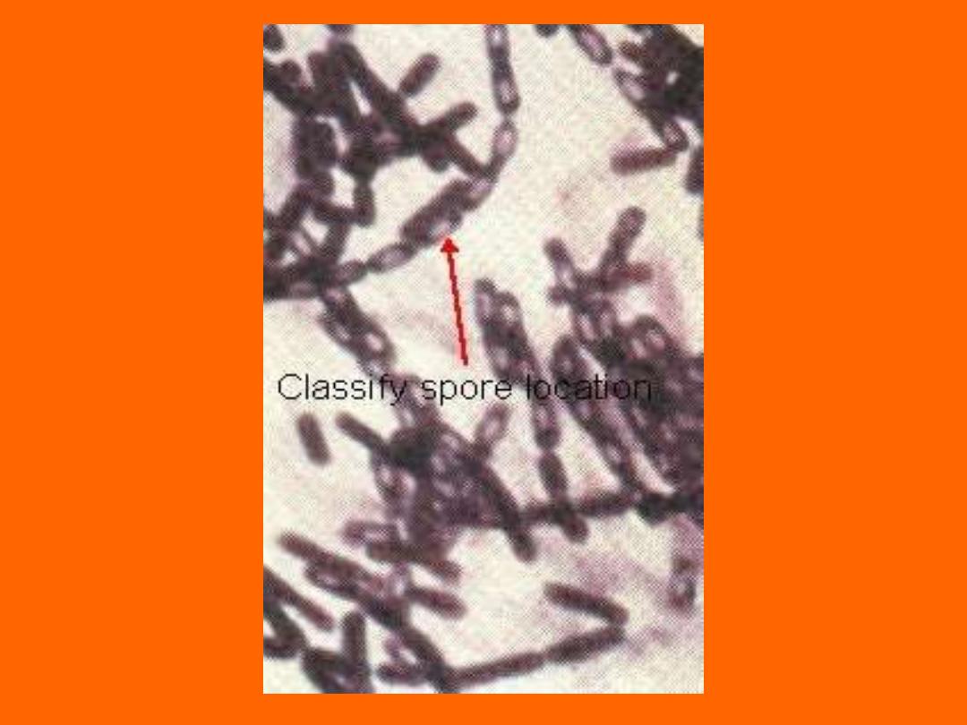
Endospores
• Resistant structure
– Heat, irradiation, cold
– Boiling >1 hr still viable
• Takes time and energy to make spores
• Location important in classification
– Central, Subterminal, Terminal
• Bacillus stearothermophilus -spores
– Used for quality control of heat sterilization
equipment
• Bacillus anthracis - spores
– Used in biological warfare
Chapter 4
Chapter 4

Chapter 4
G+ vs. G-
• G+
– Thicker cell wall
– Teichoic Acids
• G-
– Endotoxin - LPS
• Which are more sensitive to Penicllin?
• Alcohol/Acetone affects which more?

Chapter 4
Prokaryotes vs. Eukaryotes
• Cell Wall
• Teichoic Acids
• LPS
• Endospores
• Circular DNA
• Plasmids
Chapter 4
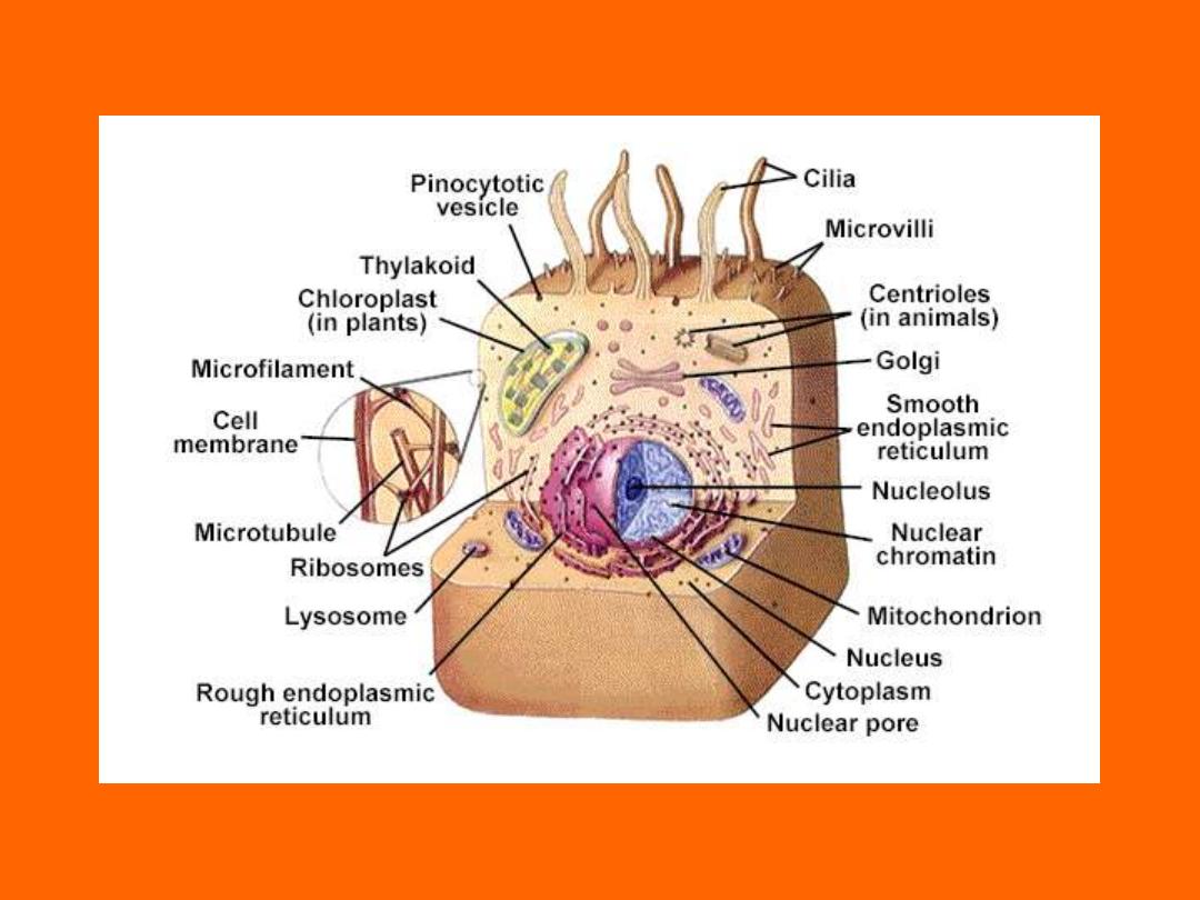
Eukaryote Cell Structure