
Genus Compylobacter
C. jejuni & C. coli ← emerged as common human pathogens.
General characteristics
-Gram negative comma , S or gall-wing shape rods, motile with
single polar flagellum & non-spore forming
-They are thermophilic (37
o
C – 42
o
C), grow on reduced O
2
(0.5%) and 20% CO
2
-The selective media is
"Skirrows media"
incorporated with
vancomycin, polymyxin B & Trimethoprim to inhibit growth of
normal flora.
-They are oxidase positive, catalase positive, urease negative,
reduce nitrate and produce H
2
S, they do not ferment CHO.
-Hippurate Hydrolysis test is used to differentiate between spp.
The antigens structure:
1-Heat stable lipopolysaccharide O-Ag with endotoxic activity.
2-Heat labile flagellar H-Ag.

Pathogenesis
Domestic animals (cattle, chickens & dogs) serve as source of
the pathogens for humans. Transmission is usually feco-oral,
large infective doses are needed (≥10
4
m.o.) because They are
sensitive To gastric acidity. The bacteria multiply in the small
intestine → invading the epithelium & producing inflammation
which result in appearance of RBC & WBC. So, localized
invasion and toxic effect are responsible for development of
enteritis.
Clinical findings:
After incubation period (1-7) days, acute onset begins as watery
foul-smelling diarrhea followed by bloody stool accompanied
by fever and sever abdominal pain. This enterocolitis usually
self-limited (5-8) days, but are susceptible to erythromycin that
shortens the duration of fecal shedding of bacteria.
Laboratory diagnosis:
A-specimen diarrheal stool.
Blood
B-culture on skirrow's media → small, gray, circular and
glistening colonies ← detected by Gram’s stain for typical
morphology.
Helicobacter pylori
This micro-organism emerged in 1990
S
, previously classified as
Compylobacters because they have common characters.
General characteristics
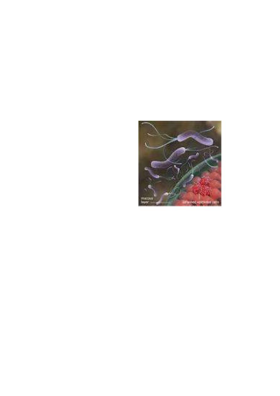
1-Gram negative, spiral shaped rods. actively motile with
multiple polar flagella.
2-Culture on skirrow’s media (3-6 days∕ 37
o
C)
3-Oxidase positive, catalase positive and urease positive (more
active than that of Proteus).

Pathogenesis of H.pylori
The natural habitat of H.pylori is the human stomach, it is
aquired by ingestion. H.pybri use to live on gastric mucosa but
not in the lumen where the pH only (1-2), while it is actively
motile when attach to epithelial surface where the pH (6-7), also
produce protease that modifies the gastric mucosa reducing the
ability of acid to diffuse through mucus.
H.pylori produce potent urease activity that cause accumulation
of large ammonia which buffer the acidity. Toxin and LPS may
damage the mucosal cells.
Gastritis is characterized by chronic and active inflammation,
even epithelial destruction and glandular atrophy may occur,
thus H.pylori may be a major risk factor for gastric cancer.
Clinical findings
After short incubation period, patient will develop acute gastritis
(abdominal pain, nausea, flatulance and bad smell breath) for 2
weeks but hypochlorhydria may persist leading to chronic
gastritis, peptic ulcer, deudenal ulcer or high risk of gastric
cancer. Those present with recurrent upper abdominal pain
frequently accompanied by GIT bleeding. No bacteremia or
disaminated disease.

Complete cure observed after elimination of the organism,
although some people can harbor the pathogans for years
without ill effect.
Labroatory diagnosis of H.pylori
*Speciment -
Gastric biopsy
-
Blood (serological Ab)
1-Histological smear
: Gastritis and peptic ulcer usually
diagnosed by gastroscopy, the taken biopsy can be stained by
Giemsa or special silver stain, those can show the curved or
spiral organisms.
2-Culture of biopsy
: usually use Skirrow
s media, it take 3-6
days in microaeropilic environment.
3-Urease test either:
-In vitro urease
test done by inoculating the biopsy material on
urea agar media, urease enzyme will split urea within 2hr
shifting the pH to alkaline changing the color of indicator
(phenol red) from yellow to pink.
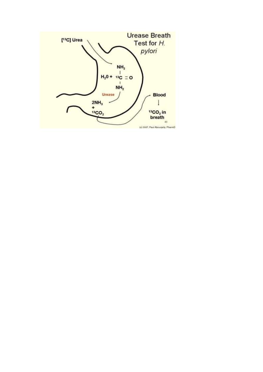
-Invivo (urea breath test)
done when C
13
or C
14
labeled urea
ingested by the patient
urease activity
labeled CO
2
will be
generated and can be detected in patients exhaled breath.
4-Serological test:
the presence of serum IgG as anti-H.pylori
Ab will reflect the infection but is of limited use in follow up.
Treatment
Triple therapy of Amoxicilin or tetracycline with
metronidazol and bismuth sulfate for 14 days, can eradicate
H.pylori in 70-95% of patients.