
Objectives;
1- Pathogenicity of E . Histolytica infection.
2- Pathological anatomy of intestinal amoebiasis.
3- Complications of intestinal amoebiasis.
4- Extra-intestinal amoebiasis
.
Pathogenicity
Factors affecting the severity of amoebic infection
depends on:
1.Host factors(stress,malnutrition,alcoholism,host
immunity).
2.Parasite factors (virulance of strain,size of
inoculum).
pH,bacterial flora).
)
3.Enviromental factors

Pathogenicity
--The first step in the pathogenicity of E. histolytica
infection includes
colonization
of the trophozoites on
Intestinal mucosa .
--The second step includes
destruction and invasion
of
Intestinal wall.
Factors affecting colonization of the
trophozoites.
1.Infective dose: Number of active Trophozoite
in contact with intestinal mucosa depend on
number of viable mature cysts ingested by
the host.
2.Amount of food: Bulky food does not give
opportunity for the parasite to be colonize.
.
3.Hypermotility of bowel (stasis) give less
chance for the parasite to become in
contact with mucosa, this explains the high
rate of colonization in cecal area because of
reduced peristalsis.
After establishment of colonies on
intestinal mucosa, the trophozoite starts to
penetrate intestinal wall.
Factors affecting destruction and
invasion of intestinal wall
1.Motility of the parasite: more active parasite give
more chance for penetration. Amoebae enter
intestinal mucosa by their pseudopodial movements
which cause displacement of the cells.
2.Bacteria: number of bacteria present in the intestine
enhanced enterance of amoebae in the wall of
intestine.
3.Enzymes: virulence strain of E. histolytica has the
ability to secret enzymes e.g. cytolytic enzymes
which cause lysis of epithelial cells.
Pathological anatomy of intestinal amoebiasis:
The trophozoites multiply and colonize in glandular crypts of
large intestine and adher to intestinal mucosa which leads to
the formation of early lesion.
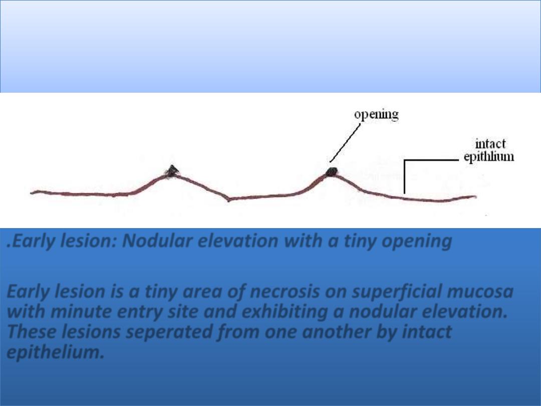
.Early lesion: Nodular elevation with a tiny opening
Early lesion is a tiny area of necrosis on superficial mucosa
with minute entry site and exhibiting a nodular elevation.
These lesions seperated from one another by intact
epithelium.
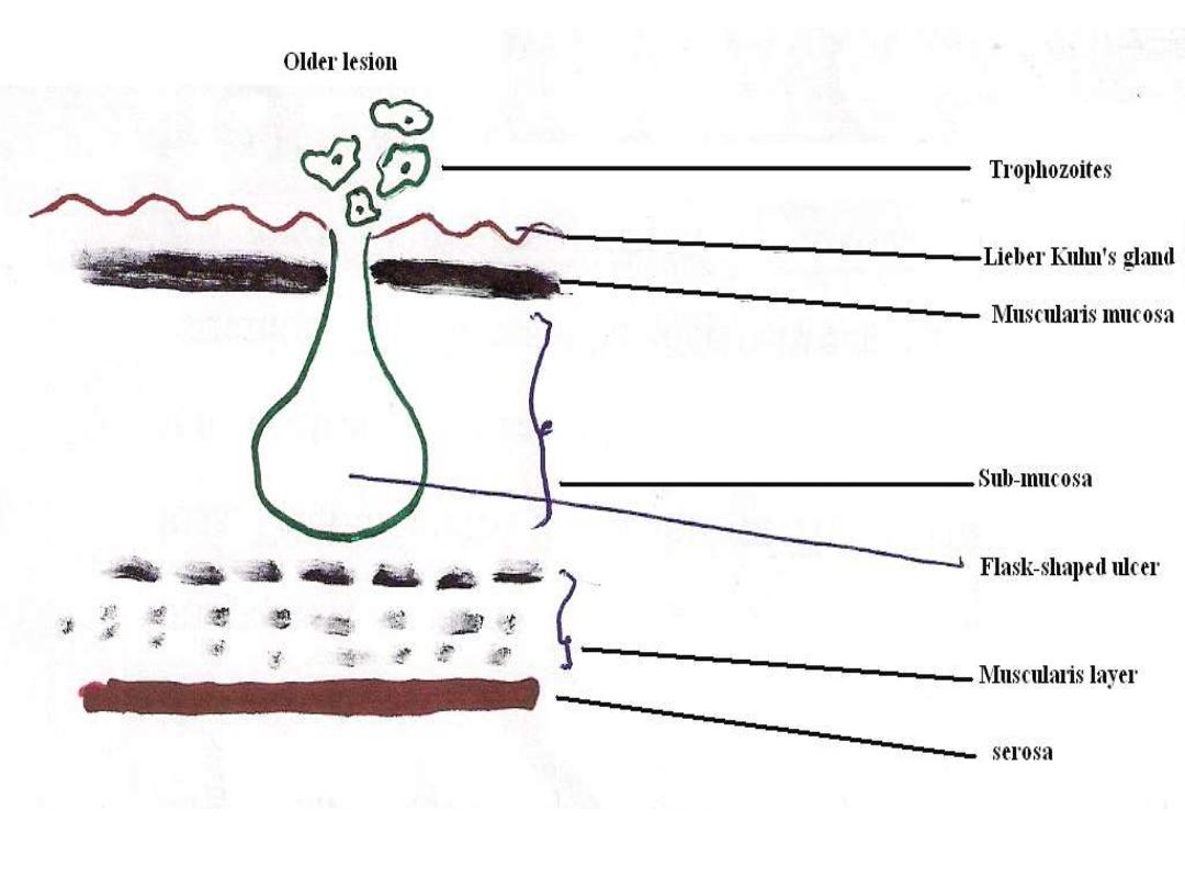
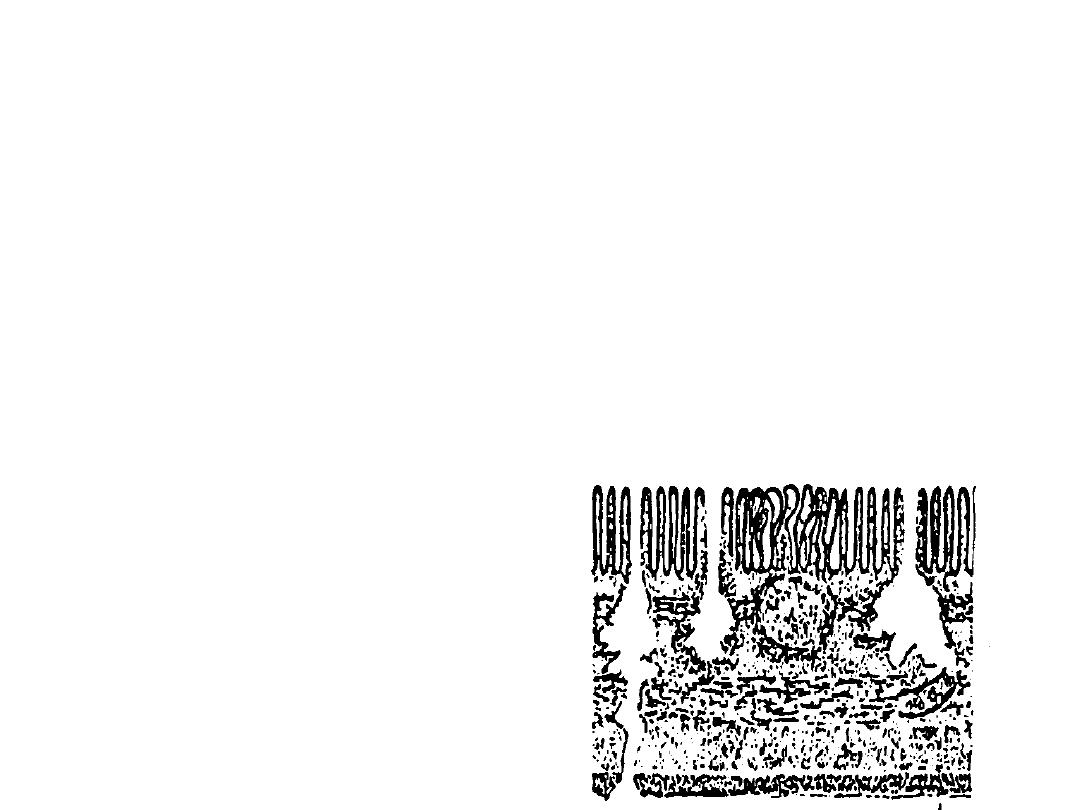
Flask Shape Ulcer
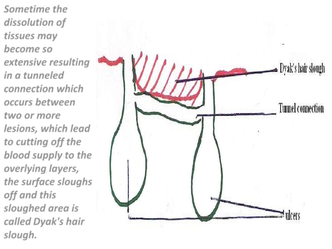
Sometime the
dissolution of
tissues may
become so
extensive resulting
in a tunneled
connection which
occurs between
two or more
lesions, which lead
to cutting off the
blood supply to the
overlying layers,
the surface sloughs
off and this
sloughed area is
called
Dyak's hair
slough.
As a result of inflammatory process, fibrous thickening
may result due to degenerative proliferation of
connective tissues which make the surface of
intestine irregular and this shape of intestine surface
is called
Sea Anemone ulcer
. Sometimes invasion of
serosa may result by further penetration which leads
to
perforation
of large intestine

Complications of Intestinal Amoebiasis
1 - Appendicitis.
2 - Perforation.
3 - Peritonitis.
4-Haemorrhage
.

5 - Amoeboma or amoebic granuloma
: a tumor-like
mass in the wall of intestine it is firm, nodular
inflammatory thickening around an ulcer occuring
mostly in cecum and may lead to intestinal
obstruction. Amoeboma may be confused with
neoplastic growth, tuberculosis or actinomycotic
granulomas, but may be diagnosed by biopsy,
serology and response to antiamoebic treatment.
6 - Extra-intestinal amoebiasis
Extra – intestinal amoebiasis
Extra – intestinal amoebiasis is secondary to intestinal
infection, this may occurs in patients with clinical
dysentery and in those with mild infections and only
trophozoites are found in infected tissues.
Usually trophozoites are disseminated by
blood stream
or by
direct extension
from intestinal lesion or through fistula.
The liver is the most frequent involved, although the
amoebae may be carried to any organ of the body.
Early amoebic infection in the liver lead to
amoebic hepatitis
and the patient complain of enlarged tender liver,
irregular fever, leucocytosis and disturbances of liver
function and occasional jaundice, this may give rise to
amoebic liver abscess
.
Amoebic liver abscess
The following three zones may be recognized grossly
and under the microscope:
. An inner necrotic center containing dead liver cells,
dead amoebae mixed with bile, fat, and RBC.
. Median zone of connective tissue strands
. Outer zone of healthy liver cells invading by
amoebae

Amoebic lung abscess
Pulmonary amoebiasis may usually result from
direct extension of hepatic abscess through the
diaphragm to the lung and less frequently from
blood stream.It is usually occurs in right lung.
Amoebic brain abscess:
This infection rarely occurs and it is very difficult
to diagnosed.
Trophozoites can reach the brain through blood
stream and cause
amoebic brain abscess
and
amoebic meningoencephalitis
.

Cutaneous amoebiasis
Cutaneous amoebiasis is a rare reported complication of
amoebic infection ,it involve abdominal wall as aresult of
syrgical interference of colostomy or amoebic liver abscess
aspirate or directly from fistulous tracts that arise from
intestinal ulcer or hepatic abscess.Also it involve anal and
perianal areas by direct extention of rectal lesion .Genital
organs may involved mainly in homosexuals.