Ascaris lumbricoides and
Ascaris suum
(intestinal roundworms of
humans and pigs)

Phylum Nematoda
Class Secernenta
Order Ascaridia
Family Ascarididae
Genus Ascaris
A. lumbericoides

Introduction
As.Lumbricoides
is the giant
roundworm of human, belonging to
the phylum Nematode. It is the largest
and most common parasitic
worm in human

It is responsible for the disease
called ascariasis
in human

One sixth of the human population is
estimated to be infected by this
parasite. Ascariasis is prevalent
worldwide and more so in Tropical
and Subtropical countries

Ascariasis can occur at all ages, but is
more
prevalent in 5-9 years old group.
The incidence is higher in poor rural
population.

Ascaris lumbricoides is one of the
largest and most common
parasites found in humans. The
adult females of this species
can measure up to 18 inches long
(males are generally shorter),

It is estimated that 25% of the
world's population is
infected with this nematode. The
adult worms live in the small
intestine and eggs are passed in the
feces.

Habitat:-
The adult worm lives in the small
intestine of man.

Morphology :-
The adult worm is the largest round
worm parasitizing the human
intestinal tract. It is elongeted,
cylindrical, and tapers both anteriorly
&posteriorly to relatively blunt
conical ends. The head is provided
with three fleshy lips .

The digestive &reproductive organs
float inside the body cavity which
contain an irritating allergic fluid .The
irritant action is due to the presence
of atoxin called a scarone or a scarase
which is probably of the nature of
primary albomenoses

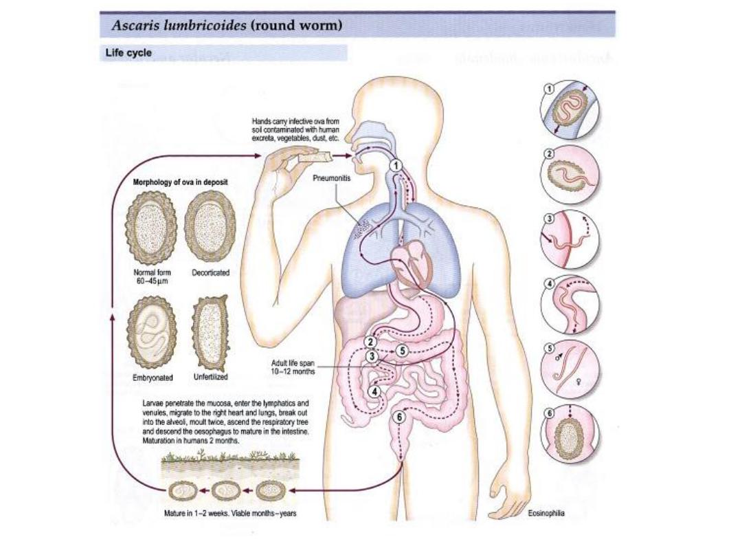
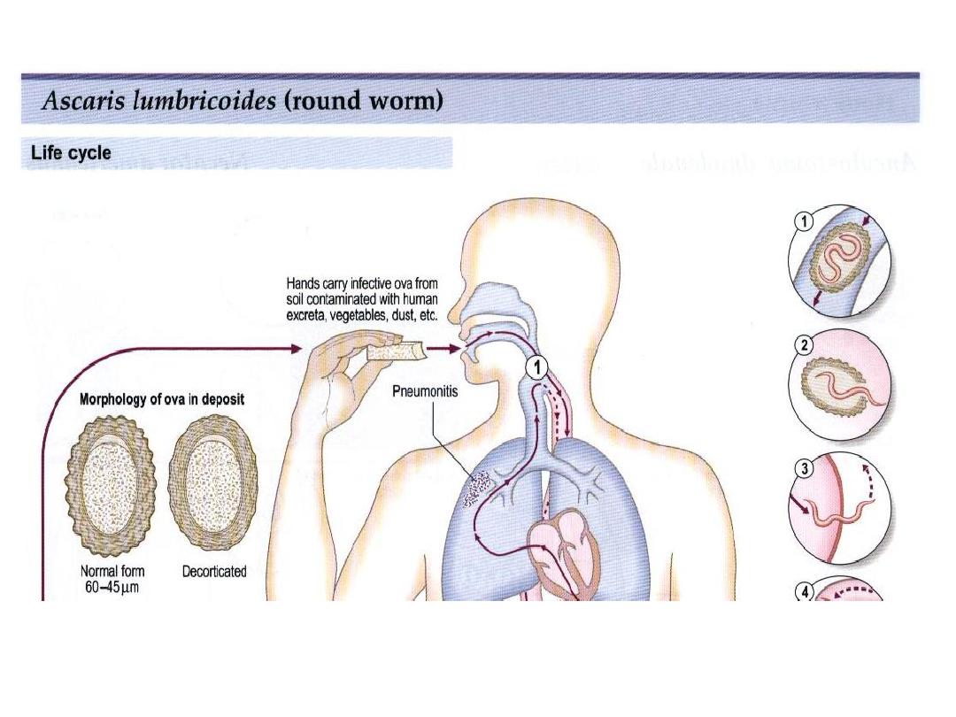
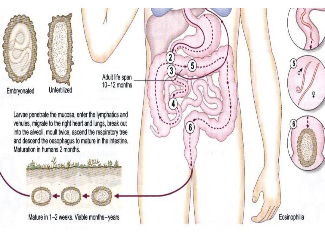
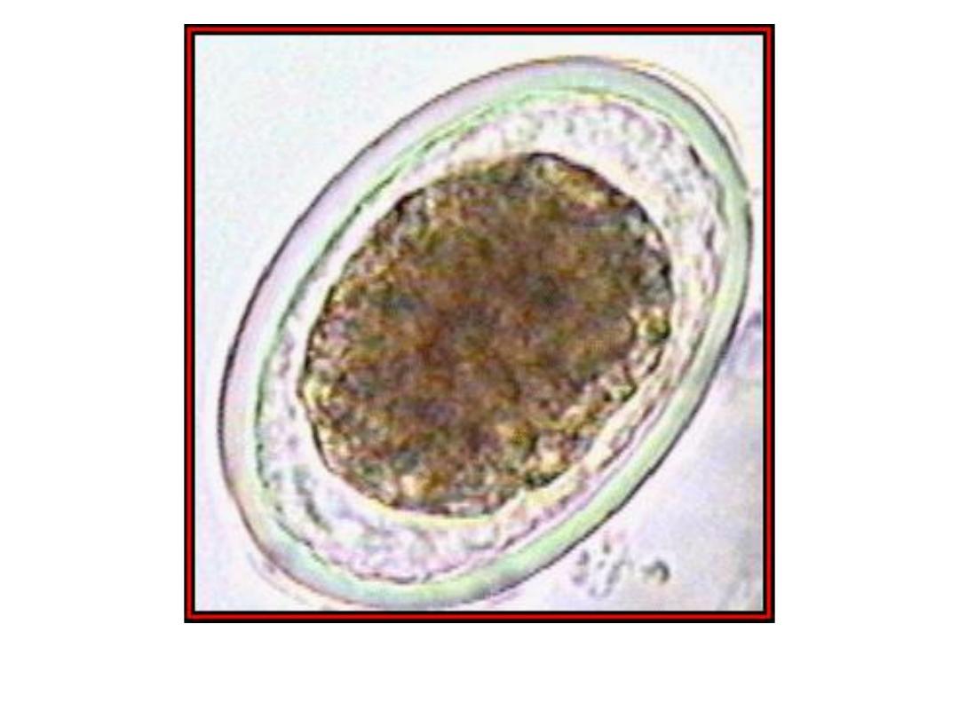
Egg:
The fertilized egg of Ascaris
lumbricoides at the time of
oviposition is spherical or sub-
spherical,measures 65-75um
by35-50um &consists of the
following observable structures

1-A coarsely granular ,spherical
ovumthat usually does not completely
fill the shell.
2-A thin innermost membrane that is
highly impermeable.
3-A relatively thick,colorless middle
layer that is smooth on both inner
&outer surfaces .
4-An outer most ,coarsely mammilated

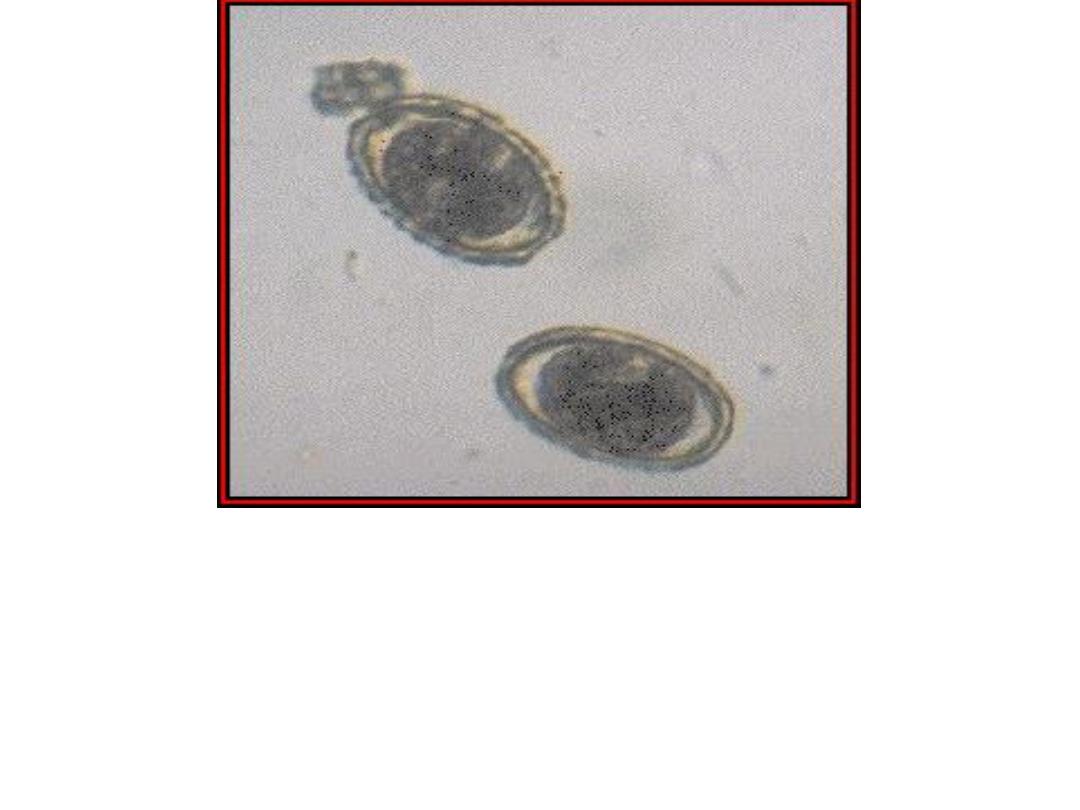
Female worms without males
produce infertile eggs that are
markedly subspherical
(88um by38-44um),internally they
contain
a mass of disorganized granules
that completely fill the shell

Life cycle
A single female
can produce up to 200,000 eggs
each day! About two weeks
after passage in the feces the eggs
contain an infective larval

or juvenile stage, and humans are
infected when they ingest
such infective eggs. The eggs hatch
in the small intestine

the juvenile penetrates the small
intestine and enters the circulatory
system, and eventually the juvenile
worm enters the lungs.

In the lungs the juvenile worm
leaves the circulatory system and
enters the air passages of the lungs.

The juvenile worm then migrates
up the air passages into the pharynx
where
it is swallowed, and once in the
small intestine the juvenile
grows into an adult worm.

Why Ascaris undergoes such a
migration through the body to only
end up where it started is
unknown.

Such a migration is not unique to
Ascaris, as its close relatives
undergo a similar migration in the
bodies of Ascaris
infections in
humans can cause significant
pathology.
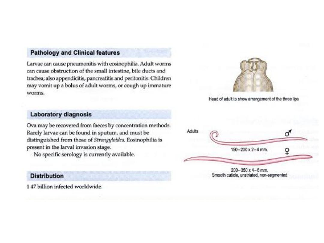

Pathology :
The migration of the larvae through
the lungs causes
the
blood vessels of the lungs to
hemorrhage, and there is an
inflammatory response accompanied
by edema.

The resulting
accumulation of fluids in the lung
results in "ascaris pneumonia," and
this can be fatal

2-The large size of the adult
worms also presents problems,
especially if the worms
physically block the
gastrointestinal tract.

Ascaris is not orious
for it reputation to migrate within
the small intestine, and when
large worm begins to migrate there
is not much that can stop it

Instances have been reported in
which Ascaris have migrated
into and blocked the bile or
pancreatic duct or in which
the worms have penetrated the
smallintestine resulting in acut and
fatal peritonitis.

Ascaris seems to be especially
sensitive to anesthetics, and numerous
cases have been
documented where

3-patients in surgical recovery
rooms have had
worms migrate from the small
intestine, through the stomach,
and out the patient's nose or
mouth

Ascaris
suum is found in pigs. Its life
cycle is identical to that of
A. lumbricoides. If a human ingests
eggs of A. suum the
larvae will migrate to the lungs and die

." Adult worms
of this species do not develop in the
human's intestine

. (Some parasitologists believe that
there is but one species of
Ascaris that infects both pigs and
humans, but any commentary
on this issue is beyond the scope of
this web site.)

Infections of Ascaris are diagnosed
by:
1-finding characteristic eggs in
the feces of the infected host
.
22
2-the presence of three large lips, a characteristic of ascarids.
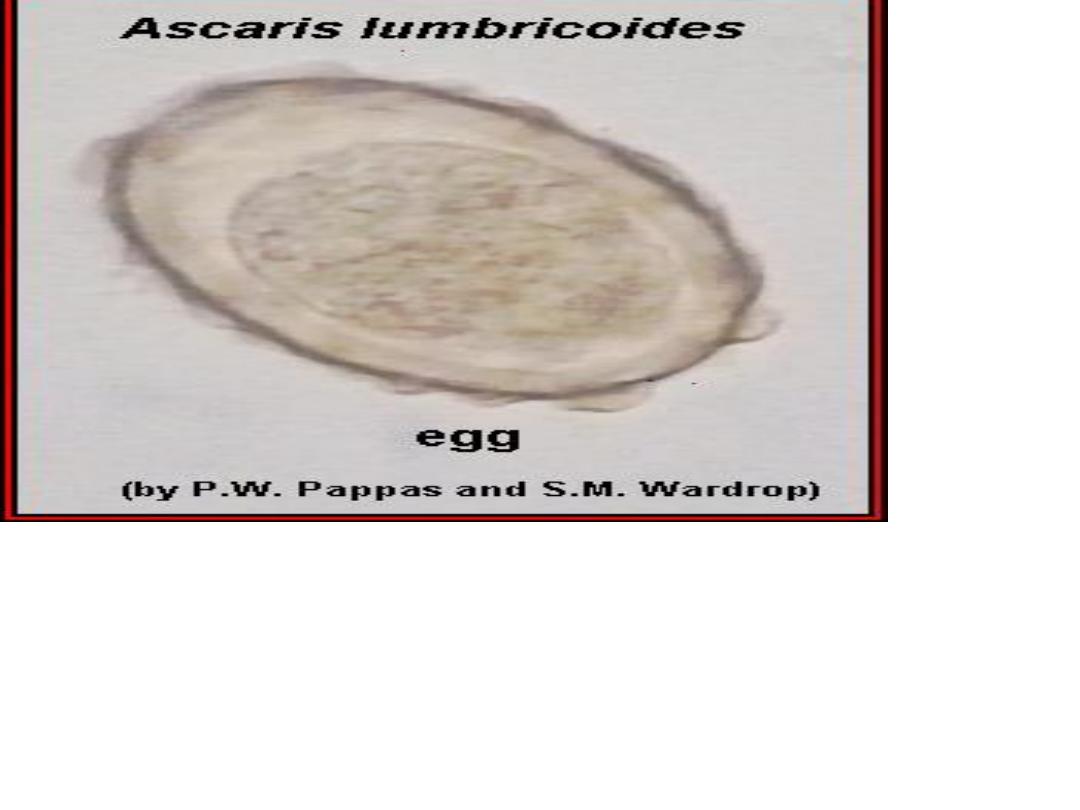
Ascaris lumbricoides, fertilized egg.
Ascaris lumbricoides,
fertilized egg.
Another example of a fertilized Ascaris
lumbricoides egg. (Original image from
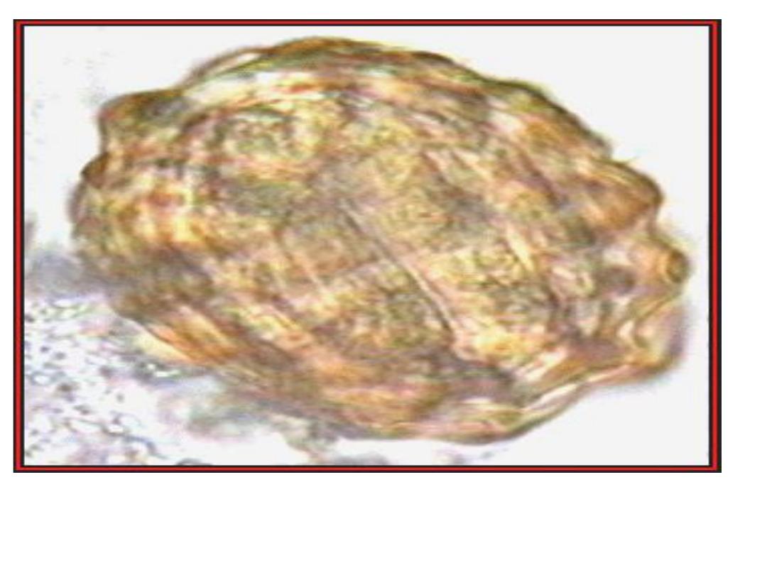
An example of an unfertilized A.
lumbricoides egg. (Original image
from:
Atlas of Medical
Parasitology
.)
An example of an unfertilized A. lumbricoides egg. (Original image from:
Atlas of Medical Parasitology
.)
A "decorticated," fertilized, Ascaris lumbricoides. (Original image from:
Atlas of Medical Parasitology
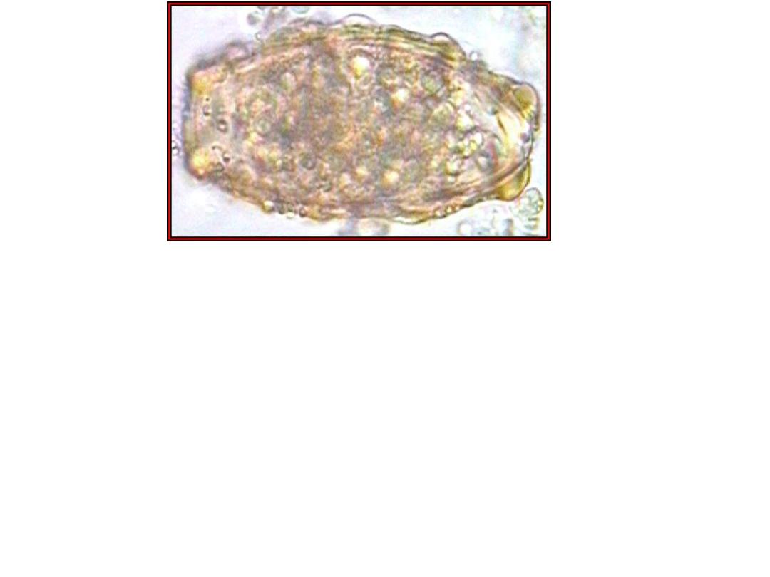
Eggs of Ascaris suum. A. suum is a common parasite of pigs.
The eggs are virtually indistinguishable from those of A.lumbricoides.
(Original image from
Oklahoma State University, College of Veterinary Medicine
.)
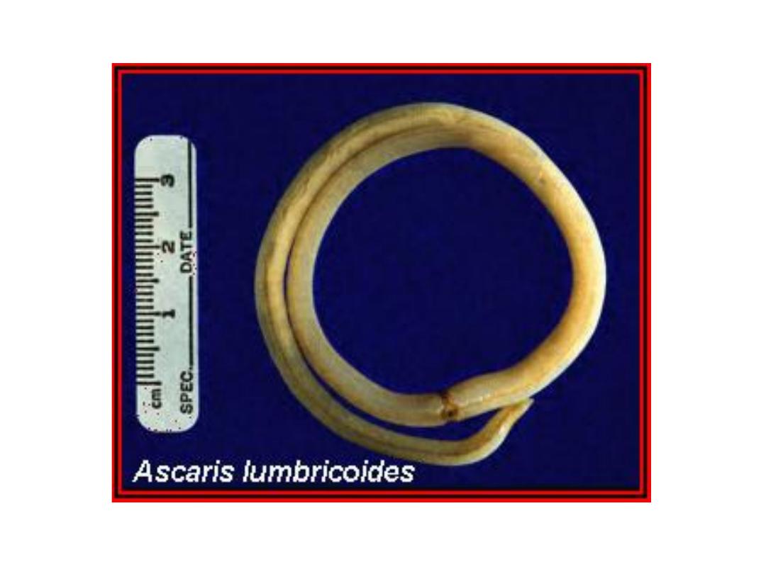
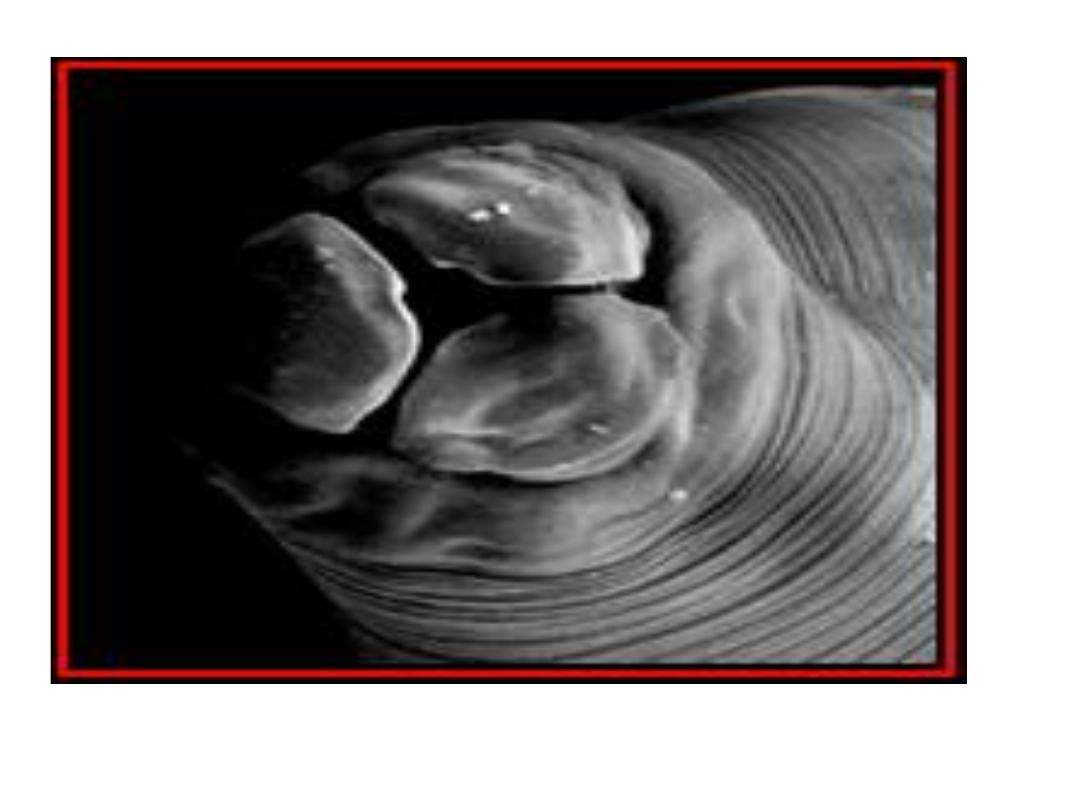
Female and male Ascaris lumbricoides; the female measures
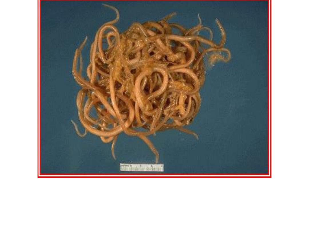
female Ascaris lumbricoides. Females of this
species can measure over 16 inches long. This
specimen was passed by a
young girl in Florida.

Note : up stream movement this
movement for A. lumbericoides
through mouth or nose.

Note:
some times the infected man may die
due to this irritation action after
changing to anaphylatic or HSR.

Lecture 2 Dr. Jabar Etaby
Hookworms
Introduction
Patients with hookworm
infection often are
asymptomatic; however,
chronic hookworm infection
is a common cause of
moderate and severe
hypochromic, microcytic
anemia

in people living in
tropical developing
countries, and heavy
infection can cause
hypoproteinemia with
edema.

EPIDEMIOLOGY
Humans are the only reservoir.
Hookworms are
prominent in rural, tropical, and
subtropical areas where
soil contamination with human
feces is common.

Although the prevalence of both
hookworm species is
equal in many areas, A. duodenale is
the predominant species in the
Mediterranean region, northern Asia,
and
selected foci of South America.

N. americanus is
predominant in the Western
hemisphere, sub-Saharan
Africa, Southeast Asia, and
a number of Pacific islands

Larvae and eggs survive in loose,
sandy, moist, shady,
well-aerated, warm soil (optimal
temperature 23°C–33°C)
[73°F–91°F]).

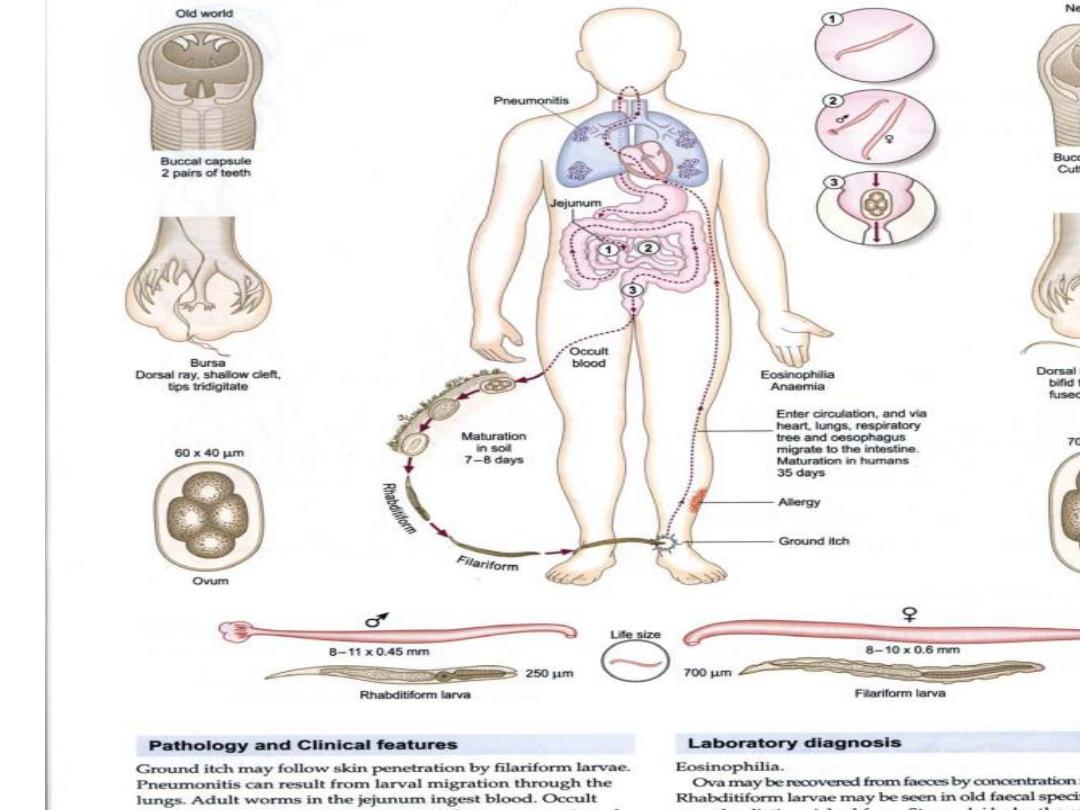
Life cycle
Hookworm eggs from stool hatch
in soil in 1 to 2 days
as rhabditiform larvae. These
larvae develop into infective
filariform larvae in soil within 5 to
7 days and can persist for weeks to
months.

Percutaneous infection occurs after
exposure toinfectious larvae.
A.duodenale transmission can occur by
oral ingestion
and possibly through human milk.

Untreated infected patients
can harbor worms for 5
years or longer.

The time from exposure to
development
of noncutaneous symptoms
is 4 to 12 weeks.

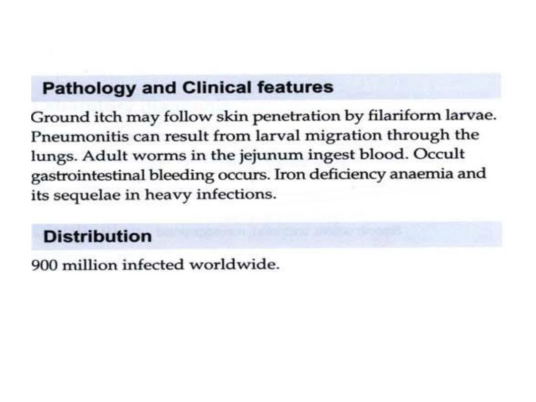
Clinical signs
Patients with hookworm infection
often are
asymptomatic; however, chronic
hookworm infection is a
common cause of moderate and
severe hypochromic

microcytic anemia in people living in
tropical developing
countries, and heavy infection can
cause
hypoproteinemia with edema

Chronic hookworm
infection in children may lead
to physical growth delay,
deficits in cognition, and
developmental delay.

After
contact with contaminated
soil, initial skin penetration of
larvae, often involving the feet,
can cause a stinging

or
burning sensation followed
by pruritus and a
papulo vesicular rash that
may persist for 1 to 2
weeks

Pneumonitis associated with migrating
larvae is
uncommon and usually mild, except
in heavy infections.

Colicky abdominal pain, nausea,
and/or diarrhea and
marked eosinophilia can develop 4
to 6 weeks after exposure

Blood loss secondary to hookworm
infection
develops 10 to 12 weeks after initial
infection and
symptoms related to serious iron-
deficiency anemia can
develop in long-standing moderate
or heavy hookworm
infections.

After oral ingestion of
infectious Ancylostoma
duodenale larvae, disease
can manifest withpharyngeal
itching, hoarseness, nausea,
and vomiting shortly after
ingestion.

ETIOLOGY
Necator americanus is the
major cause of hookworm
infection worldwide, although
A. duodenale also is an
important hookworm in
someregions.

Mixed infections
are common. Both are
roundworms (nematodes)
with
similar life cycles
.

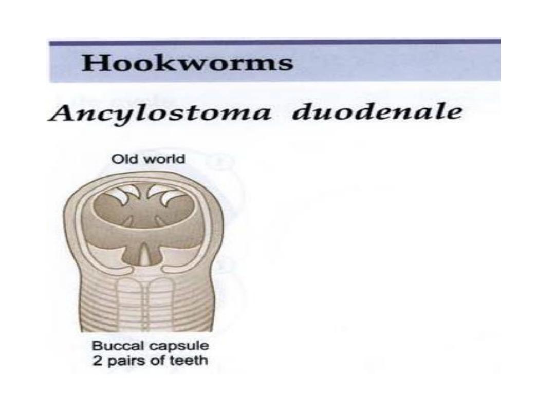
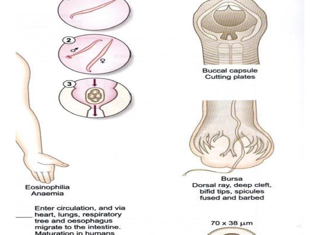
Ancylostoma spp. and Necator spp.
(hookworms)
There are many species of
hookworms that infect mammals

The most important, at least from the
human standpoint, are
the human hookworms, Ancylostoma
duodenale and Necator
americanus, which infect an estimated
800,000,000 persons,

and the dog and cat hookworms,
A.caninum and A. braziliense,
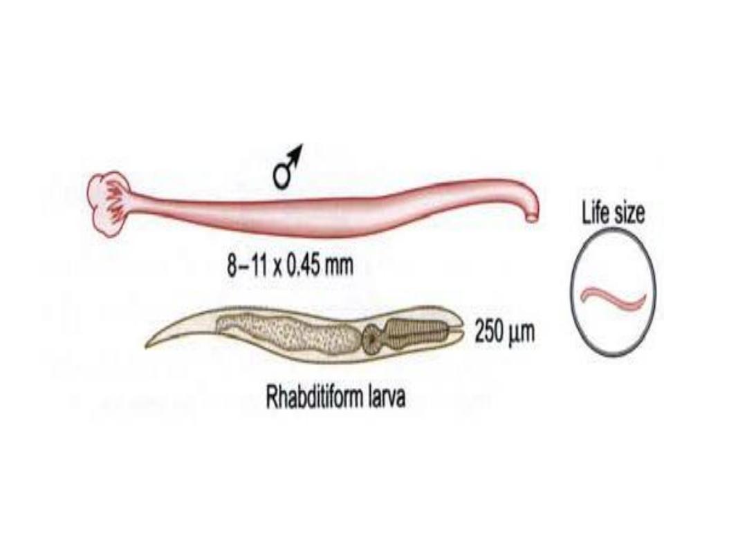
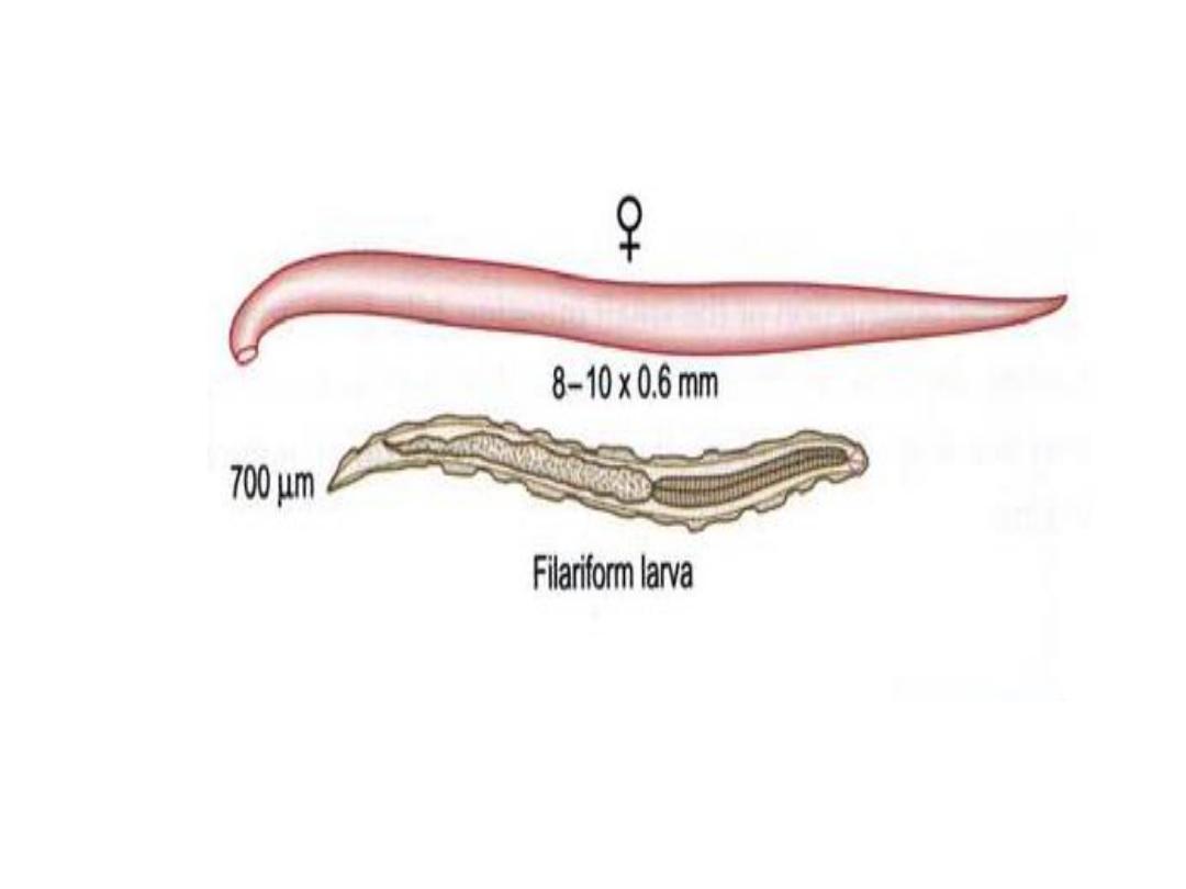
respectively. Hookworms average
about 10 mm in length
and live in the small intestine of the
host

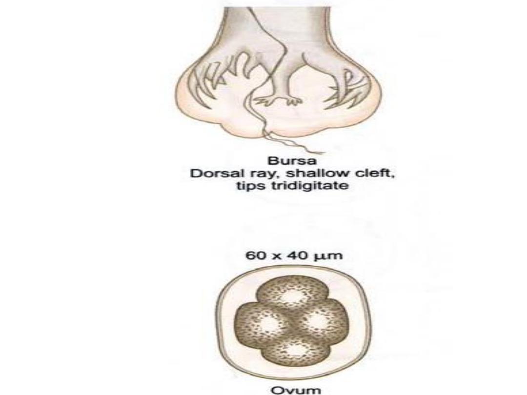
The males and
females mate, and the female
produces eggs that are passed in
the feces. Depending on the species,
female hookworms can
produce 10,000-25,000 eggs perday.

About two days after
passage the hookworm egg
hatches, and the juvenile worm (or
larva) develops into an infective
stage in about five days.

The next host is infected when an
infective larva penetrates the
host's skin.

The juvenile worm migrates
through the host's
body and finally ends up in the
host's small intestine where it
grows to sexual maturity.

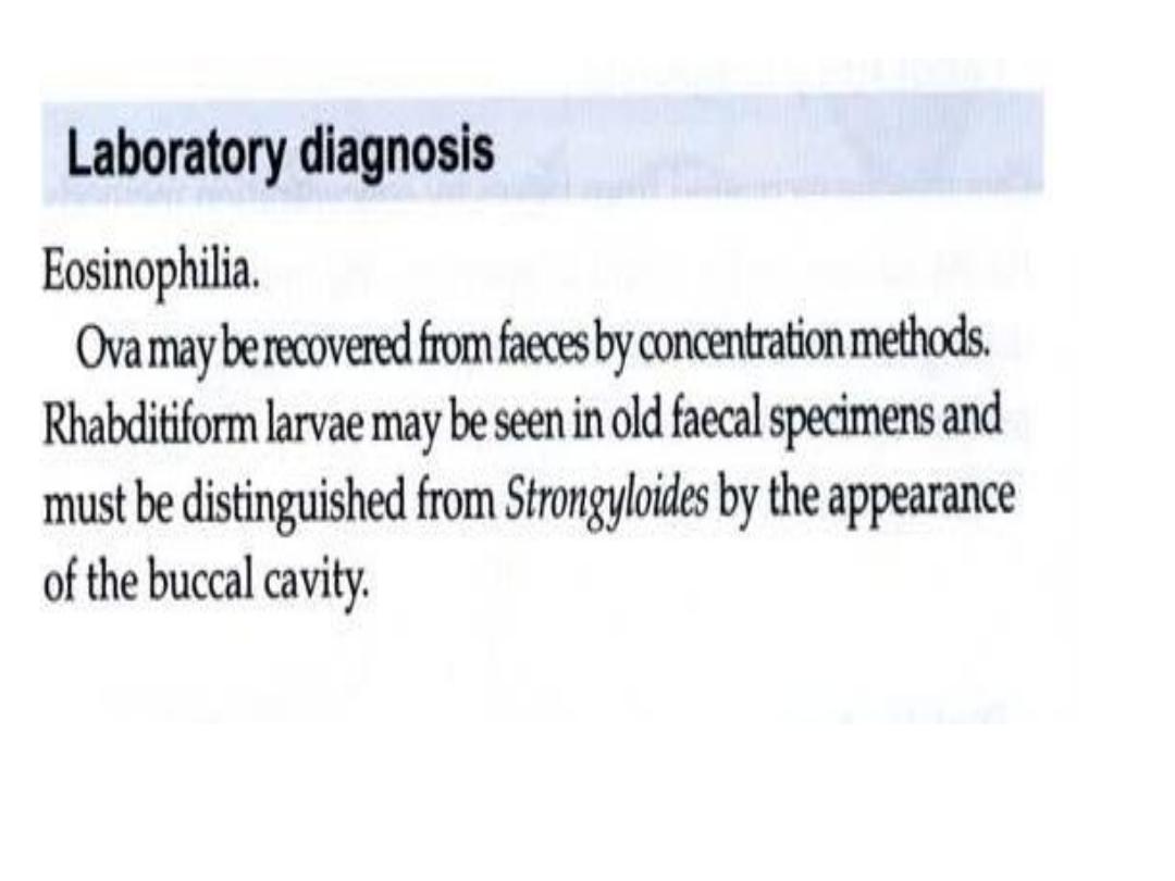
The presence of hookworms can
be demonstrated by finding the
characteristic eggs in the feces; the
eggs can not, however, be
differentiated to species

Juveniles (larvae) of the dog and
cat hookworms can infect
humans, but the juvenile worms
will not mature into adult worms.

This results in a condition known
as "cutaneous" or
"dermal larval migrans" or
"creeping eruption." Hence the
importance of not allowing dogs
and cats to defecate
indiscriminately.

The following image provides an
excellent
example of how hookworms are
attached to and embedded in
the epithelium of the host's
gastrointestinal tract.

DIAGNOSTIC TESTS

1- Microscopic demonstration of
hookworm eggs in feces is
diagnostic.
&
Adult worms or larvae rarely are
seen.

2-Approximately 5 to 8
weeks are required after
infection
for eggs to appear in feces.

3-A direct stool smear with
saline solution or potassium iodide
saturated with iodine
is adequate for diagnosis of heavy
hookworm infection;

4-light infections require
concentration techniques.
Quantification techniques

5-, Kato-Katz, Beaver direct
smear, or Stoll egg-counting
techniques) to determine
the clinical significance of infection
and the response to
treatment may be available from
state or reference
laboratories.

CONTROL MEASURES
Sanitary disposal of feces to
prevent contamination of
soil is necessary in areas with
endemic infection

Treatment of all known infected
people and screening of
high-risk groups (ie, children and
agricultural workers) in
areas with endemic infection can
help decrease environmental
contamination.

. Wearing shoes may not be
fully protective, because cutaneous
exposure to
hookworm larvae over the entire
body surface of children
could result in infection.

Despite relatively rapid
reinfection, periodic
deworming treatments
targeting
preschool-aged and school-
aged children have been

advocated to prevent
morbidity associated with
heavy
intestinal helminth
infections`
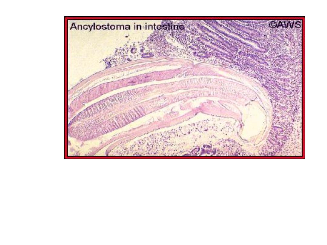
A histological section of a hookworm in the host's small
intestine. Original image copyrighted and provided
byDr. A.W.
Shostak, and used with permission