
Lecture 4
DR.
Jabar Etaby

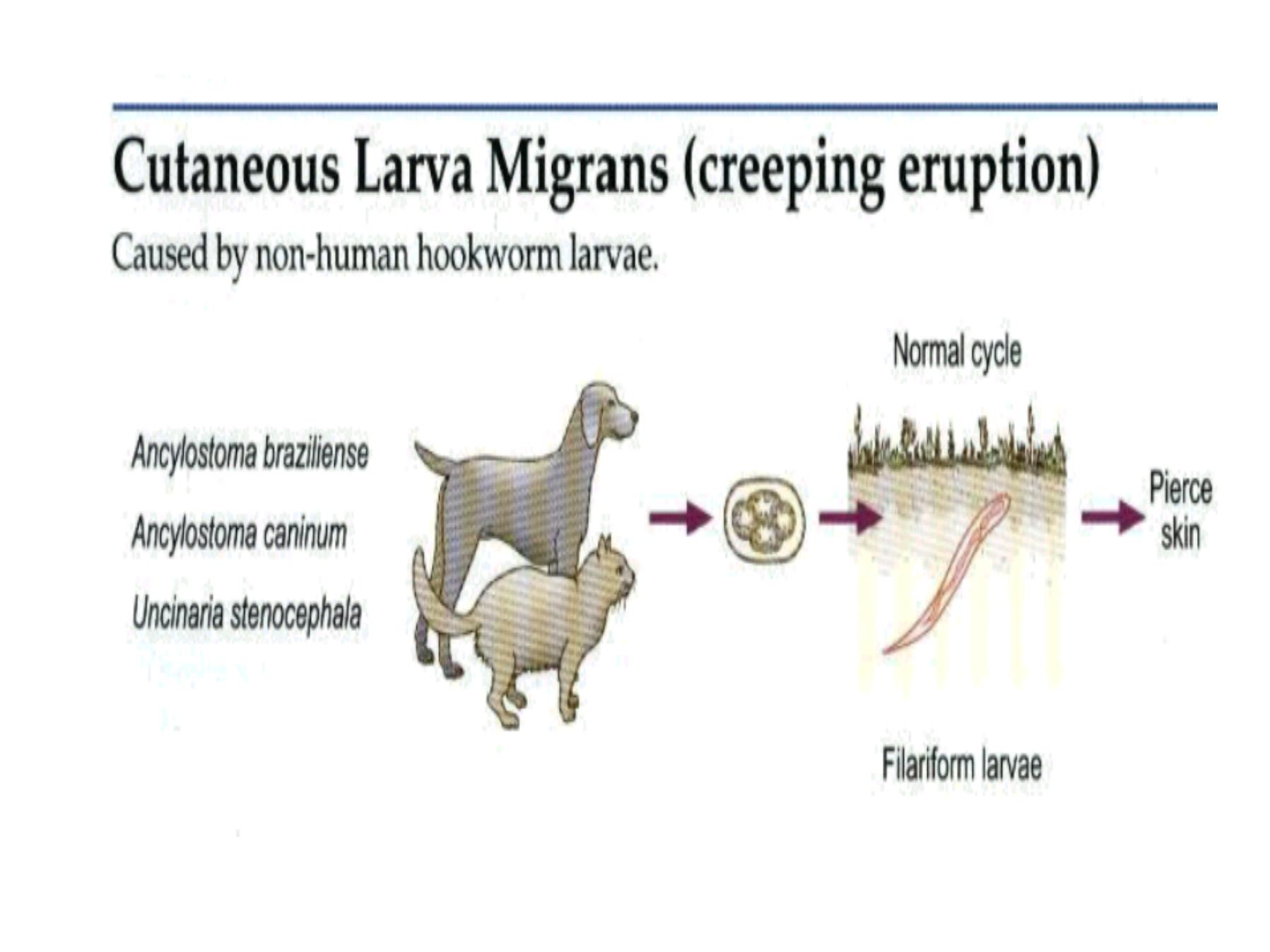
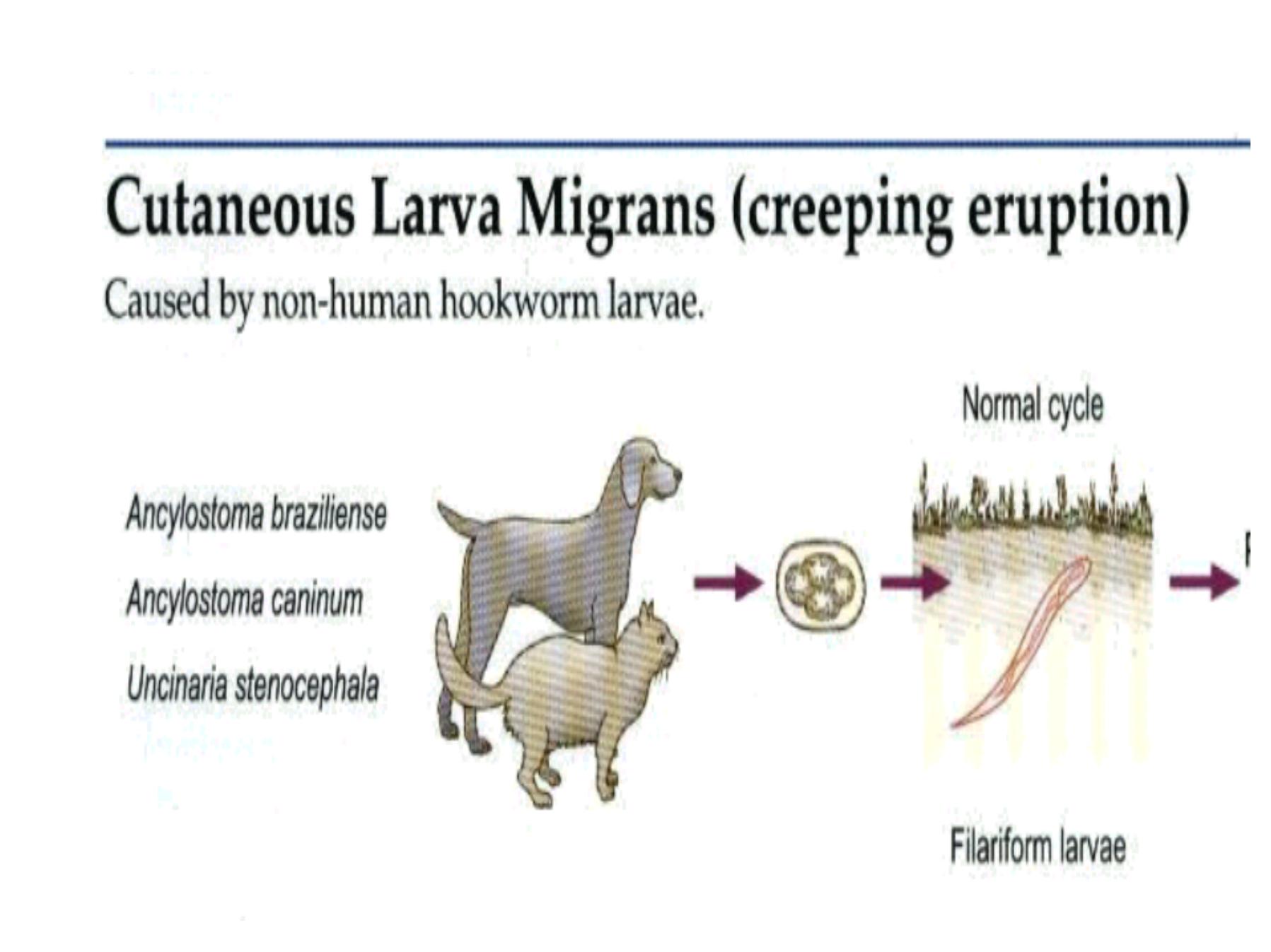
Introduction : Cutaneous larva
migrans(CLM),frequently termed
creeping eruption,is a parasitic skin
infection that is caused by the
filariform larvae of various animal
hookworm nematodes.

Distribution
CLM has a worldwide distribution
wherever have had skin contact with soil
contaminated with infected animal feces.
The disease most commonly occurs in
subtropical &tropical regions, but may
also occur in temperate climates
particularly during the summer months
&during rainy seasons.

Caustive agents
A ncylostoma braziliense
ahookworm of wild &domestic dogs
&cats is the
most commonly identified etiolgic
agent of CLM
Cutaneous (dermal) larval migrants
There are several examples of parasites that are
normally found in pets but can be transmitted to humans.

For example, acommon tapeworm of
dogs,
Dipylidium caninum
, can be
transmitted to humans.

Immature forms of the common
roundworm of dogs,
Toxocara
canis
can also be found in humans,
causing a disease known as
visceral
larval migrans
.

Immature forms of both cat and
dog hookworms can also infect
humans, and this results in a
disease called cutaneous or dermal
larval migrants (CLM or DLM).

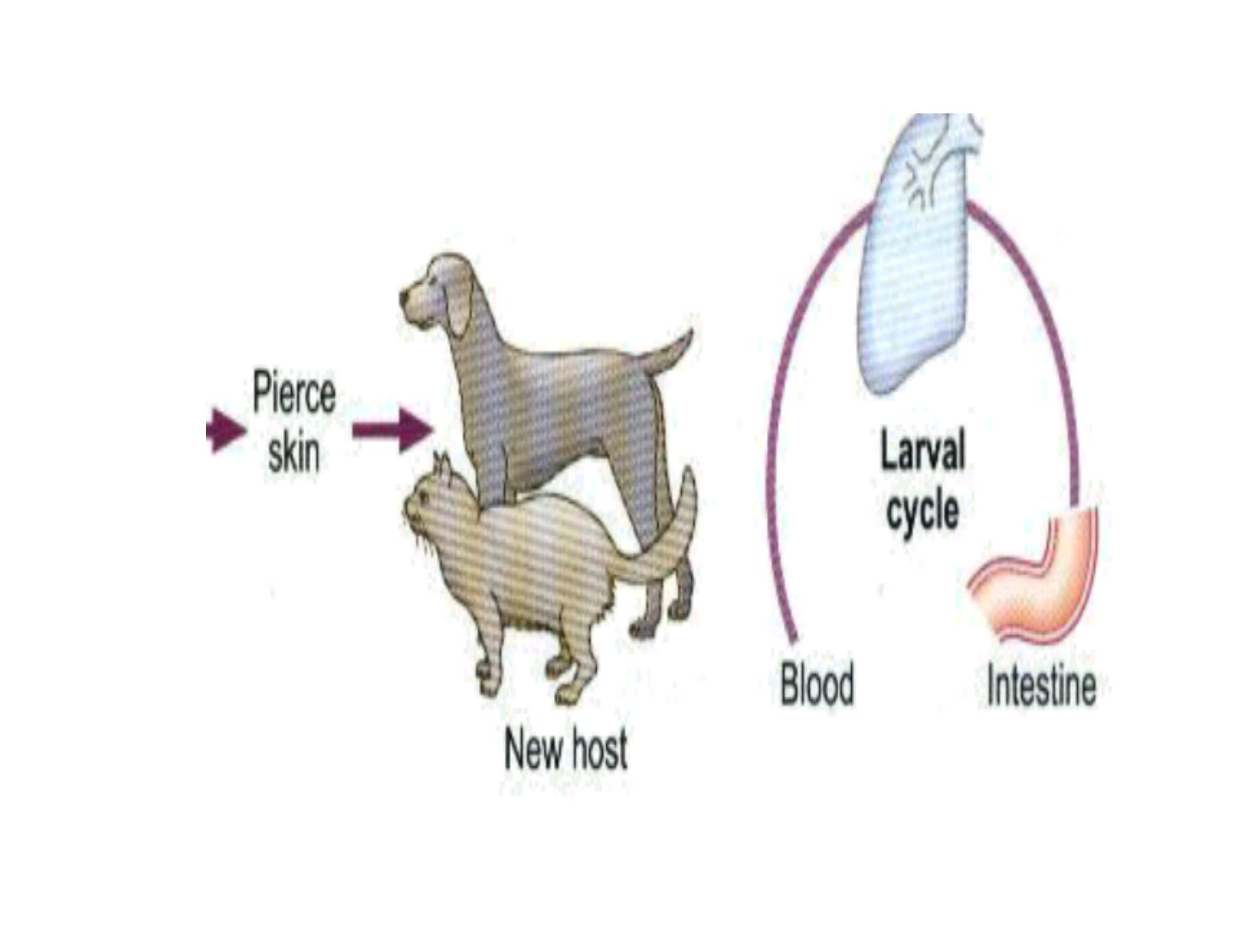
The eggs of dog and cat
hookworms hatch after being
passed in the host's feces, and the
next host is infected when these
larvae penetrate the host's skin.

Unfortunately, these larvae can not
tell the skin of one animal from
another, so they will penetrate
human skin if they come in contact
with it.

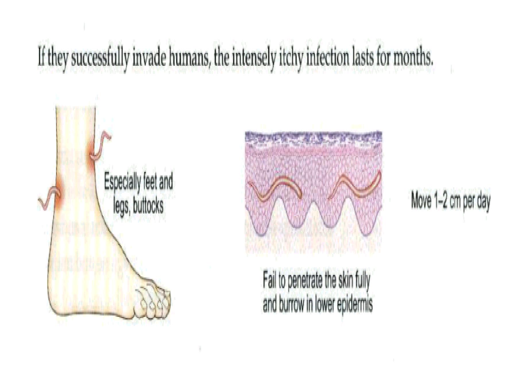
However, a human is an unnatural
host, so the larvae do not enter the
blood stream as they would in a
dog or cat.

Rather, they remain in the skin for
extended periods of time (weeks or
months in some instances) and
finally die.

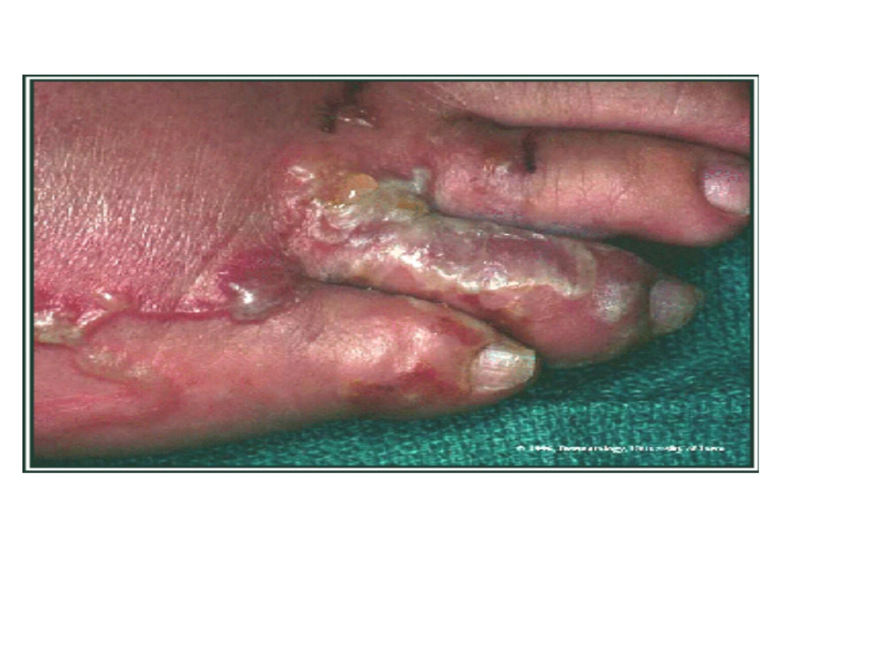
Disease Signs&Symptoms:
The most common portals of entry
by the larvae are the exposed areas
of the body such as the dorsum of
the feet, lower legs, arms and hands,
thighs and abdomen also may be
involved probably due to lying
directly on contaminated sand.

2-Visceral larval migrants( VLM)
larvae of specific or non specific
host migrates by after skin
pentration or directly to the gut
then hatching there ,then it will be
gone to the viscera, liver, lung,
muscles or even brain e.g A.
lumbericoides ,T. cains. T. cati &
Ascaris. equi

S o, there be irritation &
eosinophila(5 % or some times even
7 0 % or 8 0 % ) but still it is not
diagnostic features as we knew .
W eather it is CLM or V LM there is
some sort of attraction to words the
anterior part of the body (by
migration through blood vessels or
lymph vessels

This migrate be due to the presence
of high tension of O2 in the anterior
part of the body espically in the
viscera

Note:
T. canis cause kid blindness &painfultumor.
Toxocara spp. Cause infection of the CNS
during the 1
st
day of infection, the eggs in the
gut will hatch into larvae so the curve is high
then it will decline with migration of the
larvae to other tissues & organs. So liver
from 1
st
day of infection they start to
increase gradually till the 1
st
week then
decline

Lung :
the decrease in the liver will
syncronise with increase in the lung
due to migration of the larvae from
the liver to the lung .

Brain may be from the beginning of inf.
the larvae reach the brain directly by
remaining only for some times in the
lung then migrate & stay in the brain
because the brain tissue is soft
&represents enriched media for the
parasite which remain

Not encysted as thy are away from
the immune response &it will
increase during the 3
rd
week& at the
same time decrease in lung &liver .

The infective stage in CLM is 3
rd
stage larvae while in V LM is 2
nd
stage larvae

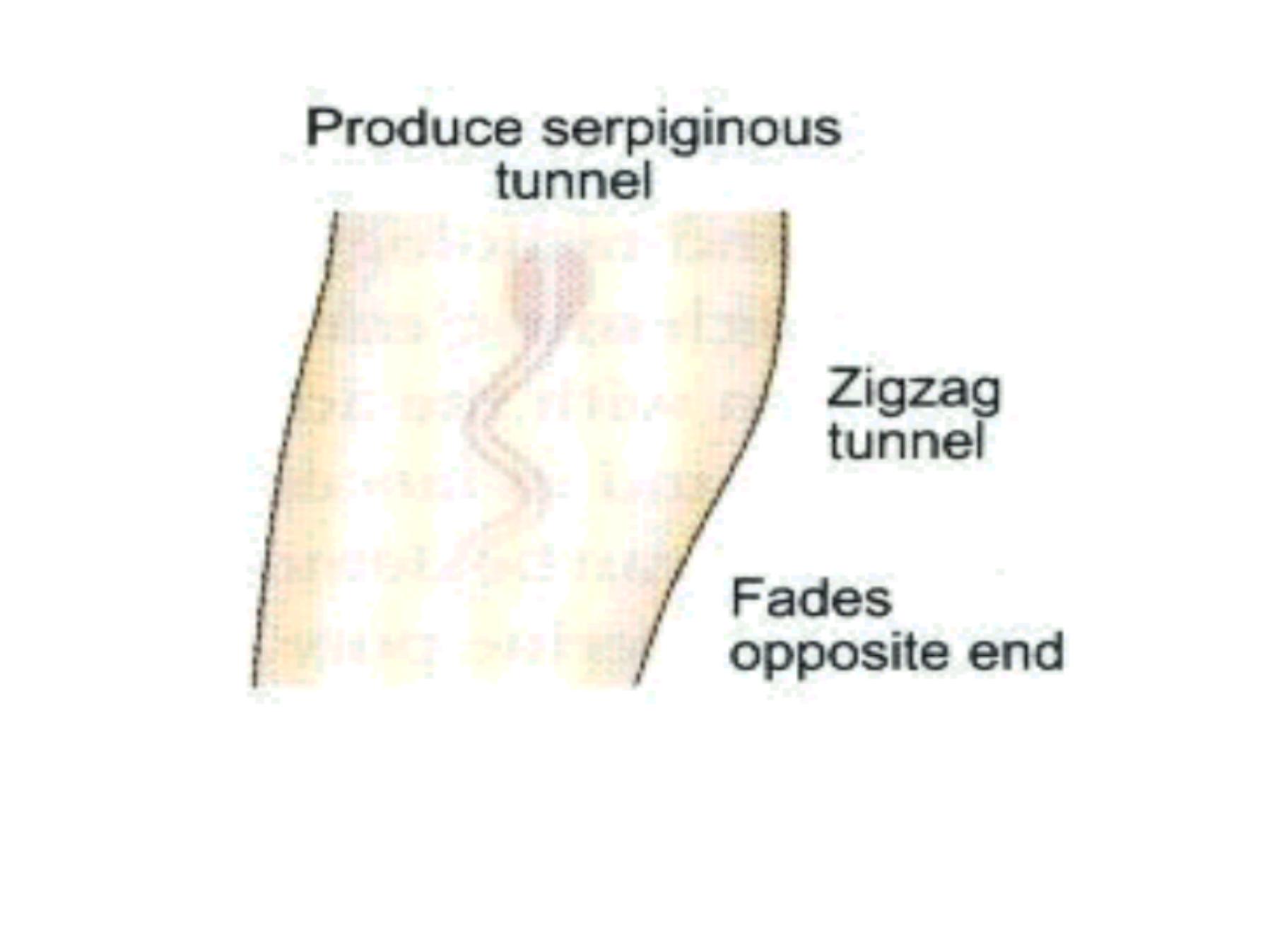
As the larvae migrate through the skin and
finally die, there is an inflammatory response,
and the progress of the larvae through the skin
can actually be followed since they leave a
tortuous "track" of inflammed tissue just under
the surface of the skin

Treatment of such infections requires surgical
removal of the migrating larvae. Considering the
location of larvae, just under the skin, in light
infections this can be done under local anesthesia
and is a relatively simple procedure.

Infections involving large numbers
of larvae can be very
uncomfortable, and treatment
(removal) might require general
anesthesia and supportive
treatment with anti-inflammatory
drugs.

How do humans come in contact
with the larvae of dog and cat
hookworms? A common source of
infection in developed countries is
probably sandboxes.

If you have a sandbox in your
backyard, it is almost certain that
cats in the neighborhood are using it
as a large litter box.

Moreover, the sand provides a
nearly ideal environment for the
hookworm eggs to develop and
hatch and for the larvae to survive.

Diagnosis:
dignosis of CLM is based on a
history of exposure and clinical
appearance of the skin eruption.
Allergic dermatitis, secondary
bacterial infection

Peripheral eosinophilia and increase
IgElevels are found in a minority of
patients. S kin biopsies are usually not
effective at establishing the diagnosis.

Histologic examination
may lead to edge of the track may
contain a larva trapped in follcular
canal, stratum cornea or dermis.
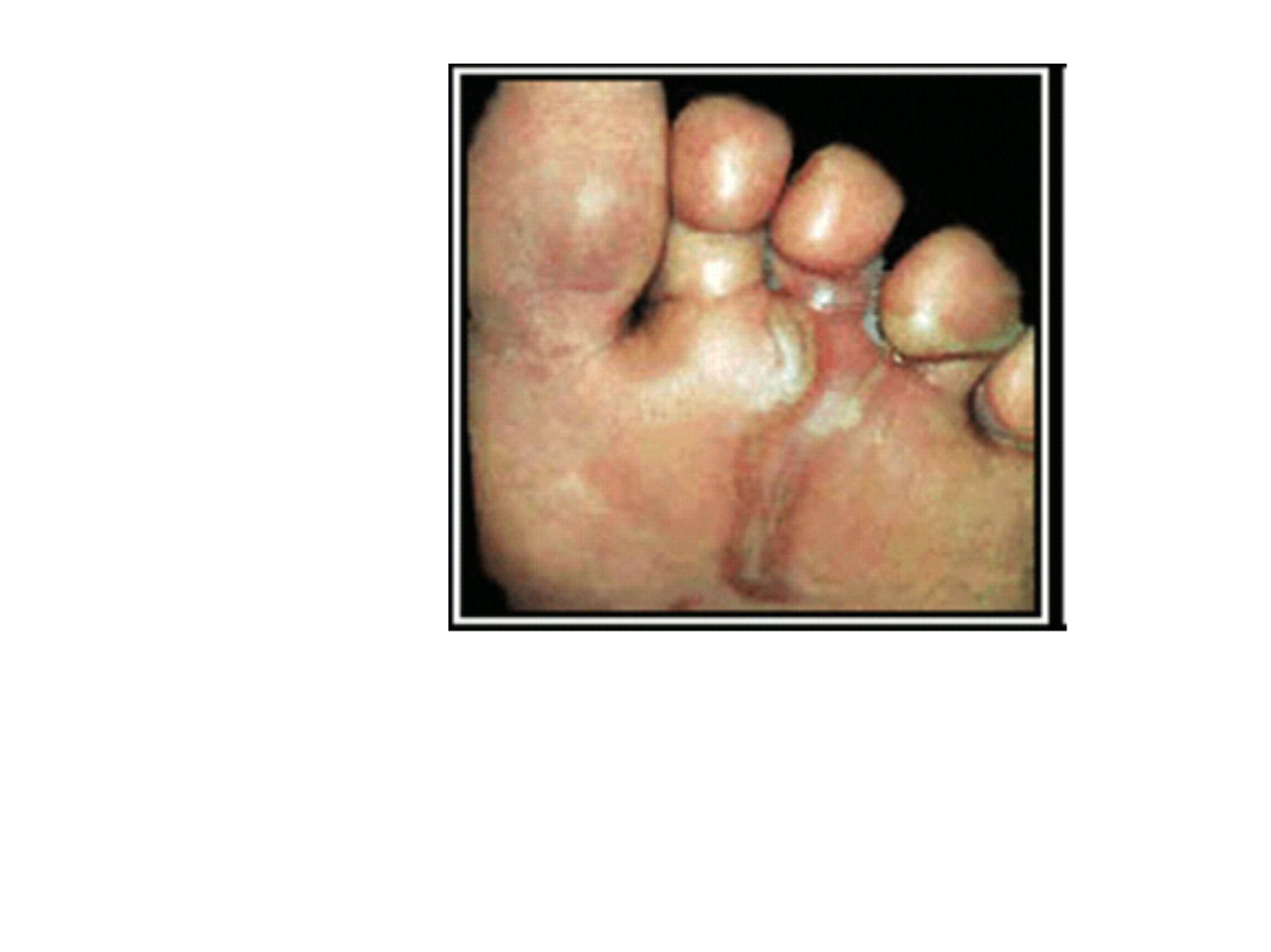
CLM of the foot.
(Original image from:
Companion Animal Surgery
.")
CLM of the foot.

Thus, keeping sandboxes covered
to prevent cats from defecating in
them is a worthwhile "ounce of
prevention."
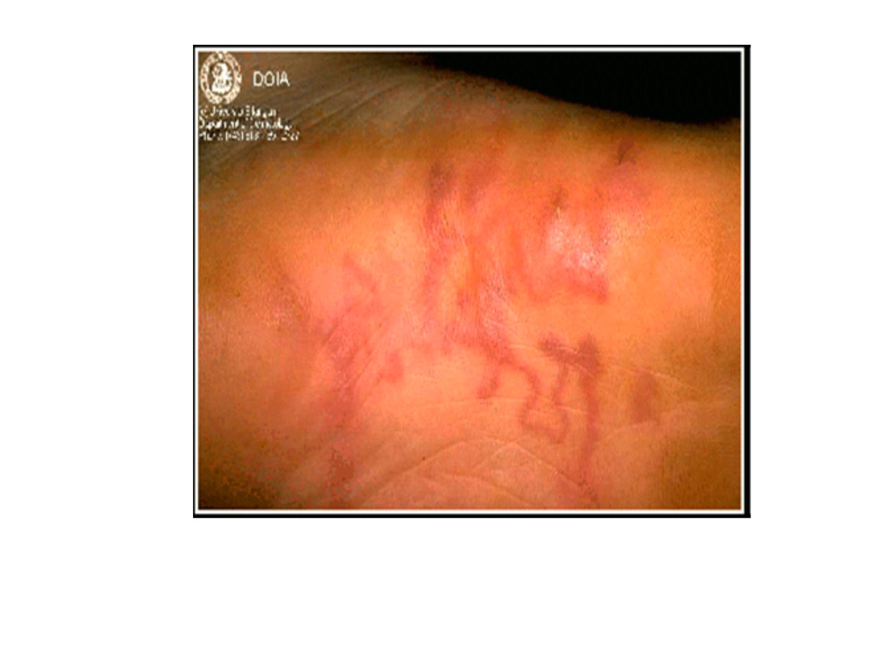
CLM (Original image from and copyrighted by
Dermatology Internet Service,
Department of Dermatology, University of
Erlangen
.)
(Original image from and
copyrighted by
Dermatologic
Image Database, Department of
Dermatology, University of Iowa
College of Medicine
).

Other places where cats might
defecate are also possible sources
of infection, including flower beds
and vegetable

Garden Dogs are much less
fastidious about where they
defecate, so it is more difficult to
control dog feces as a possible
source of infection

If you own a dog two measures that
you should take are (1) keep you
dog free of hookworms and(2)
make sure that you clean up the
dog's feces on a regular basis

Also, if you "walk" your dog in a
park or playground, and in
particular in my front yard, make
sure that you pick up and dispose
of any fecal material the dog might
leave behind