Classification of Genetic disorders:
I. Classical Genetic Diseases:
1. Chromosomal (Cytogenetic) disorders.
2. Single
gene
(or
unifactorial)
disorders
(Mendelian
Disorders).
3. Multifactorial disorders.
II. Non-Classical Diseases "or the single gene disorders with
atypical pattern of inheritance":
i. Diseases caused by mutations in mitochondrial genes.
ii. Triplet repeat mutations.
iii. Uniparental disomy / Genomic imprinting.
iv. Gonadal mosaism.
Added to that, is the group of
congenital malformations (Birth
Defects)
.

2. Defects of Single Genes with Large Effect
(Unifactorial or Mendelian Disorders)


The number of known Mendelian disorders has grown
to more than 5000. Although individually some are
rare, altogether they account for about 1% of all
adult hospital admissions and about 6-8% of all
pediatric hospital admissions.
They are caused by a mutation in a
single
gene.
A mutation
is a disturbance in the sequence of the
nucleotide arrangement in the DNA molecule
, or it
is simply
a permanent change in the DNA
.
Mutations affecting the germ cells are transmitted to
the progeny and
may
give rise to inherited
disorders.
Those occurring in the somatic cells are important in
the causation of cancers and some congenital
malformation.
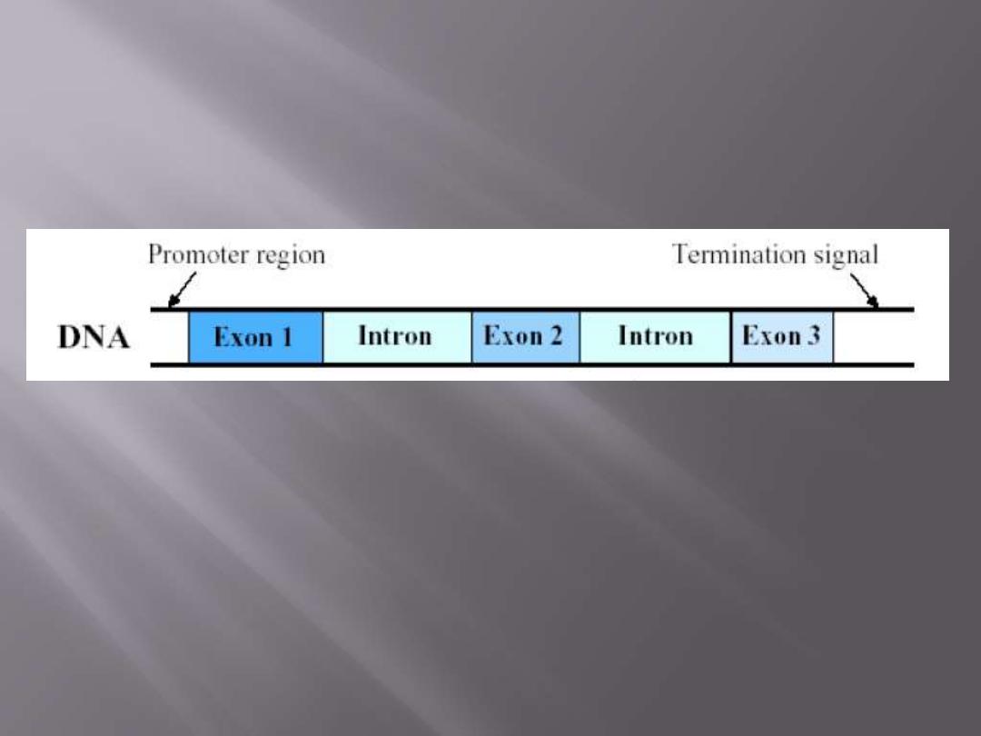
A gene is that part of the DNA that codes for a
polypeptide chain (or RNA).
30,000 genes (in contrast to what was previously though of
about 100,000).
Exons
Introns (Intragenic or intergenic)
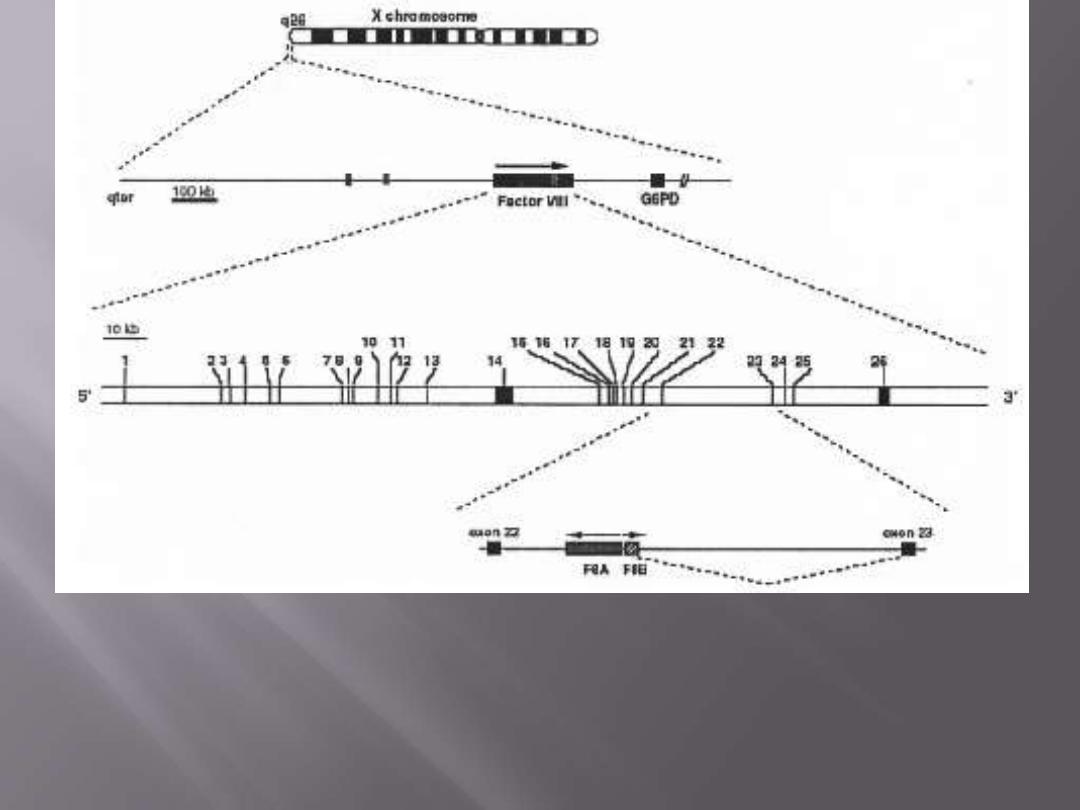
Localization and structure of the factor VIII gene, located about
1000kb from the Xq telomere (Xqter). The gene is 186kb long
and contains 26 exons .

Dominant genes
Recessive genes
Autosomal
X-linked
Codominant

Pleiotropy
Genetic heterogeneity
.

Marfan syndrome is a connective tissue disorder, so
affects many structures, including the
skeleton, lungs,
eyes, heart and blood vessels.
The disease is
characterized by unusually long limbs, and is believed
to have affected Abraham Lincoln and Ausama Bin
Laden.
Marfan syndrome is an AD disorder that has been
linked to the
FBN1 gene on chromosome 15.
FBN1
encodes a protein called
fibrillin
, which is essential for the
formation of
elastic fibres
found in connective tissue.
Without the structural support provided by fibrillin,
many tissues are weakened, which can have severe
consequences, for example, ruptures in the walls of
major arteries.

ANY QUESTION?

NOTHING COMES EASILY
A tree needs to be nurtured for a long time
to become green, strong and fruitful

Why some genes act in a dominant
manner while others behave in a
recessive one?

A single gene is responsible for the formation of a single type of
polypeptide
The types of proteins are varied; they could be
structural
proteins
, like fibrous tissue, elastic tissue; they could be
immunoglobulins
; they could be
signal proteins
; they could
be
receptors, enzymes, hormones
, … etc.
Therefore, the action of the gene being dominant or recessive
is
determined by the type of protein it produces
and its
function.
(not by the size or structure or no. of exons of the gene)
Gene = Protein
X
Gene = Polypeptide (true to some extent)

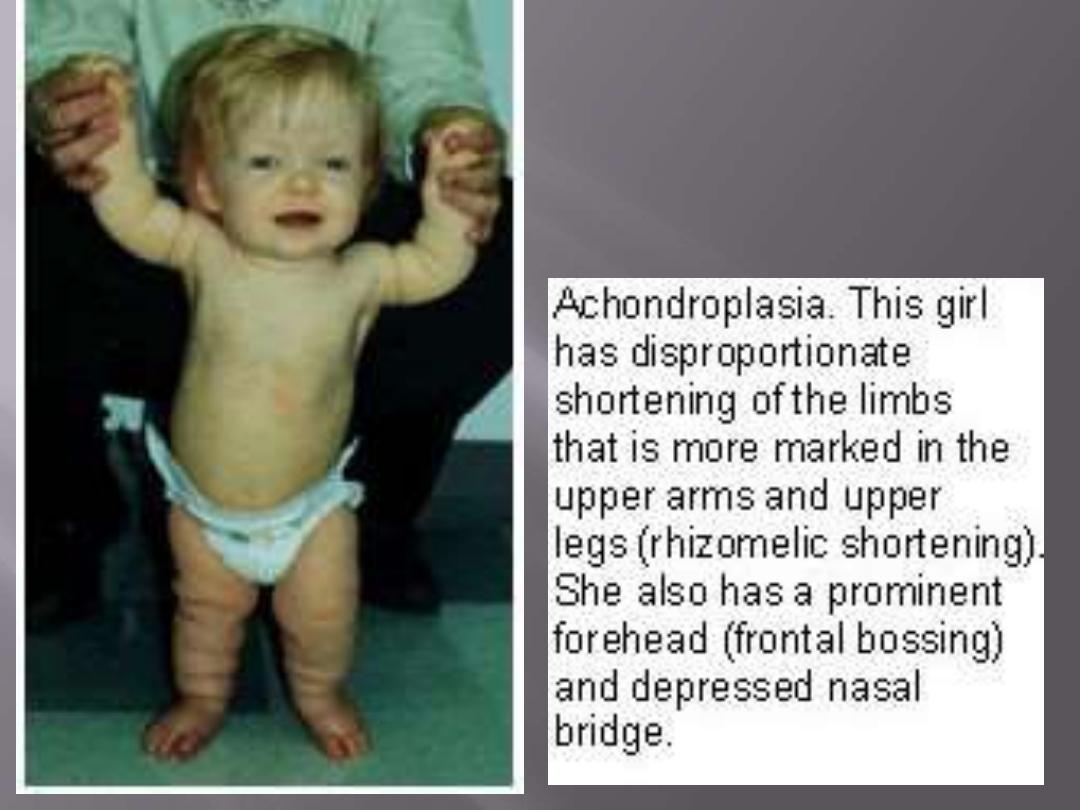
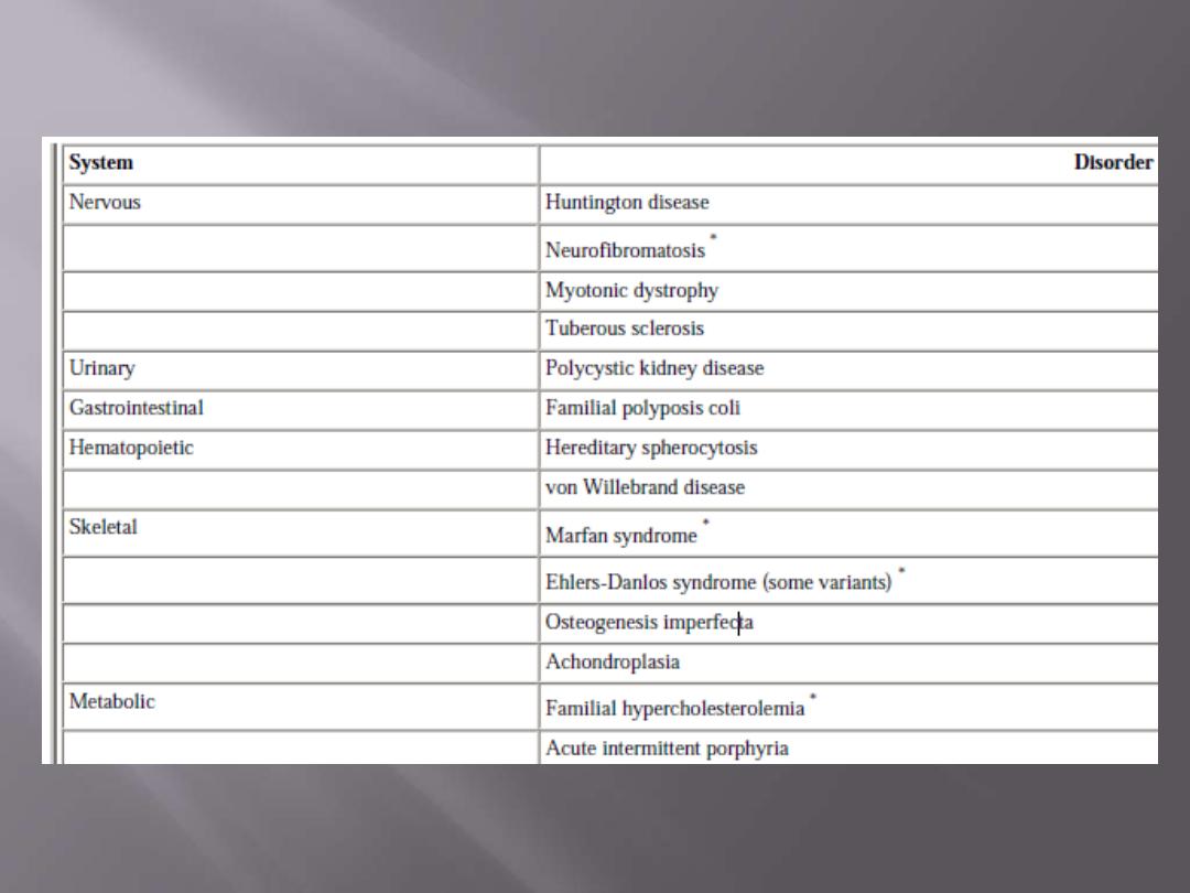
Dominant
genes usually produce two types of proteins,
either:
1. Major structural (or key non-enzymatic) proteins
(e.g.
collagen,
spectrin,
etc.);
examples
are
cases
of
achondroplasia & Ehler Danlos syndrome (lax joints and
skin).
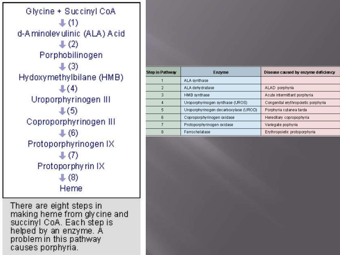
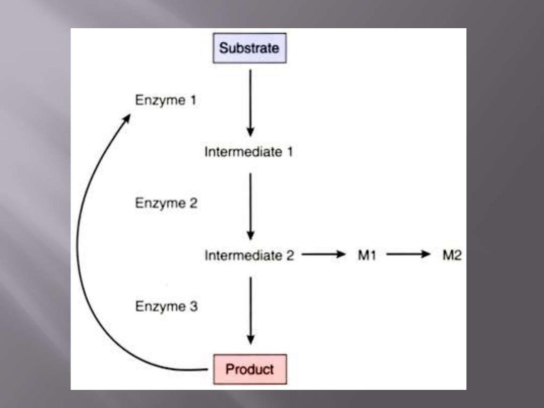
2. A key enzyme in a complex metabolic pathway usually
under feedback control
(AD porphyria)
3. a
membrane receptor regulating a metabolic pathway or a
membrane
transport
protein
(AD
familial
hypercholesterolemia).
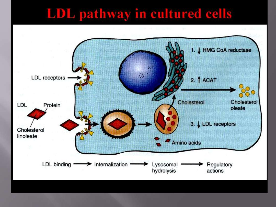
HMG-CoA reductase (3-hydroxy-3-methylglutaryl coenzyme A reductase), forms cholesterol from
fatty acids,
ACAT (acyl-CoA:cholesterol) transferase, hydrolyzes cholesterol into ester rendering it inactive

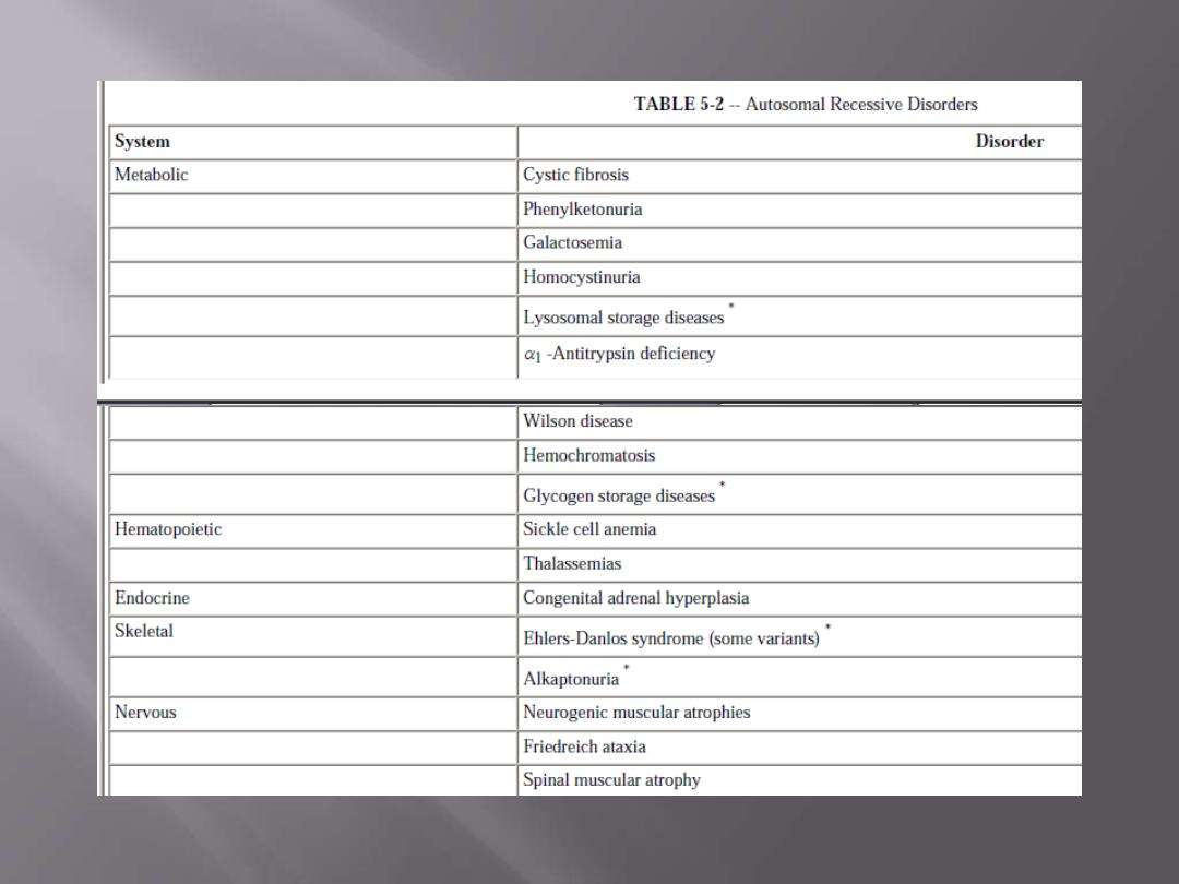
As for recessive genes
, they code for
proteins, enzymes
which usually share in catabolic pathways and when
both alleles are defective, there is no protein, i.e. no
enzyme and therefore the catabolic pathway is
obstructed with the accumulation of the biochemical
substrate.
Accumulation of the substrate in sensitive tissues will
results in disease state.
Examples of those diseases are some of diseases of
mucopolysaccharidosis, lipidosis, phenylketonuria
(PKU), albinism, and many others.
Thyronine
Diet Phenylalanine
Tyrosine L-DOPA
Melanin
phenylpyruvic acid
(excr. In urine in PKU)
Acetoacetic
acid
1
2
4
3
1 = Phenyl alanine hydroxylase PKU
2 = Tyrosinase albinism
3 = Homogenistic acid oxidase Alkaponuria (dark urine)
4= enzyme deficiencies that interfere with thyroxine biosynthetic pathway
Cretinism
Homogenistic
acid

ANY QUESTION?
ALL HAS WEAKNESSES & STRENGTHS
A butterfly is fragile but beautiful, useful and free

Sex-linked diseases
“X”-linked [both AD & AR]
Lyon’s hypothesis deviate from expected pattern
for AD or AR (female manifesting carriers)

Lyon’s hypothesis states that
in a female’s autosomal
cells, all the “X” chromosomes will be inactivated
during interphase except one which remains active
.
• It takes place
early in the post-fertilization period
,
19-20 days P.F.
• It is random
.
• All the daughter cells that descend from the inactive
X, the same “X” will remain inactive.
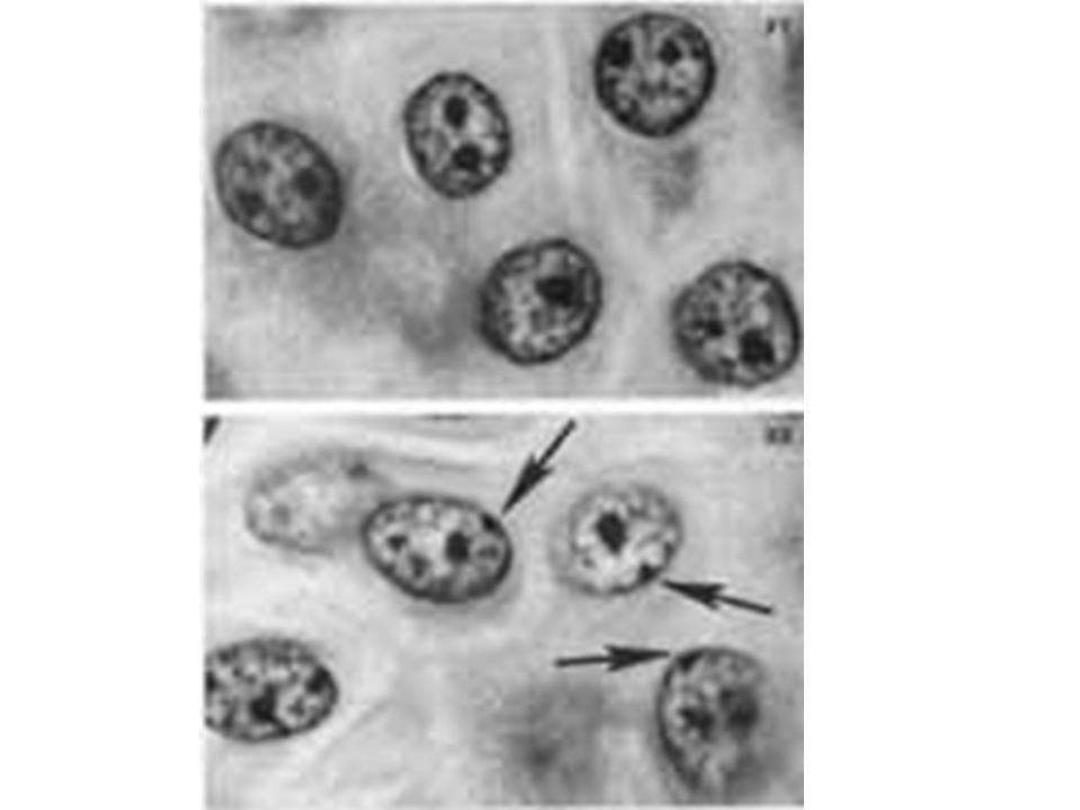

This means that about 50% of the “X” chromosomes are
inactivated. Therefore, the body of the female is a mosaic
concerning the active “X” functioning. So, the female is
considered heterozygote in regard to the origin of the X-
chromosome.
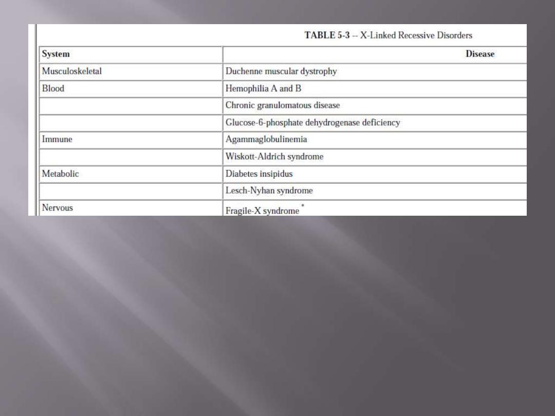
X-linked dominant disorders:
Vitamin
– D resistant rickets

ANY QUESTION?

Although
SHE
sits in the dark, but
SHE
looks
to
the light

Etiology:
All single gene diseases are due to mutations, which are
of different types
:
1. Single point mutation.
2. Addition – Deletion mutation.
3. Unequal crossing over.
1. Single point mutation
, which is the commonest type.
They usually result from a change in one of the
nucleotide bases that form the trios (three bases),
each of which codes for a specific amino acid in the
protein molecule.
The sequences are the following
:
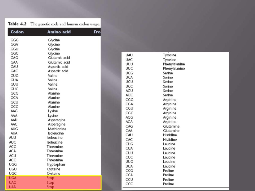
20 amino acids
4 bases in triplets (4
3 =
64 possible codes)
3 of them are stop codons
61 possibilities for only 20 a.a.
= genetic code redundancy

1. Silent mutation
(
Redundancy of the code)
2. Neutral mutation
3. Missense mutation: new code coding for a different amino
acid
in the protein changing its character and behaviour
resulting in disease, e.g. some cases of thalassemia, sickle
cell disease, PKU.
4. Mutation of the
termination codon
,
1. e.g. Constant Spring type of haemoglobin (Hb
CS
), where
the termination codon of α-polypeptide of Hb is mutated
resulting in addition of 31 amino acids to the original 14
amino acids of the normal chain
2. Non-sense mutation.
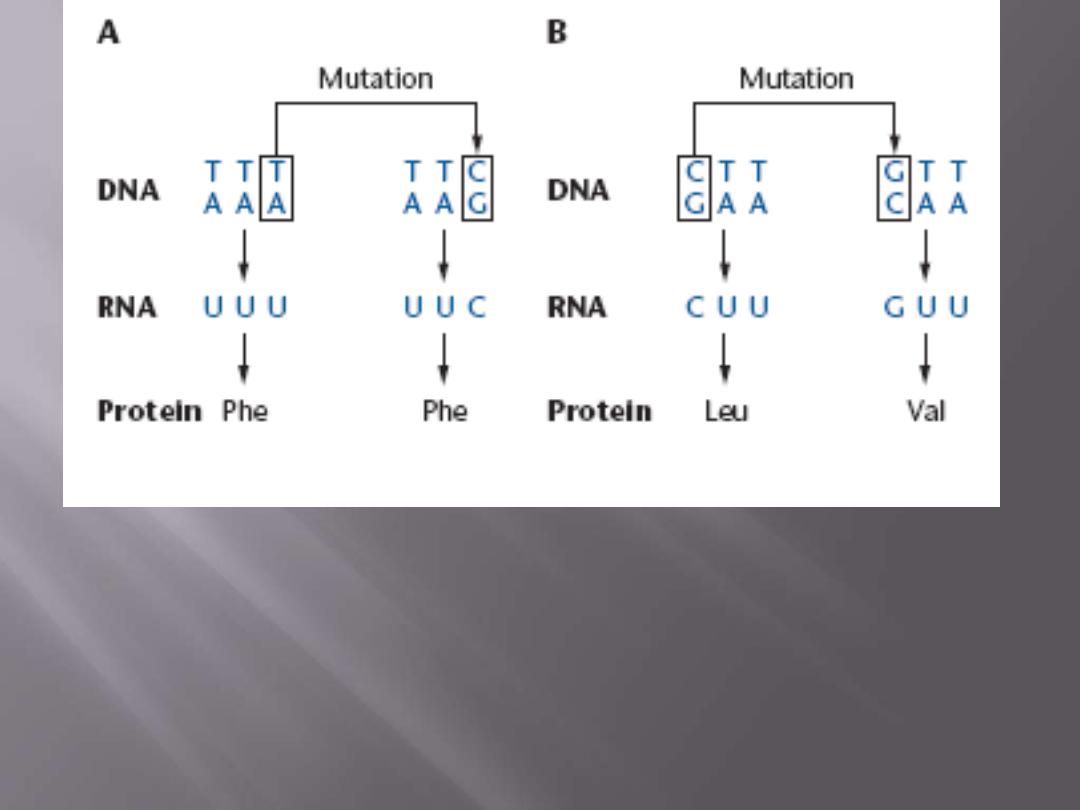
Consequences of base pair substitutions
(A) Silent mutation:
The base pair substitution in the DNA codon
does not change the coding specificity.
(B) Neutral mutation.
The base pair substitution changes the amino
acid specified by the DNA codon, but the replacement amino acid
has physicochemical properties similar to the original one.
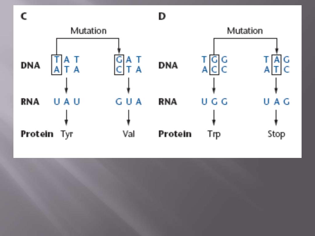
(C) Missense mutation
. The base pair substitution changes the
amino acid specified by the DNA codon.
(D) Nonsense mutation.
Base pair substitution changes a DNA
codon that codes for an amino acid into one that specifies a stop
codon.

2. Addition – deletion
mutations. They could be one of
four types:
a. Addition or deletion of a single base.
This will result in a shift in the reading frame
changing the whole reading of the trio creating a
new type of protein or sometimes it creates a
termination code in the center of the molecule and
the resulting polypeptide chain is shorter or called
truncated proteins. These types of mutations are
called
frame-shift mutations
.
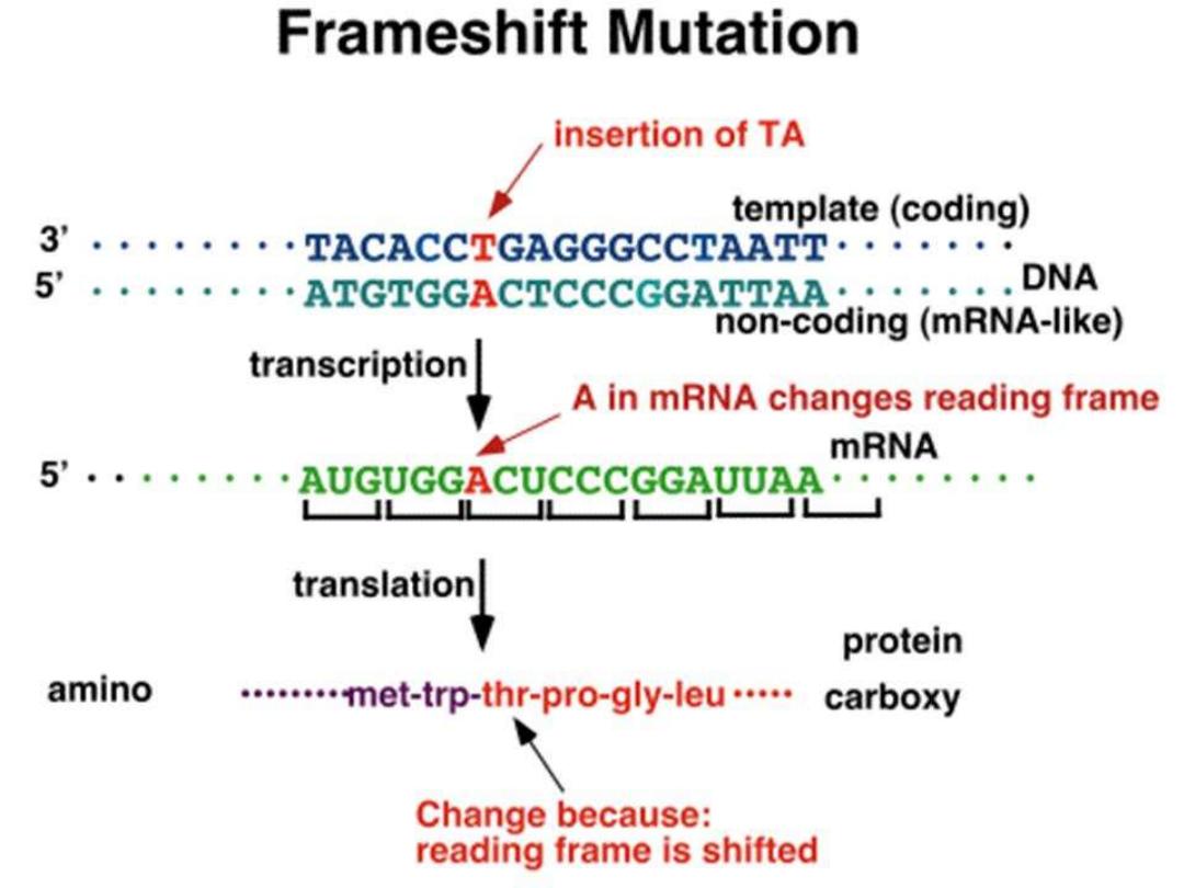
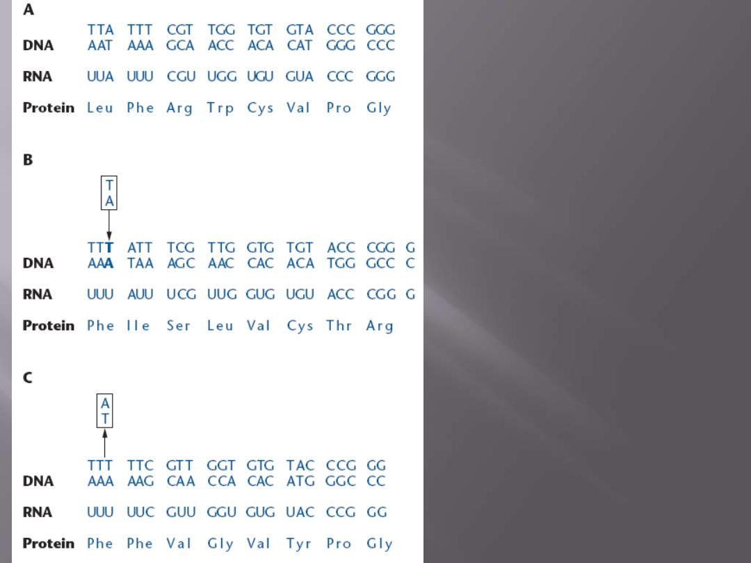
Consequences of
frameshift mutations
(A) A portion of a coding
region of a structural gene
with
the
expected
transcribed and translated
sequences.
(B) The insertion of a base
pair
(bold letters)
after the
second nucleotide site of the
DNA sequence presented in a
changes the reading frame.
(C) The deletion of the base
pair at the third nucleotide
site of the DNA sequence
presented in A changes the
reading frame.

b. Much less commonly addition / deletion of two
bases
same consequences
.
c. Addition or deletion of 3 bases or the multiple of 3
,
i.e. 6, 9, 12, 15 … etc. This will lead to addition of
1, 2, 3, 4, … etc amino acid(s) in the protein
molecule leading to abnormal protein, i.e. Frieberg
Hb, where 5 amino acids (i.e. 15 bases) is added
between amino acids 78-79 sequence in β-Hb
polypeptide.
d. Addition of deletion of a
large piece of DNA
inside
the gene (intragenic) or in between the gene
(intergenic). Again this creates variability and may
lead to a disease state but it is used for genetic
testing and diagnosis of some genetic diseases.

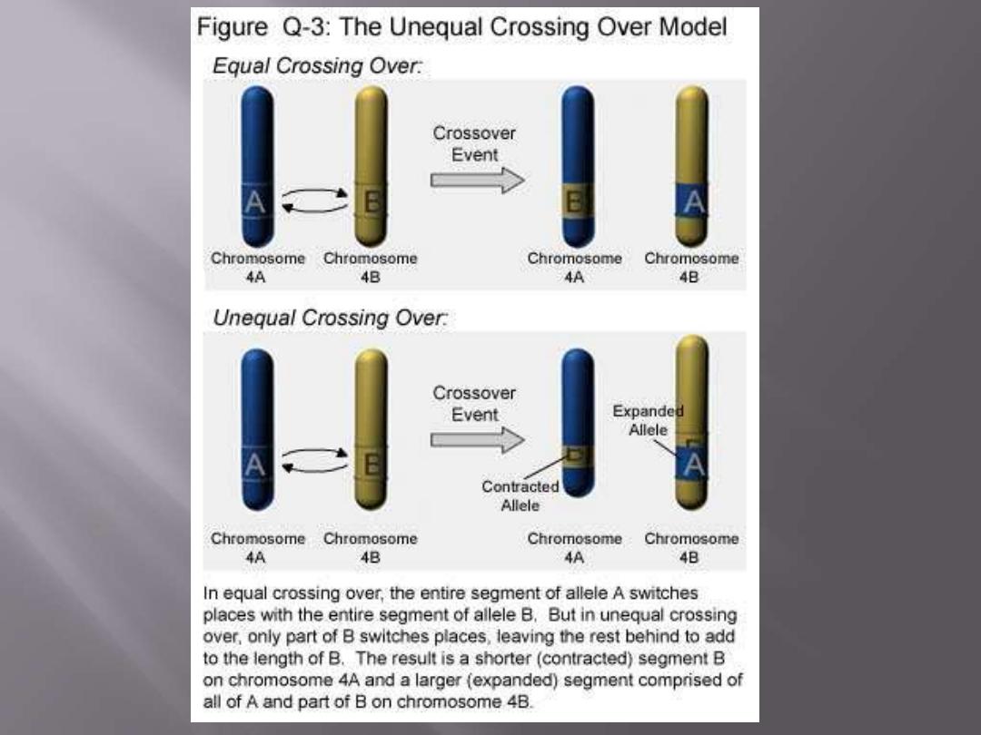
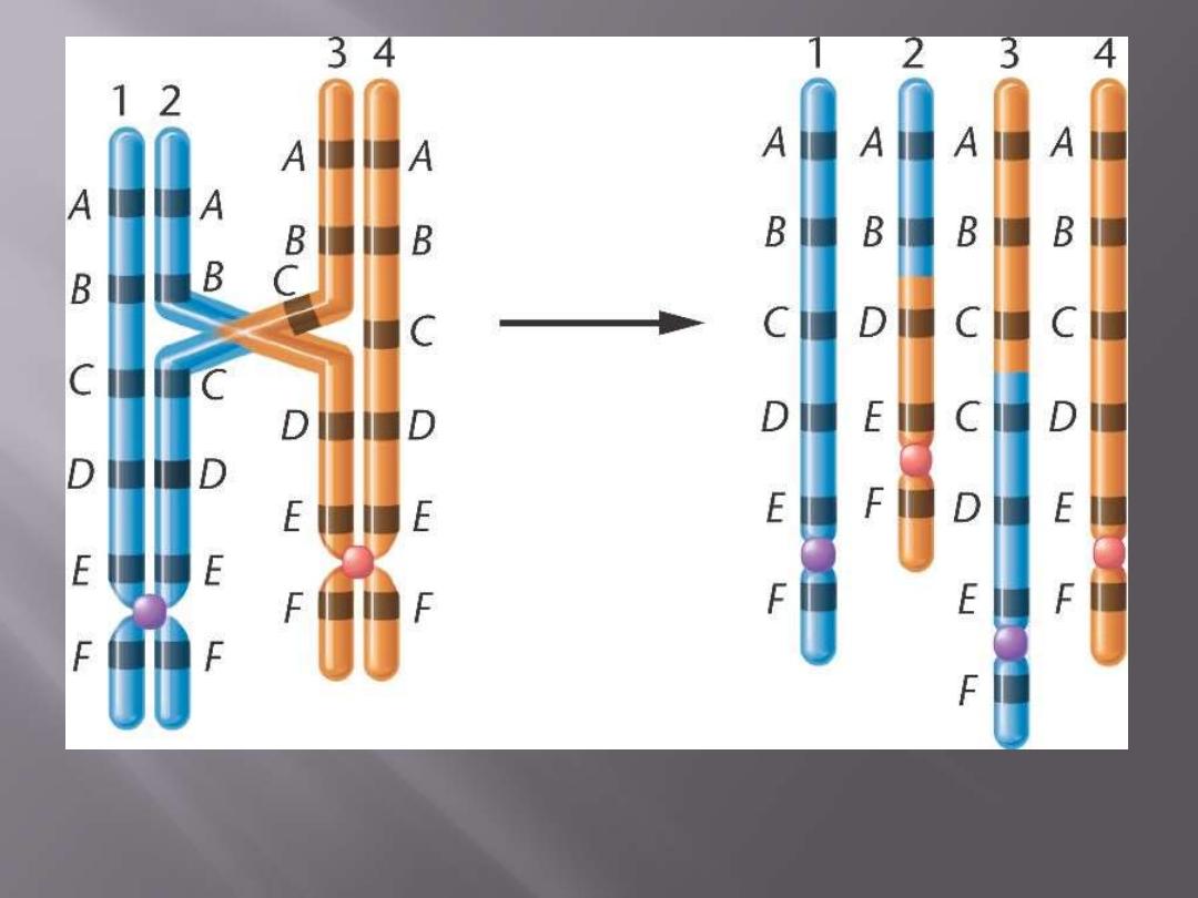
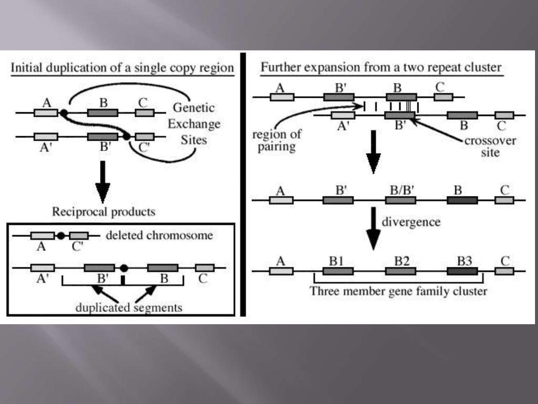
3. Unequal crossing over:
This case takes place in sites of the DNA where there are
grouping of genes of similar DNA structure with very
slight variation so one gene is mistaken for a different
gene as being its allele and if crossing over occurs.
Then a defect will result leading to the formation of
two unbalanced homologues, one containing more
genes and another less genes and both containing a
hybrid (mixed) new gene.

ANY QUESTION?

Be like our home planet, active and alive
although your surrounding is not !!

Multifactorial Inheritance (MFI)

Multifactorial (also called polygenic) inheritance is
involved in many of the physiologic characteristics
(e.g. weight, height, blood pressure, hair color, etc.).
A multifactorial physiologic or pathologic trait may
be defined as a
trait governed by the additive effect
of two or more genes of small effect but
conditioned
by
environmental,
non-genetic
influences.
Even monozygotic twins reared separately may
achieve different heights because of nutritional or
other environmental influences.

This form of inheritance is believed to underlie such
common diseases as diabetes mellitus, hypertension,
gout, schizophrenia, bipolar disorders and certain
forms of congenital heart disease as well as some
skeletal abnormalities.
It is of value to mention that
multifactorial inheritance
differs from
congenital malformation
.
In the latter, environmental factors, genetic causes
(chromosomal disorders, single gene disorders, or
multifactorial inheritance), physical agents (heart,
pressure, radiation, etc.), maternal disorders, prenatal
infection,
etc.
all
are
causes
of
congenital
malformations, but each one is a sole cause at a time,
and the disease is not the result of the additive effect
of more than one of those factors.

In multifactorial inheritance, it is the additive effect of
more than one gene
of small effect
PLUS
a
suitable
environment
causes such disorders.
In single gene disorders, individuals in regard to the
abnormal gene are one of 3 groups: a
heterozygote
(carrying one mutated and one normal gene and thus
affected in AD and not affected in AR disorders), a
homozygote
for the mutated gene (and thus affected
in all cases), or a homozygote normal. There is no
gradient in between these 3 groups.
In MFI, we could group individuals in a community
into many different grades, which have a normal
distribution curve (Gaussian distribution) with a
threshold point, which when exceeded, the disorder
is expressed.
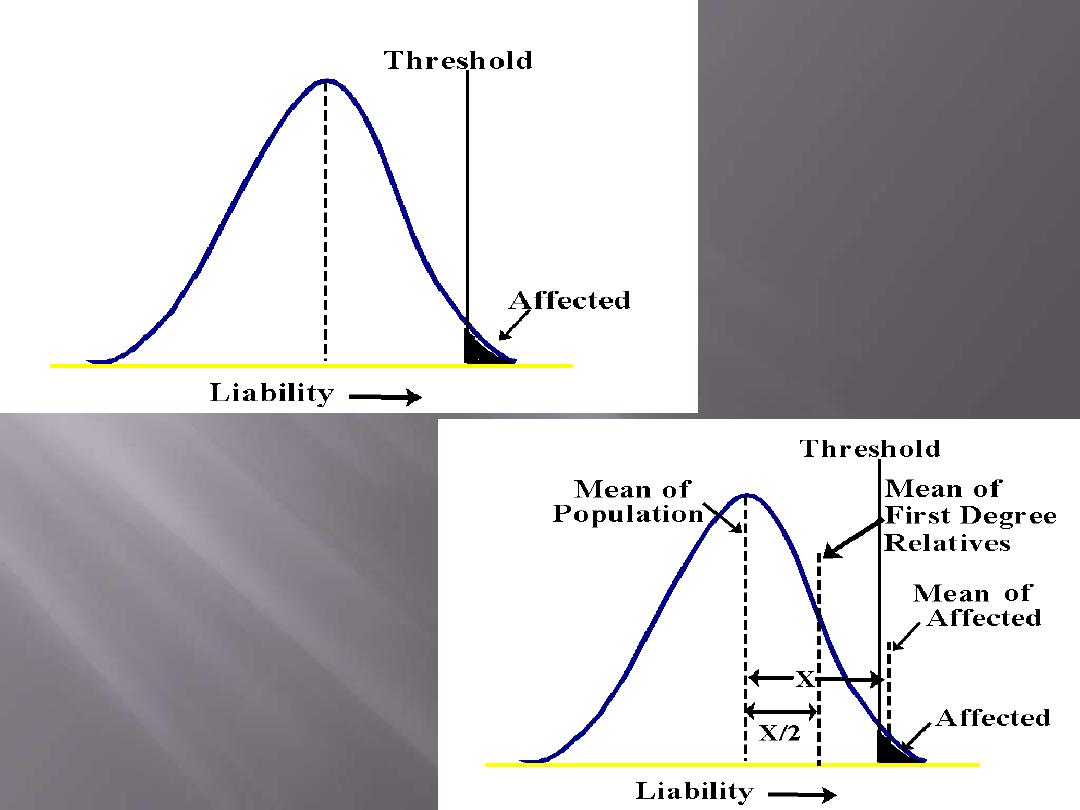

The facts that MFI are affected by many genes (not
just one) and that the additive effect of both genes
and the environment determine the expression of
MF disorder], are called Genetic Liability (or
genetic predisposition) of the individual and this
liability can be measured.
So, MFI is only
partially genetic
(unlike other types of
inheritance) and needs environmental factors to act
for the disorder to appear.

Some examples of MFI most relevant to the clinical geneticist:
1. Neural tube defects.
2. Cleft lip and/or palate.
3. Heart defects (PDA, VSD, ASD, etc.).
4. Pyloric stenosis.
5. Late-onset conditions such as hypertension, diabetes
mellitus, schizophrenia, Alzheimer disease, ...etc.

ANY QUESTION?

An eaten apple is a symbol of
a powerful worldwide corporation