
Autosomal chromosome -
disorders
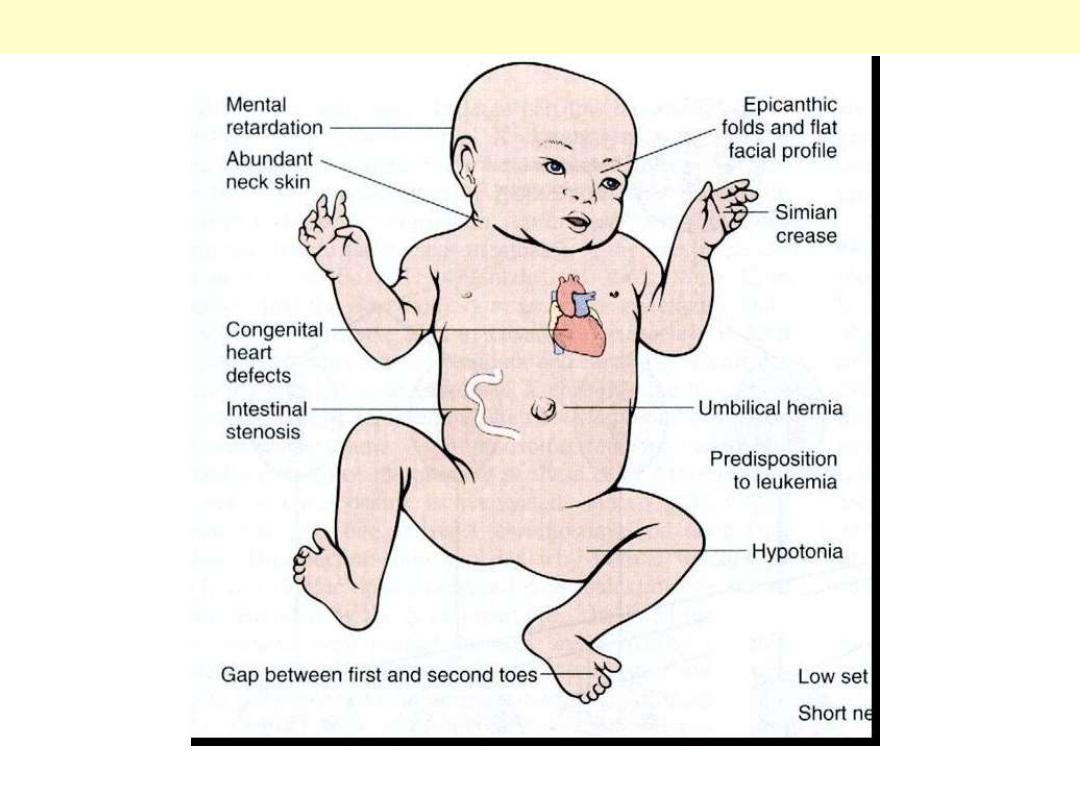
Clinical features of Down’s syndrome are summarized in this depiction.
Autosomal trisomy of chromosome 21 (Down’s syndrome)
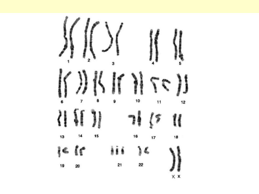
Karyotype of a girl with Down’s syndrome. Notice the 47 total count, the 3 copies of chromosome 21
and the 2 X chromosomes.
Autosomal trisomy of chromosome 21 (Down’s syndrome)
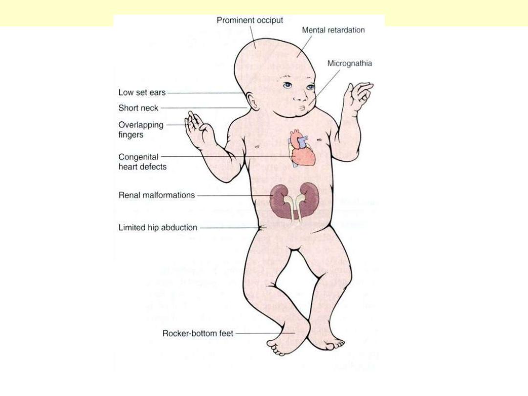
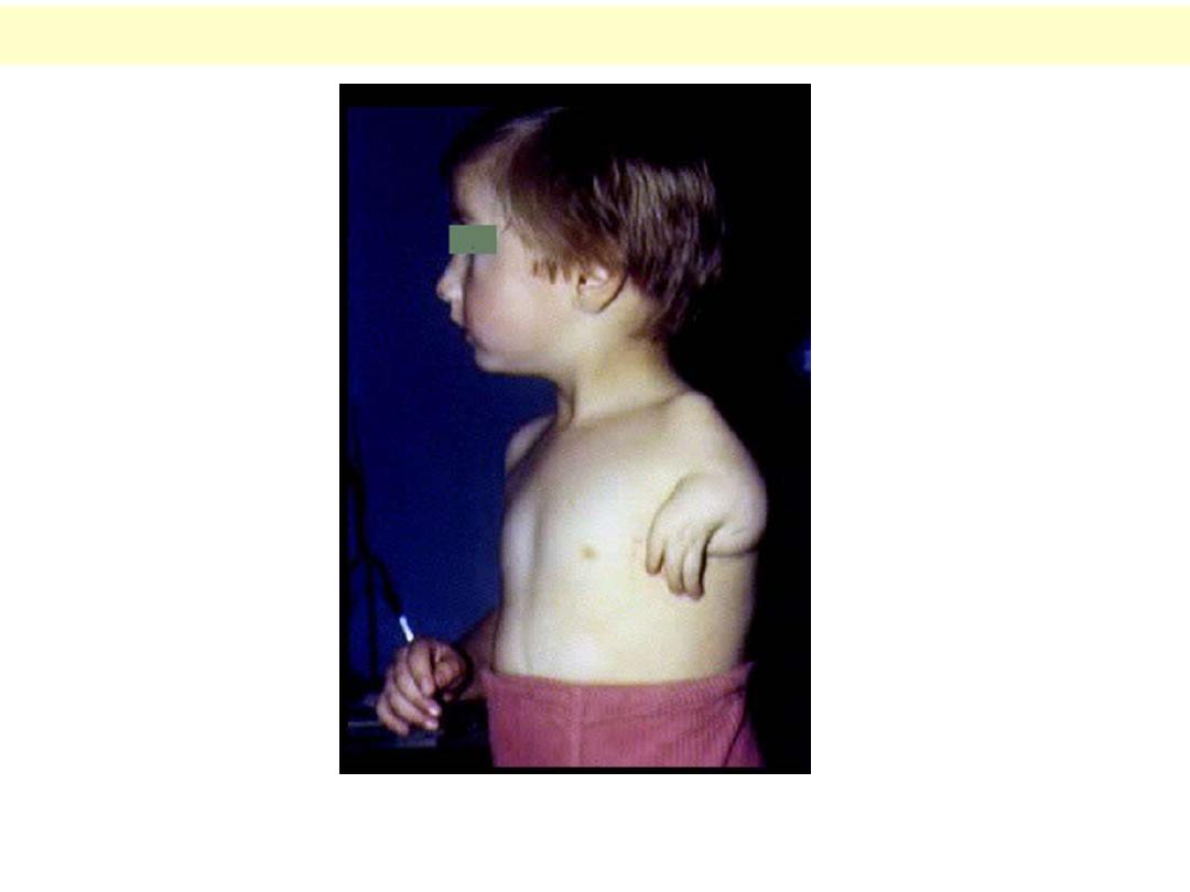
Clinical features of Edward’s syndrome are seen here in this photograph.
Autosomal trisomy 18 (Edward’s Syndrome)
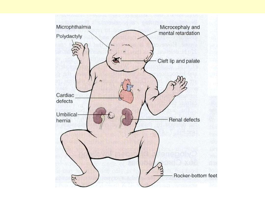
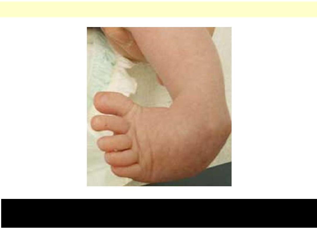
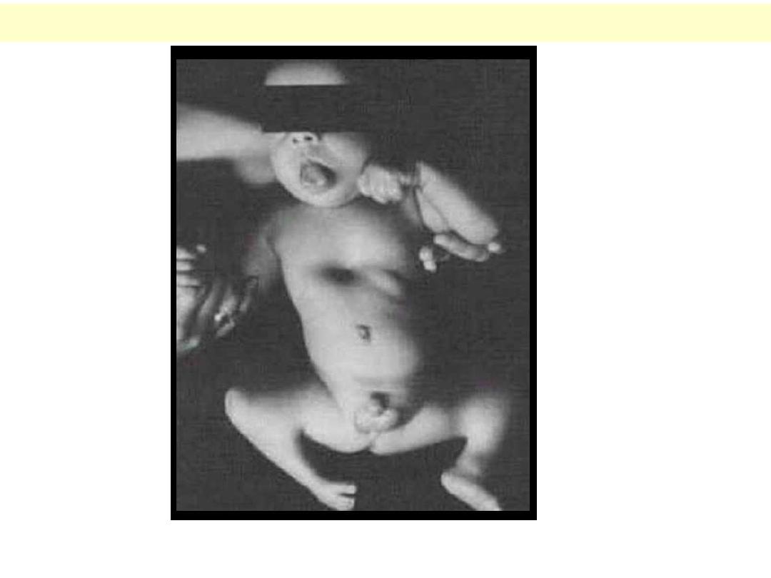
Clinical features of Patau syndrome are seen here.
Autosomal trisomy 13 (Patau Syndrome)
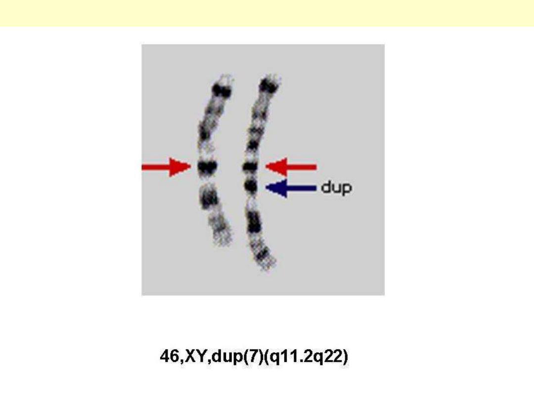
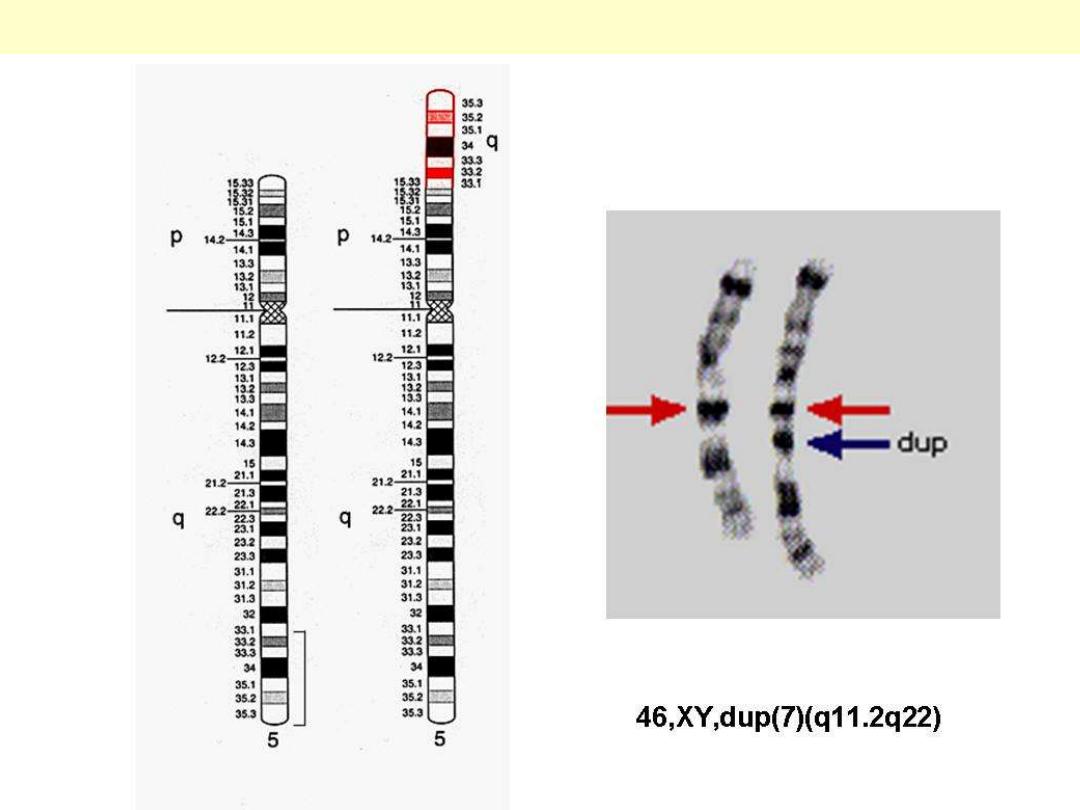
Chromosome 7 showing the duplicated piece and the karyotype writing for this abnormality.
Duplication of chromosome 7

Barr body
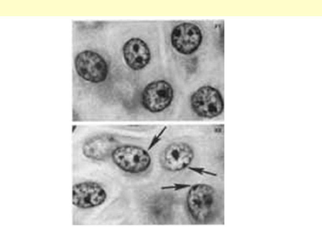
The condensed mass attached to the nuclear membrane of some female cells are called Barr Body.
Barr Body

Chromosomal abnormalities -
Examples
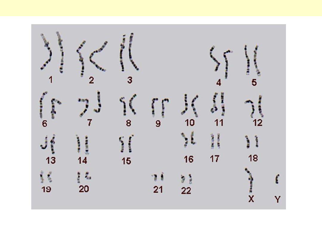
All chromosomes are normal in number and shape. Notice the X and the Y chromosomes.
A normal male karyotype
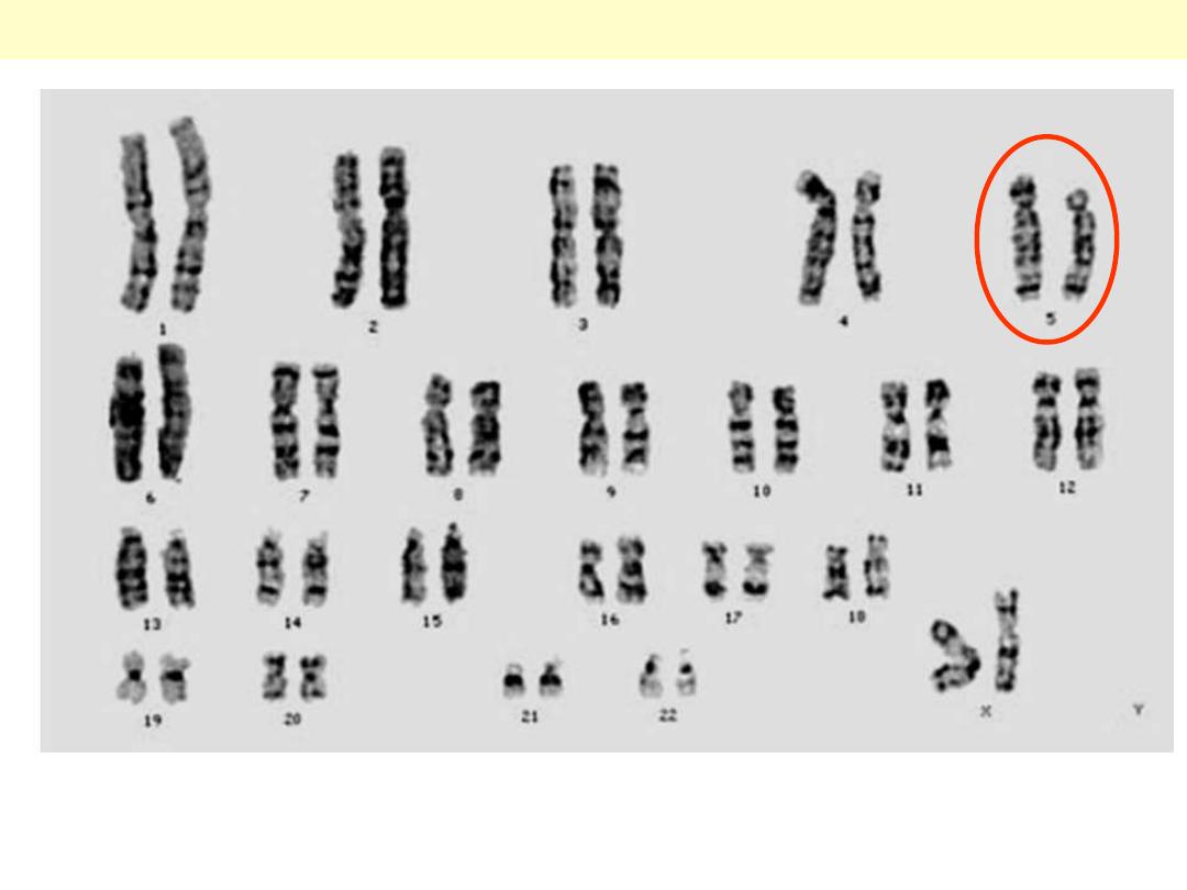
Cri du Chat (5p-) [Cat-cry syndrome]
All chromosomes are normal in number and shape except for the deleted upper segment of 5p in a
female. The baby has a distinctive low pitched cry like mewing of a cat.
Guess what?
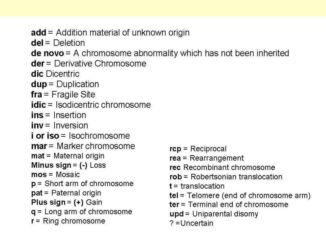
Some commonly used nomenclature in karyotype writing
d
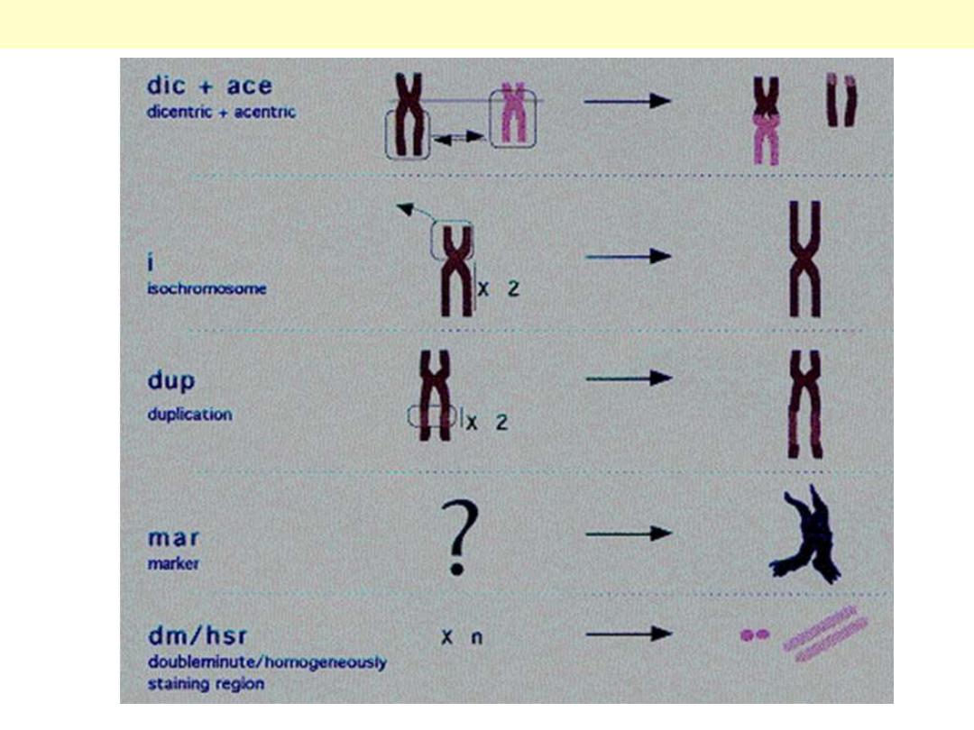
Some of the structural chromosomal anomalies
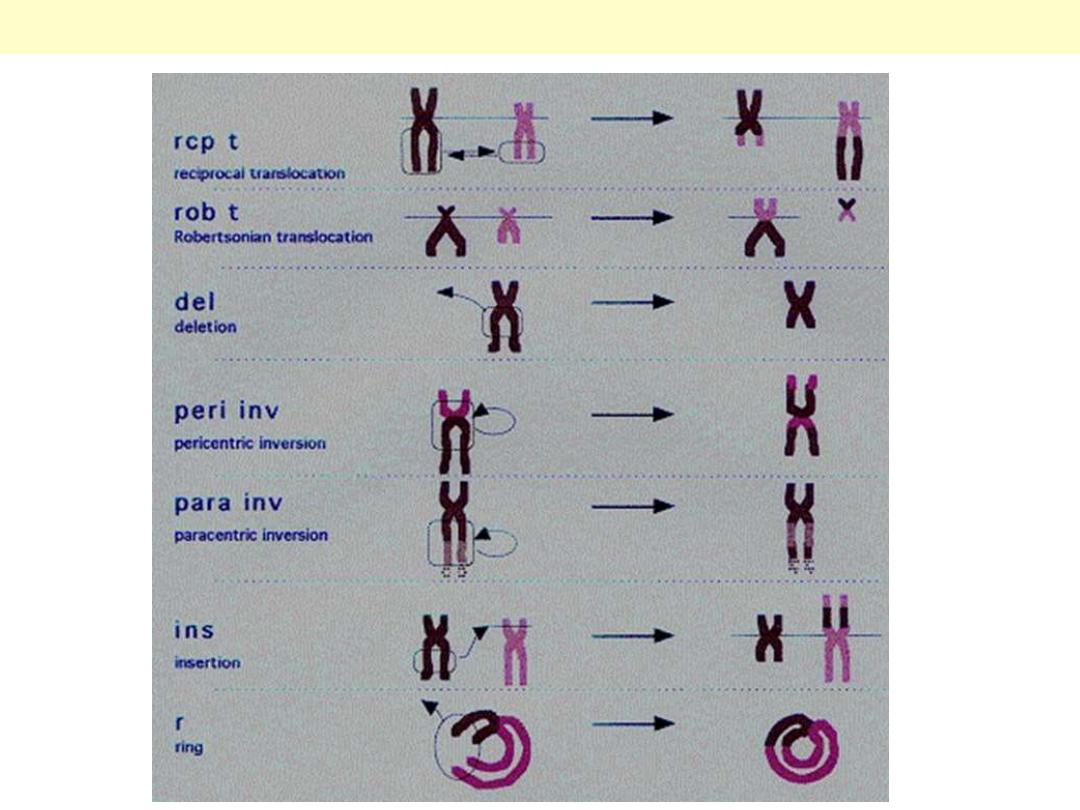
Other structural chromosomal anomalies
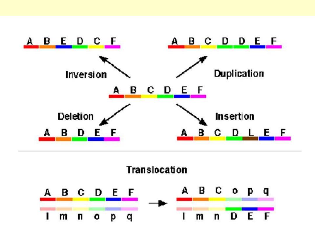
Inversion, Deletion and translocation
Duplication
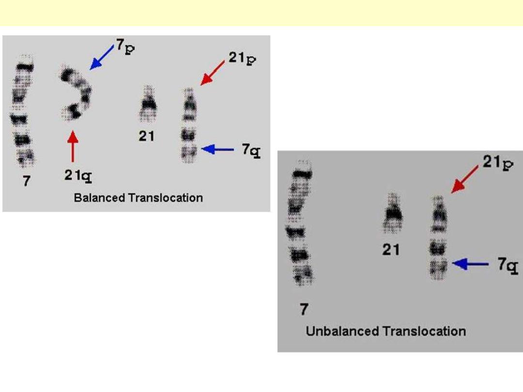
In the balanced translocation, although mislocated, there is net loss or gain of any piece, while in the
unbalanced type, there is a loss of a piece from the genome (proximal segment of chromosome 7).
Comparison between balanced and unbalanced translocation

Congenital malformations +
Teratogens
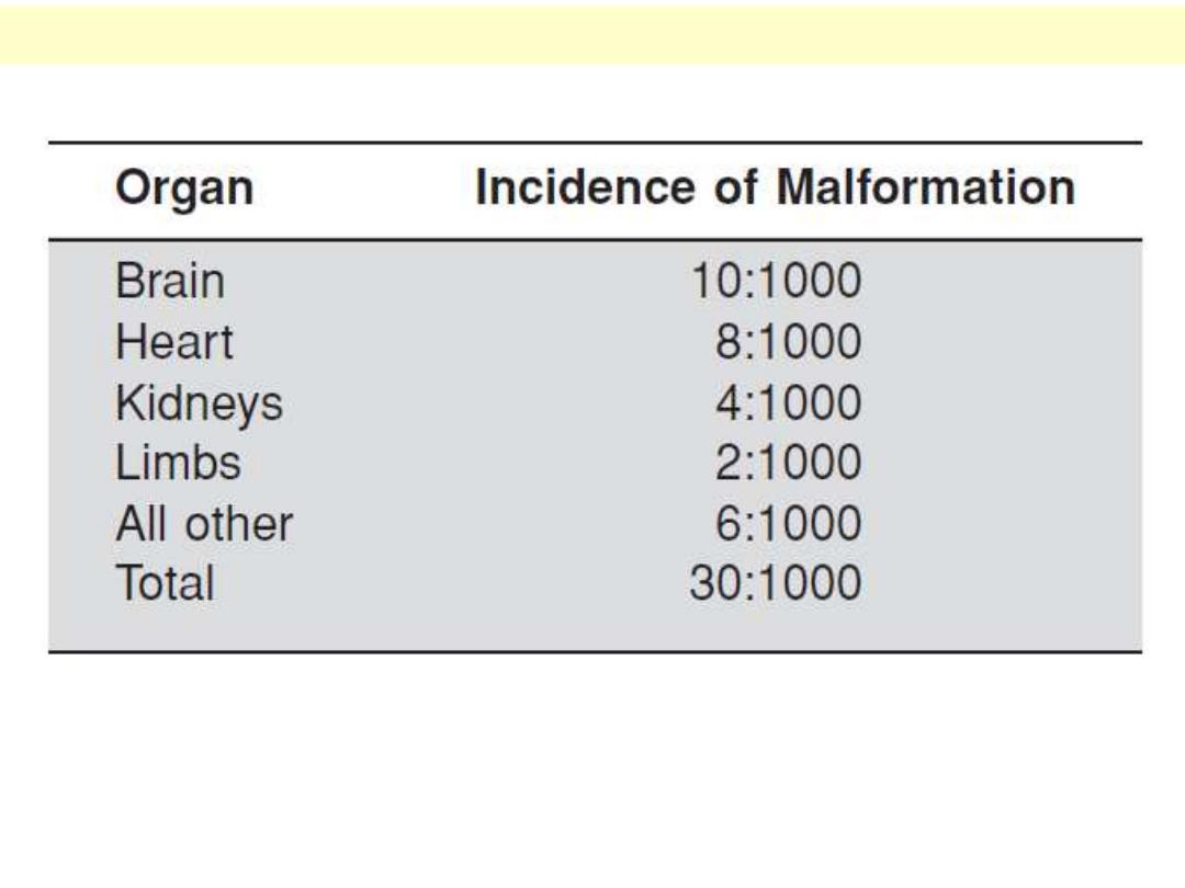
The brain constitutes the most affected organ by birth defects (i.e. the most sensitive organ),
followed by the heart and then the kidneys. Limbs and other organs (GIT, facial, etc.) are less
commonly affected organs, yet still important in clinical practice.
Incidence of Major Malformations in Human Organs at Birth
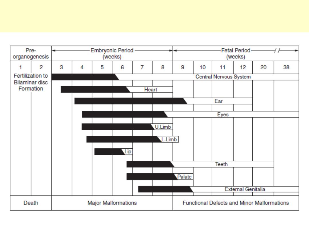
Major malformations occur earlier during the in-utero life; the more advanced the gestation, the less
susceptible the body will be to teratogenic effect.
Susceptibility to teratogenesis for different organ systems. Solid bar
indicates highly sensitive periods.

The impact of each category is variable and difficult to assess. While a genetic agent causes about
10-30%, it participates as well with the multifactorial and the unknown categories in the etiology of
birth defects. With the advances in technology and methods of diagnosis, the unknown would be less in
favour of other categories.
Approximate percentage of each teratogenic agent in the causation
of congenital malformation
Unknown
Multifactorial
Environmetnal
Genetic
30-45%
20-35%
5-10%
10-30%
Older
17%
>35%
Newer
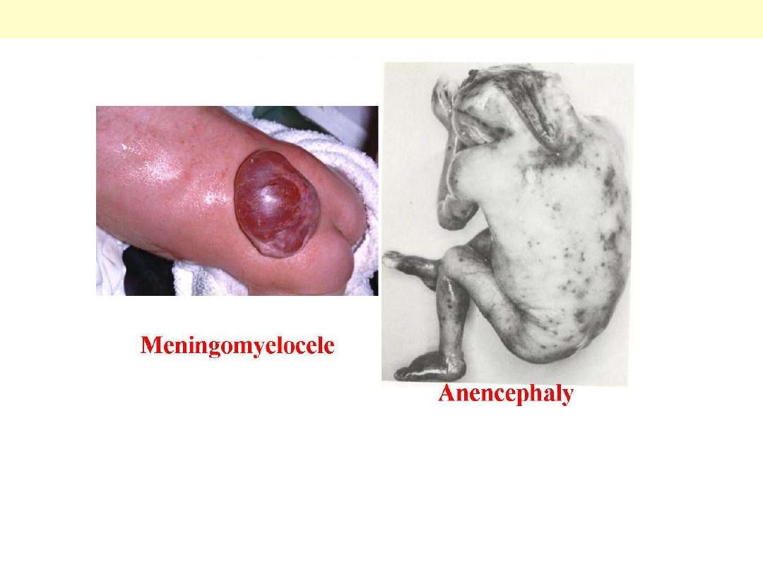
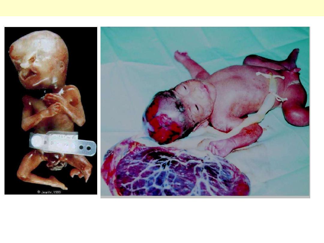
Failure of the neural tube to close normally at the end of first month leads to an open neural tube,
which is either partial (cystic) or complete, it may occur at any site along the neural tube, and could be
fatal (anencephaly) or compatible with life and amenable to surgery (meningocele).
Examples of malformations : Neural tube defects
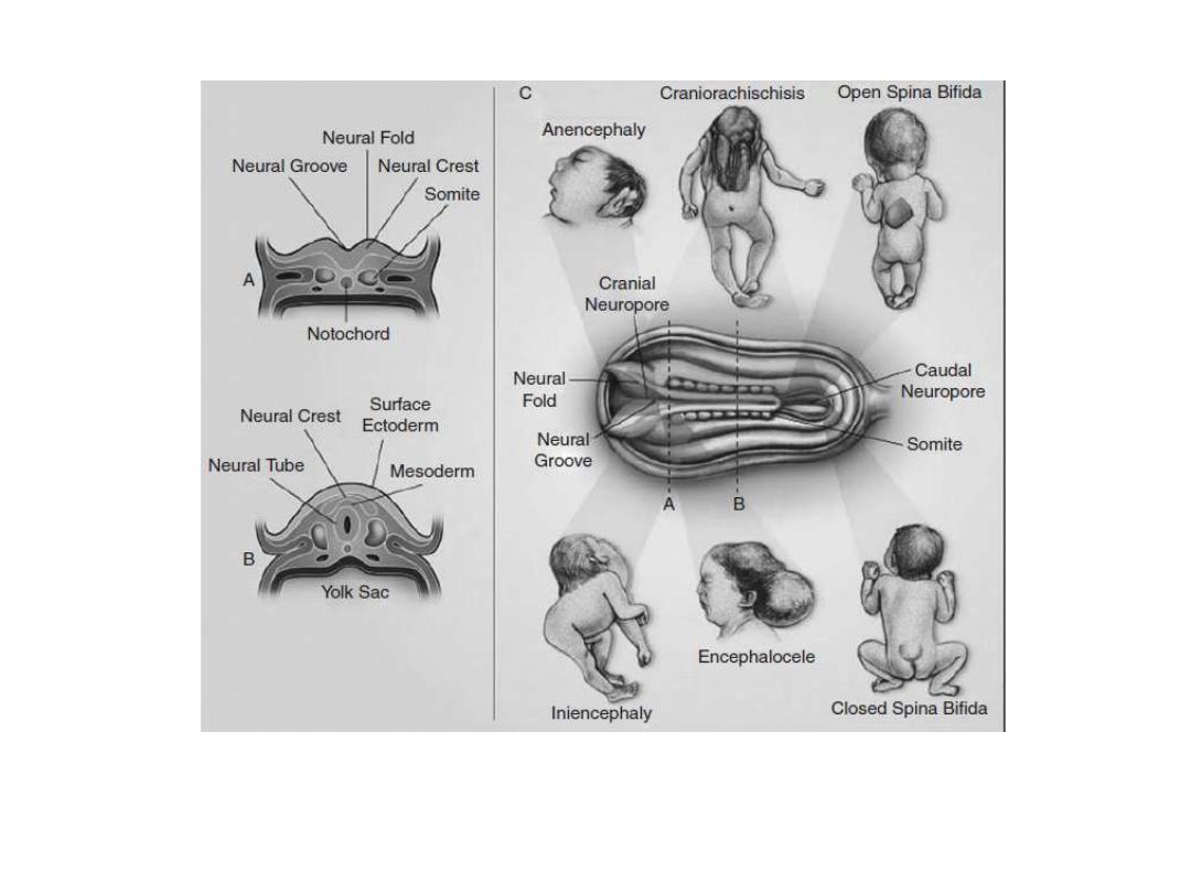
Features of neural tube development and neural tube defects
The features of the main types of neural tube defects. The diagram in the center is a dorsal view of a
developing embryo, showing a neural tube that is closed in the center but still open at the cranial and
caudal ends. The dotted lines marked A and B refer to the cross sections shown in panels A and B.
Anencephaly, spina bifida and encephalocele or craniorachischisis, are examples of a defective neural
tube.
Examples of disruption : Amniotic band sequence
Amnion disruption with mechanical disruption of fetal structures by amnion adhesion or fibrous
amniotic bands
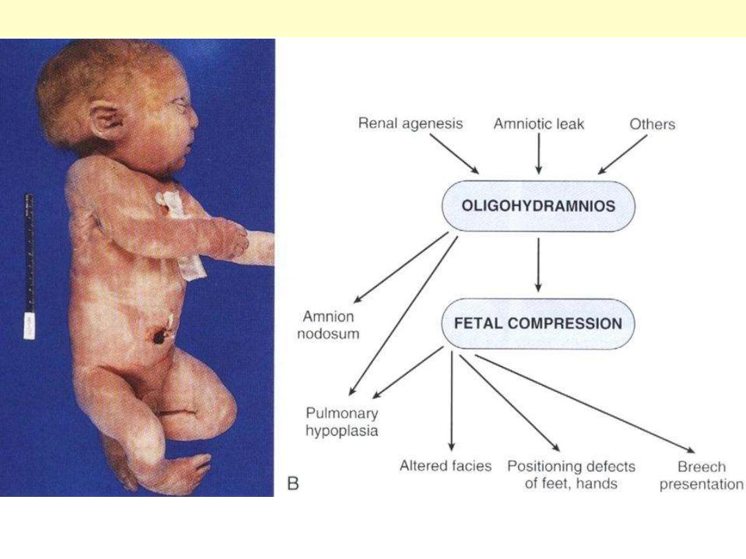
Examples of deformation : Oligohydramnios
On the right is the possible causes of oligohydramnios and their consequences. On the left is a
clinical effect of fetal compression.
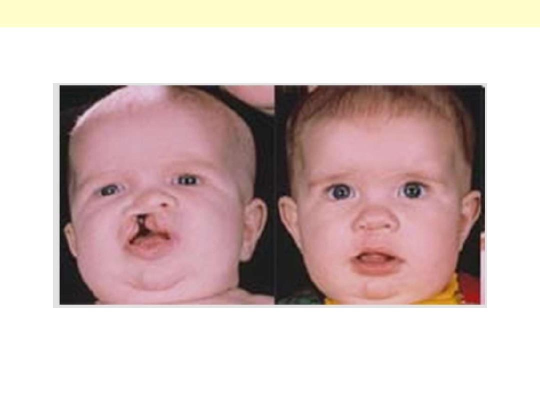
Examples of a single system defect : cleft lip and palate
A pre-surgical repair photo (Left) shows a unilateral clift in the lip and palate; (Right) is a post
surgical repair photo with an excellent result, both cosmetically and functionally.
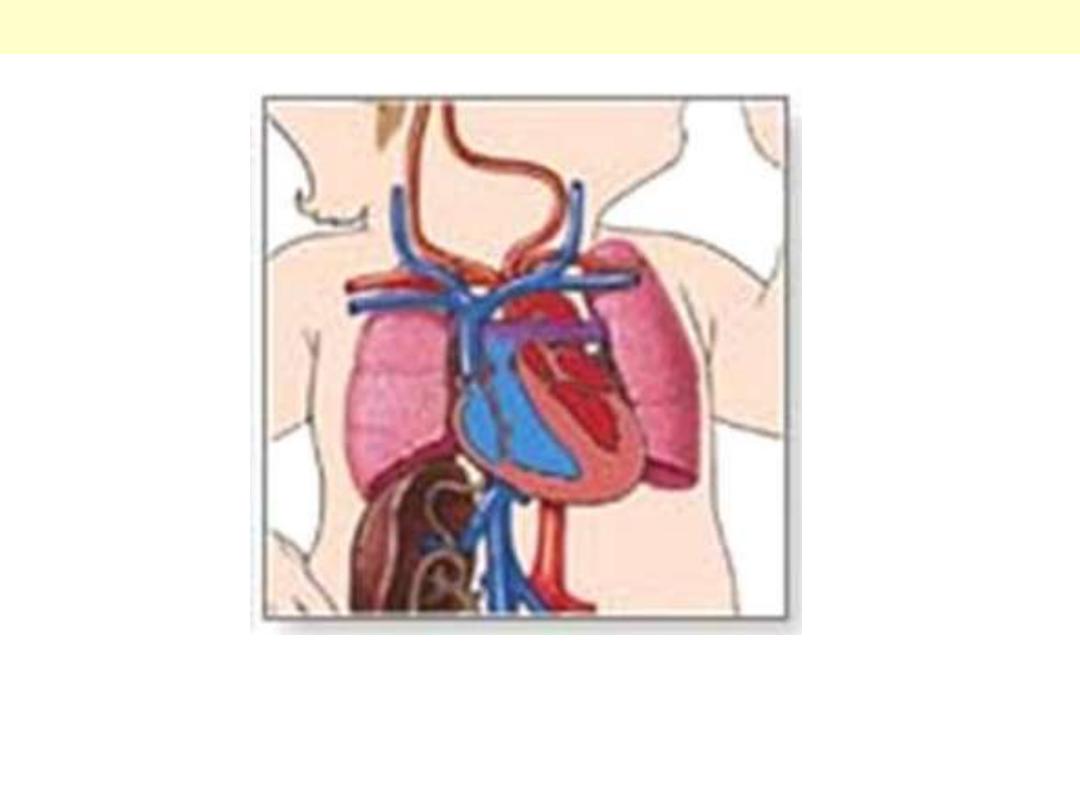
Examples of a single system defect : congenital heart defect “PDA”
A ‘PDA’ is a patent ductus arteriosus, a congenital acyanotic cardiac defect, where the ductus
arteriosus remains patent after birth disturbed normal blood flow.
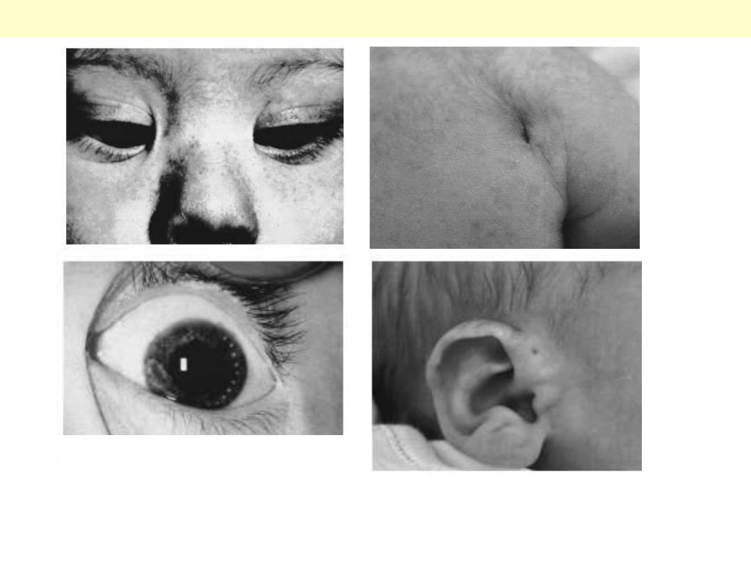
Examples of minor malformations
A. Inner epicanthic folds (top left)
B. Sacral dimple (top right)
C. Brushfield spots (lower left)
D. Preauricular pit (lower right)
Micrognathia, very small chin
Micrognathia is commonly seen as part of a syndrome, as more than 60 syndromes having
mandibular hypoplasia as a component have been described. Most commonly are Pierre Robin
syndrome, oculo-auriculo-vertebral (OAV) spectrum and Treacher Collins syndrome.
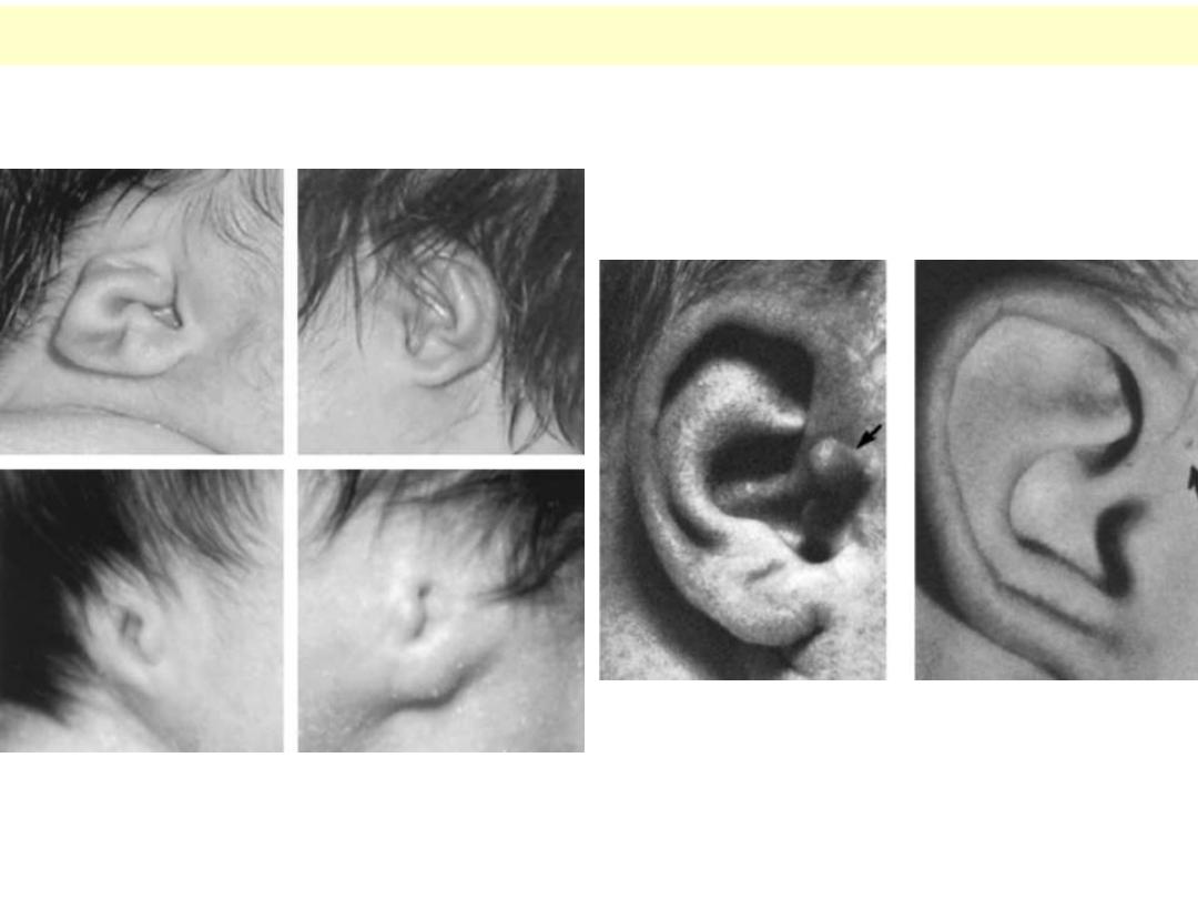
Preauricular skin tag (left) and pit (right)
A variable degree of microtia
(small deformed ear lobe)
Miscellaneous ear deformities
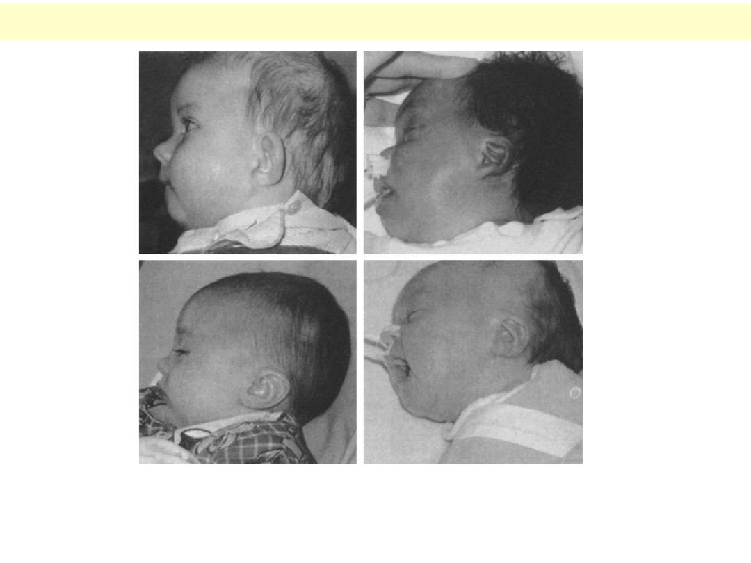
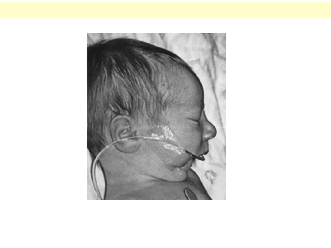
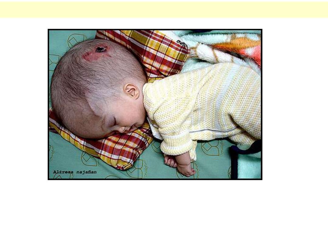
Low-set ears (abnormal positioning of the ear)
A very common and important clinical finding in clinical genetics and pediatric practice. Here is
shown variable degrees of low-set ears. At least part of the ear lobe should be above a line drawn
in line with the lateral canthus.
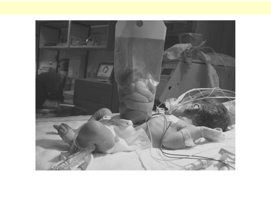
Omphalocele (abdominal wall defect)
The primary defect is in the abdominal wall, leading to herniation of the intra-
abdominal organs. Here is shown an infant with omphalocele with umbilical cord
attached at the apex of the sac.
gastroschisis (abdominal wall defect)
A severe form of abdominal wall defect allows much of the intra-abdominal organs to
herniate out, which leads to death from sepsis if not treated immediately.
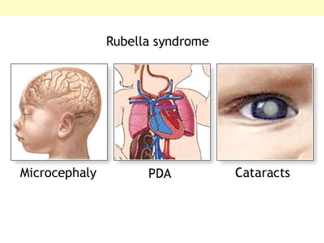
Examples of in utero viral infection – German measles
Examples of the clinical effects of Rubella infection, congenital heart defect (mainly ASD, VSD,
PDA), microcephaly mental retardation, blindness, cataract, in addition to deafness.
Microcephaly : an end result of many different teratogenic agents
Causes of microcephaly could be genetic (AR disorder), part of a syndrome, sporadic, or could be
non-genetic as in in-utero viral infection, exposure to toxic drug effect, etc.
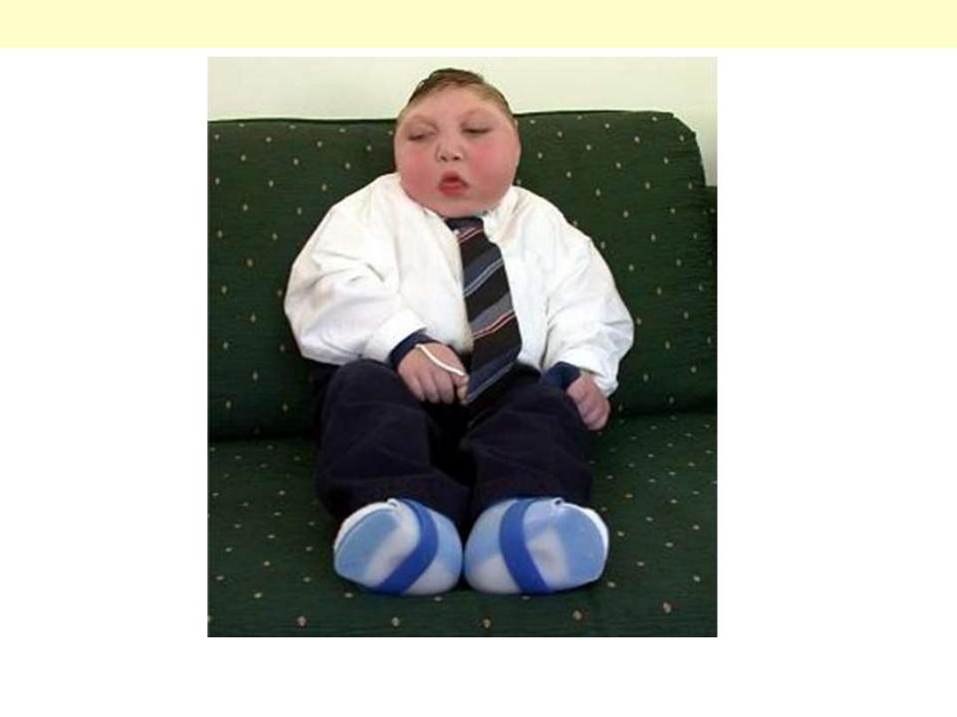
Hydrocephaly : an end result of many different teratogenic agents
Hydrocephaly results from an abnormal collection of CSF and thus dilatation of all or part of the
ventricular system due to an obstruction, over-production or under-absorption of CSF. It could be
the only manifestation or part of other associated anomalies. It causes pressure atrophy on the
brain mental retardation. Fortunately, early surgery to drain the excess CSF succeeds in
correcting the problem.
Phocomelia : very short and abnormal upper limb
A documented effect of thalidomide (used for its anxiolytic and its anti-emetic effects) is to produce
variable limb anomalies, including amelia and phocomelia.
Club foot “Talipes equino varus”
Club feet are commonly encountered problem, usually accompanied by other skeletal and/or
neurological manifestations, mainly neural tube defects. Surgical or conservative measures may correct
the defomity but not the treat the cause.
Infant of a diabetic mother
A baby born to a diabetic mother with macrosomia (body weight > 4 Kg at birth), with skeletal
deformities of lower limbs bones (femoral hypoplasia and caudal regression)
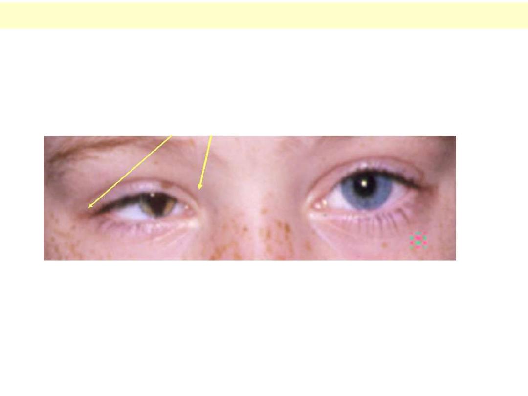
Eye anomalies
Here is shown three abnormalities; namely microphthalmia (small eye), heterochromia (different eye
colour, and coloboma (abnormal fissuring of the iris) (left eye).

Deletion - Chromosome
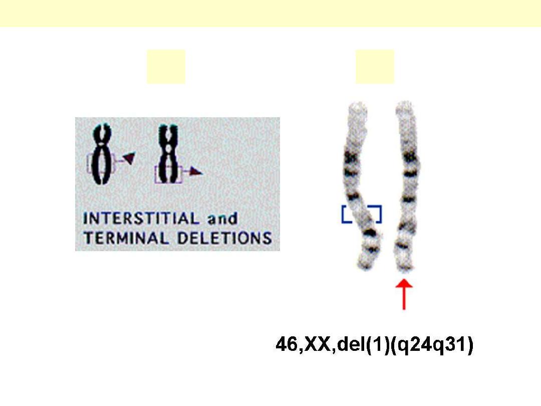
A. Types of deletion. B. Interstitial deletion of a piece of long arm on chromosome 1.
Types of Chromosomal Deletion
A
B
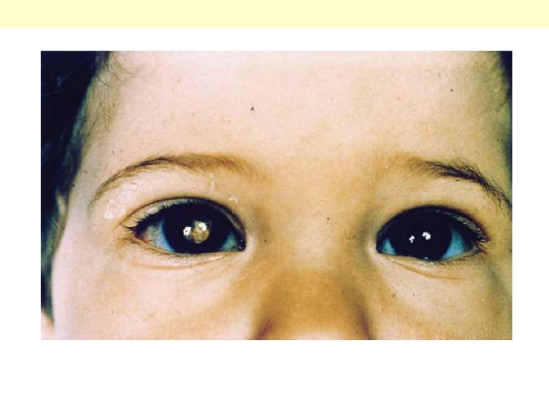
Clinical features of retinoblastoma in a child
A pediatric malignancy of the retina, retinoblastoma occurs in hereditary & sporadic forms. Shown is a
patient with a unilaterla tumor (Rt. eye).
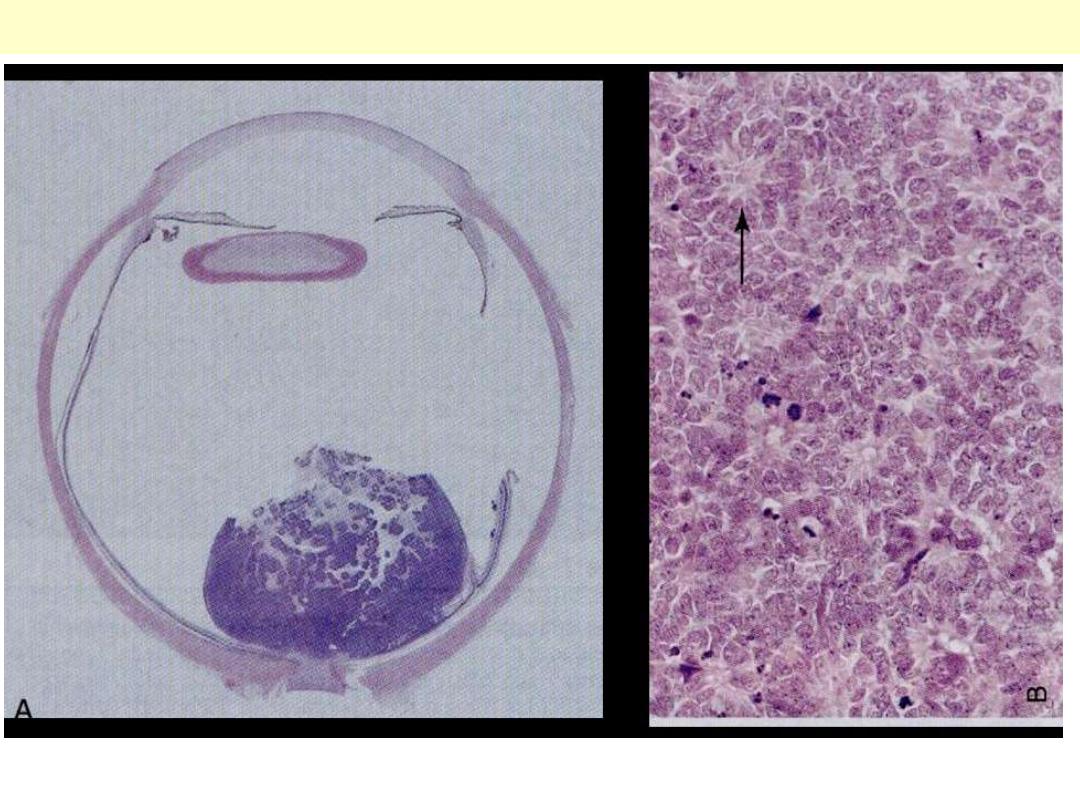
A. Gross specimen showing the retinal mass. B. Histopathological section within the tumor.
Retinoblastoma
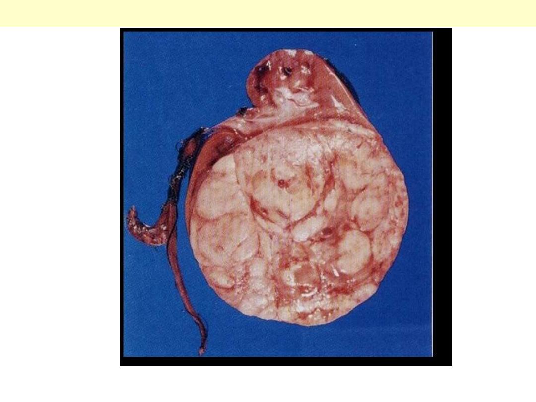
A huge well defined rounded mass is shown in the lower poly of the kidney, compressing the rest of
renal tissue.
Wilm tumor : Gross

Inversion – Chromosome
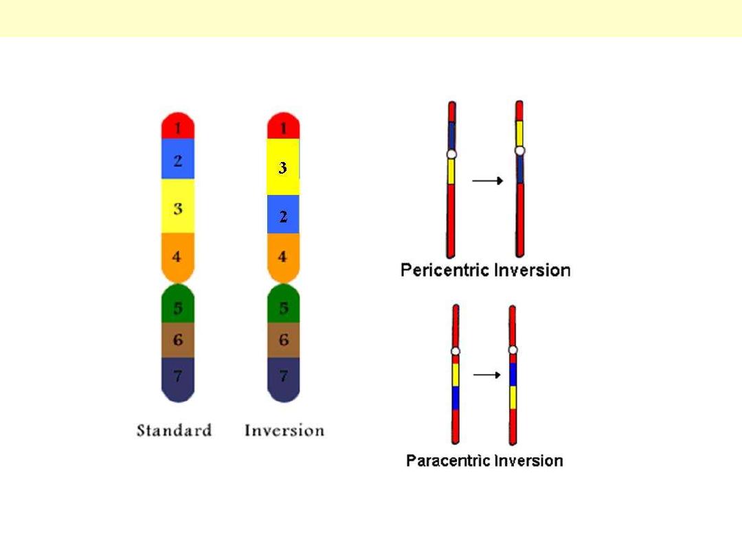
Two types of inversion are seen here; Pericentric (centromere involved) and paracentric (at one side of
the centromere).
Chromosomal inversion

Iso - Chromosome
A horizontal division of the centromere (instead of the normal vertical one) results in 2 short arms and
2 long arms.
Isochromosome

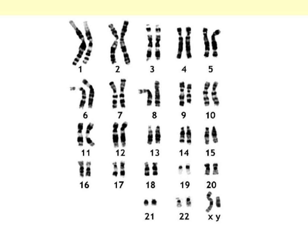
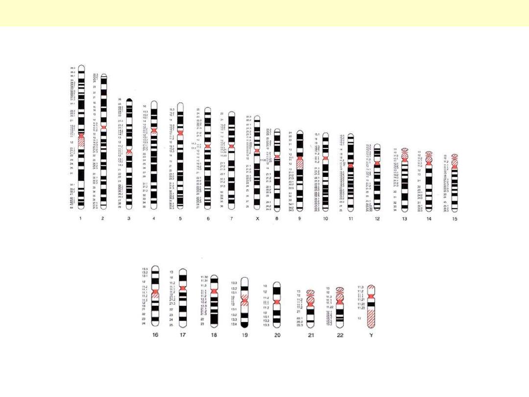
Karyotype – Human
chromosome
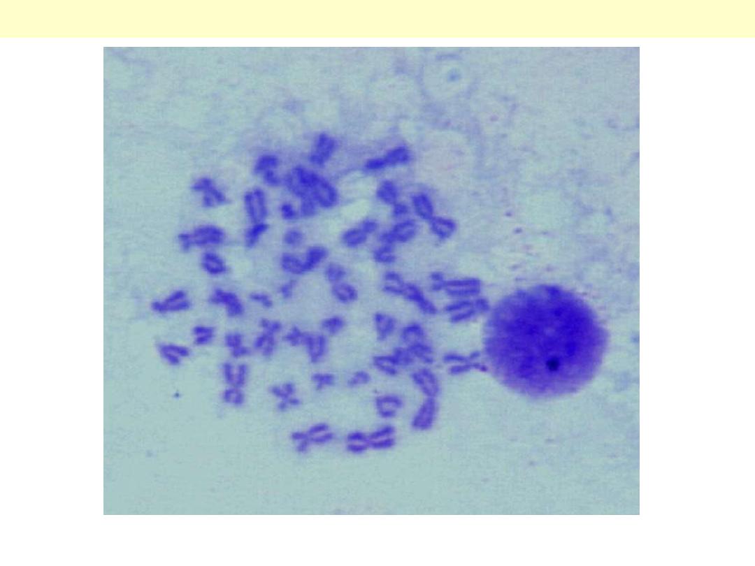
A total 46 chromosomes are seen, varying in size and location of the centromere. Also seen is
‘unruptured’ nucleus of another cell (rounded mass).
Human chromosomes as seen under the microscope
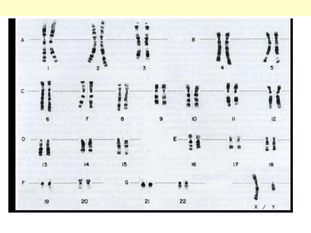
In this karyotype, each chromosome is seen consisting of 2 pairs and the last pair consists of X and Y
chromsome (i.e. male model).
Normal male karyotype
As seen here, the short arm (p) and the long arm (q), each consists of many regions; each region is
divided into multiple bands; each band is further subdivided into sub-bands.
Human Chromosomes arranged according to their sizes
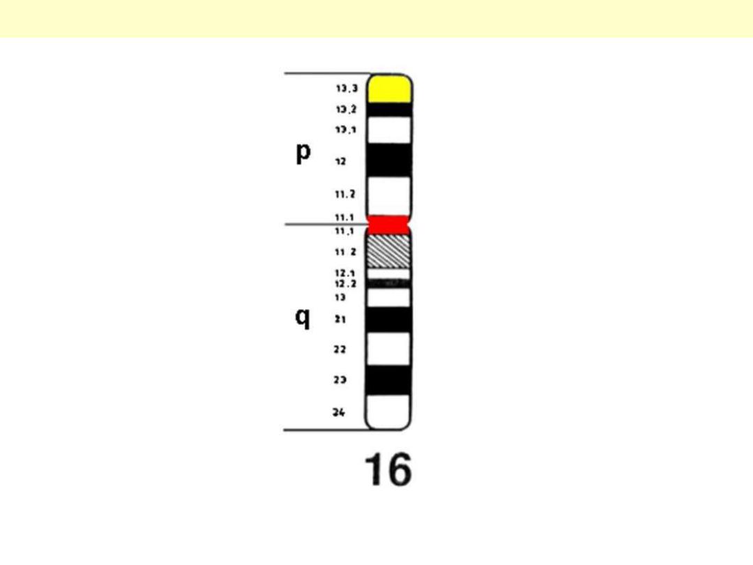
As seen here, the short arm (p) and the long arm (q), each consists of many regions (1 for p, 2 for q);
each region is divided into multiple bands (3 for p, 4 for q); each band is further subdivided into sub-
bands.
Details of chromosome 16

LDL pathway
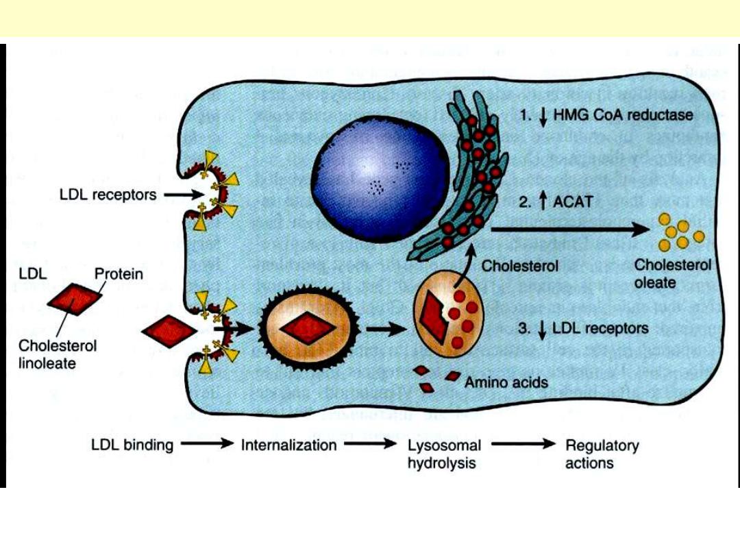
Notice the importance of the LDL receptors in regulating LDL in the circulation
LDL pathway in cultured cells

Multifactorial inheritance
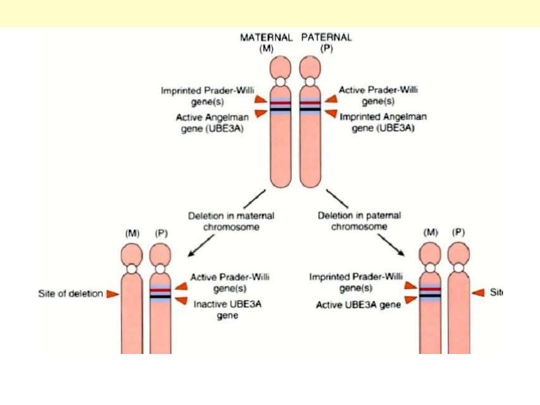
If maternal 15q12 was deleted Angelman Syndrome; on the other hand, if the paternal 15q12 was
deleted Prader Willi syndrome
Clinical Effect of Deletion of 15q12
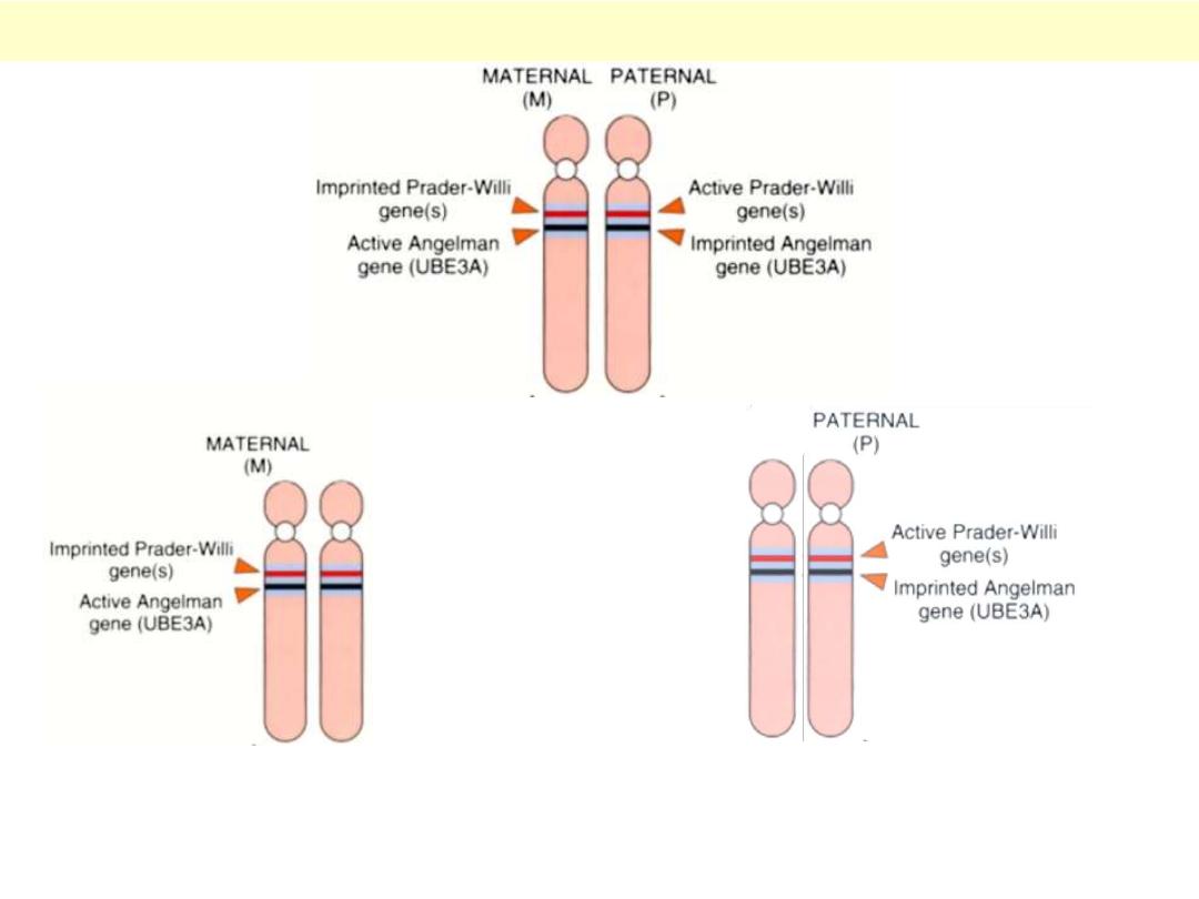
Angelman’s syndrome ( Paternal UPD)
Prader Willi syndrome ( Maternal UPD)
If maternal PW gene was imprinted, paternal side does not have an active PW gene PW syndrome.
The same is said on Angelman’s syndrome only in case of paternal UBE3A is being imprinted.
Effect of Genomic imprinting / Parent of origin

Mutations – Frame shift
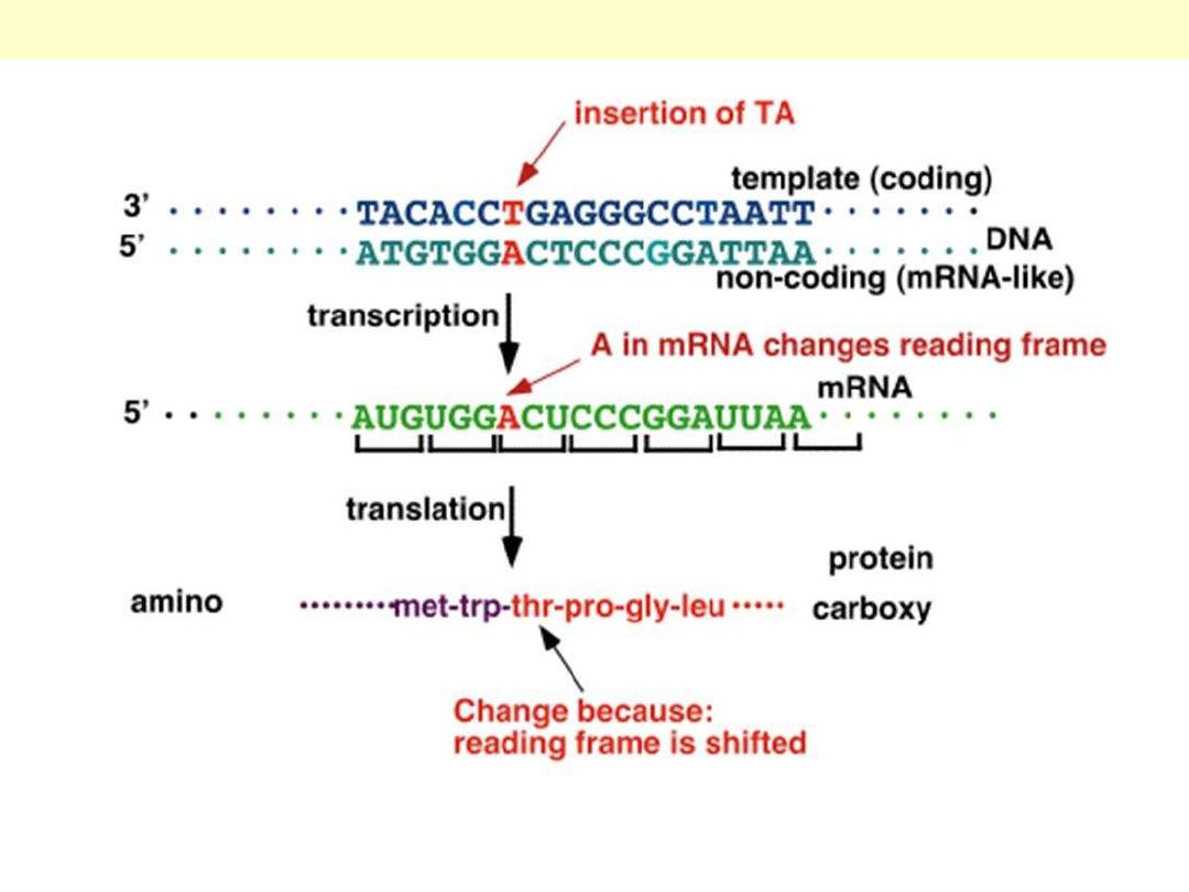
Once a base is inserted (or removed), it will change the reading sequence of the codons shift and
change of all codons that follow change the whole polypeptide chain that follows more severe
effect
Frame shift mutation

RFLP - Analysis
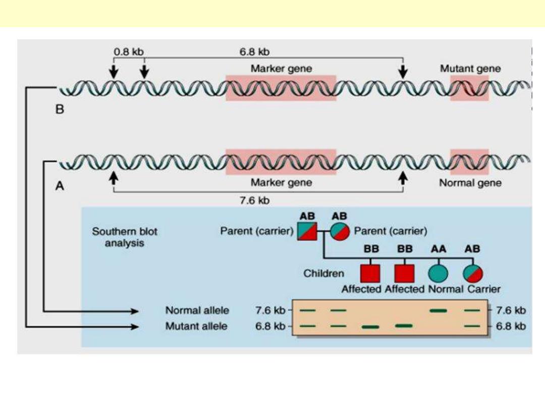
The principle of RFLP for diagnosis of genetic disorders is shown in this illustration.
Restriction fragment length polymorphism (RFLP) analysis

Ring chromosome
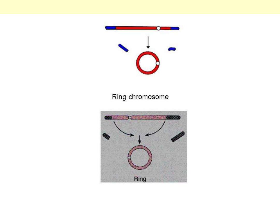
Two cuts at both ends of a chromosome leads to loss of the distal pieces and the ‘sticky’ ends fuse to
form a ring.
Ring chromosome

Sex chromosome disorders
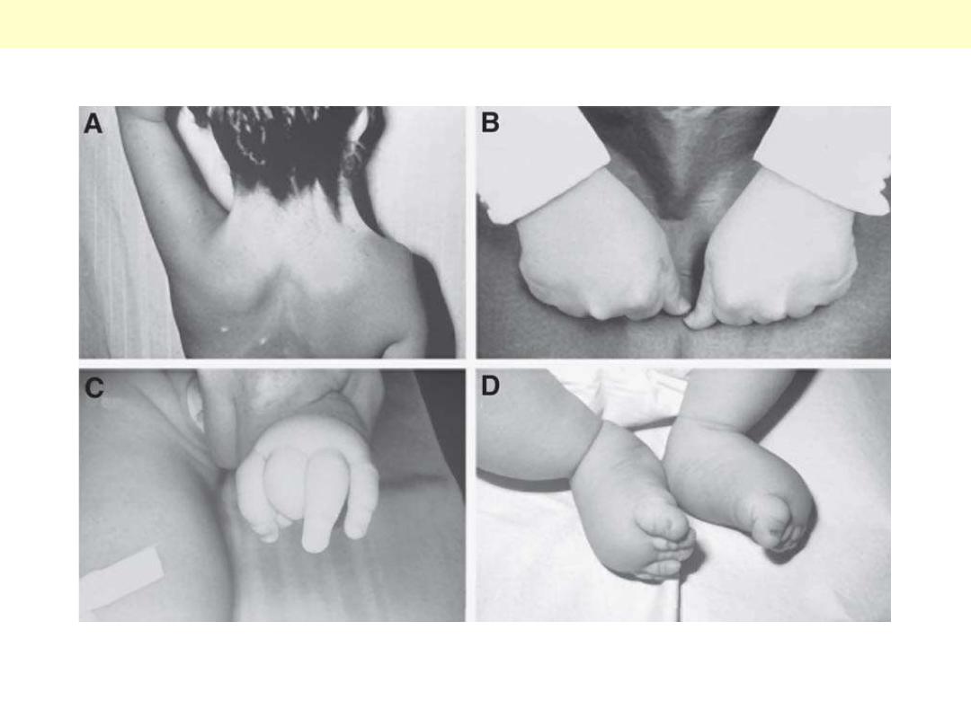
Child with Turner syndrome and low posterior hairline and webbed neck (A) and short fourth
metacarpals (B). Infant with Turner syndrome with lymphedema of the hand (C) and feet (D).
Some clinical features of Turner’s Syndrome
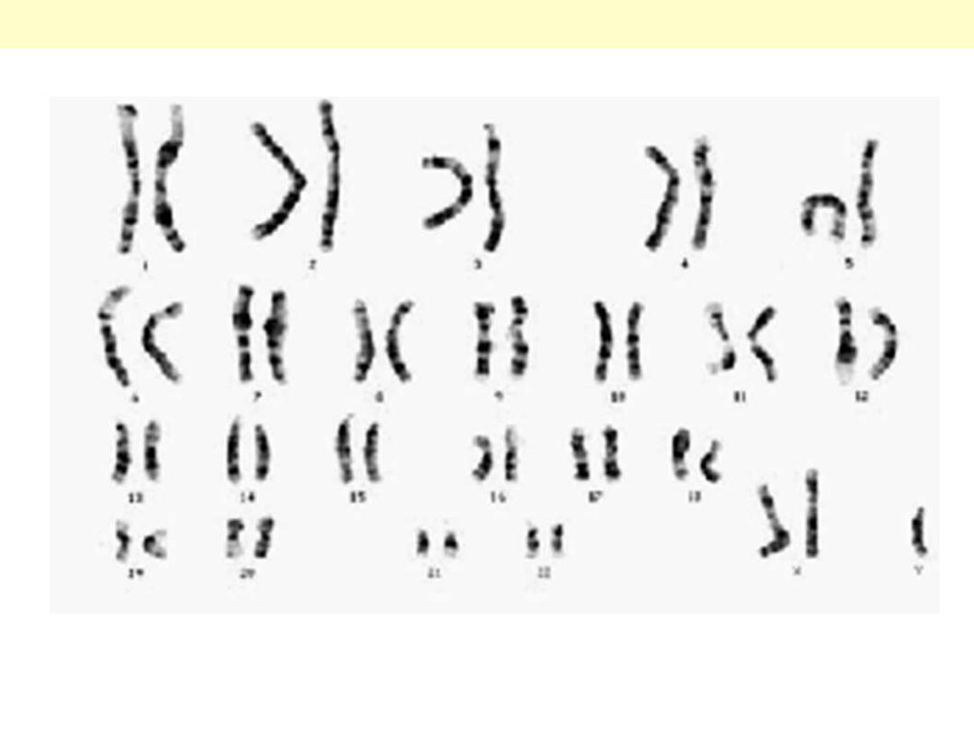
Note in this karyotype, 2 X chromosome in addition to a Y-chromosome.
Karyotype of Classical Klinefelter Syndrome

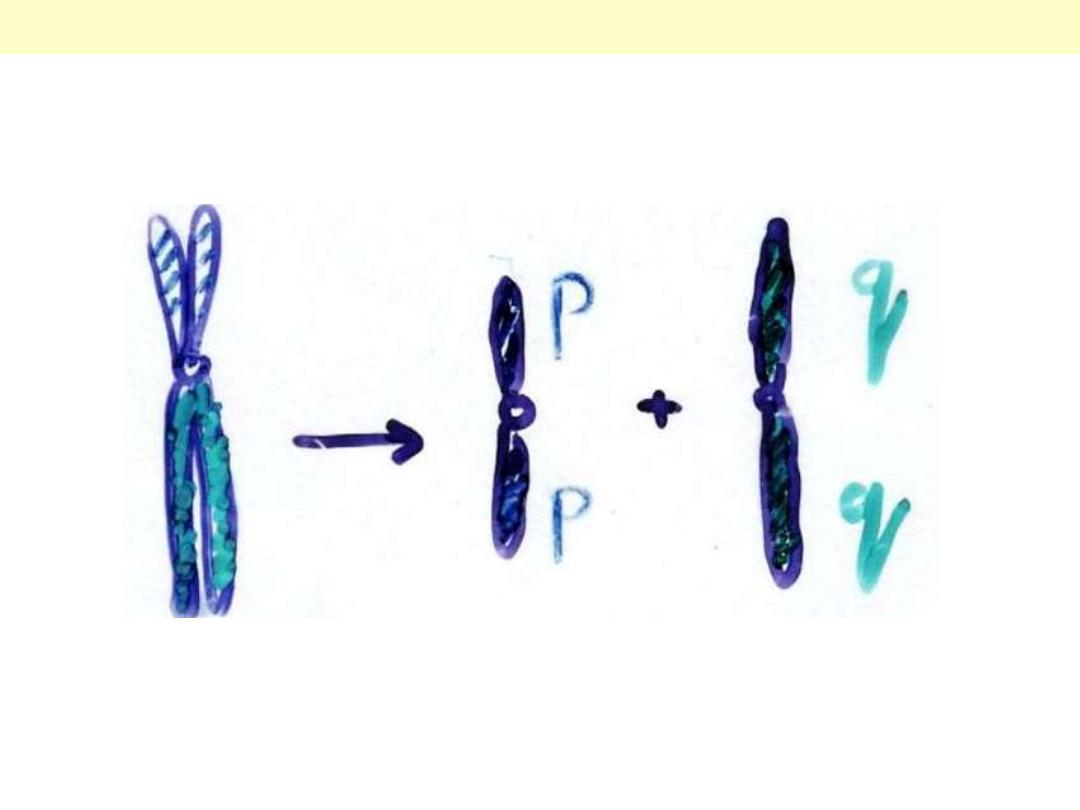
Translocation - Chromosome
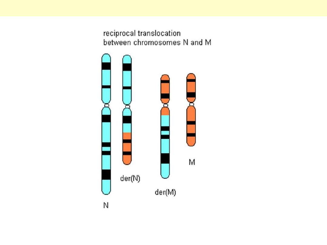
The original ‘normal’ chromosomes [N & M] are seen on the periphery. After a reciprocal
translocation, both chromosomes carry a piece from the other (hybrid) and has lost a piece
(translocated to the other).
Reciprocal Translocation
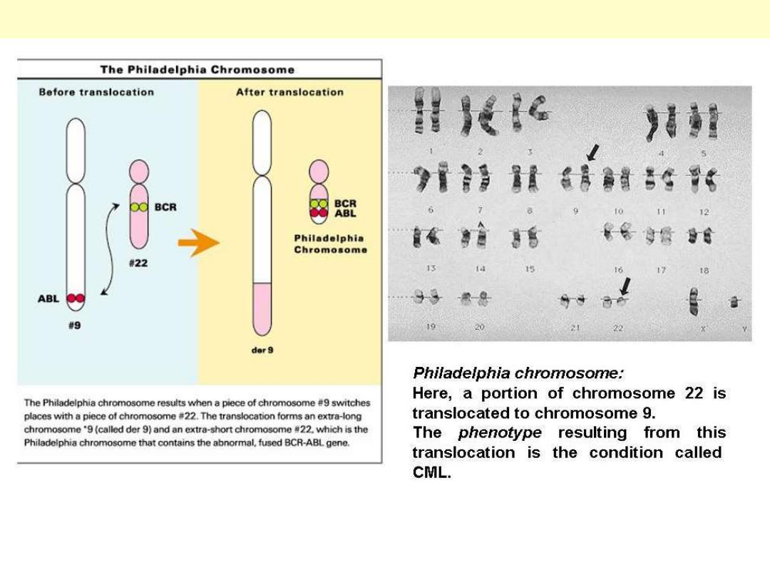
A clinical example of the effect of translocation is chronic myeloid leukemia (CML). Some cases are
found to have a special type of translocation resulting in a hybrid chromosome called Philadelphia
chromosome. The carriers of this translocation are found uniquely responsive to certain drug therapy.
Reciprocal translocation Philadelphia chromosome
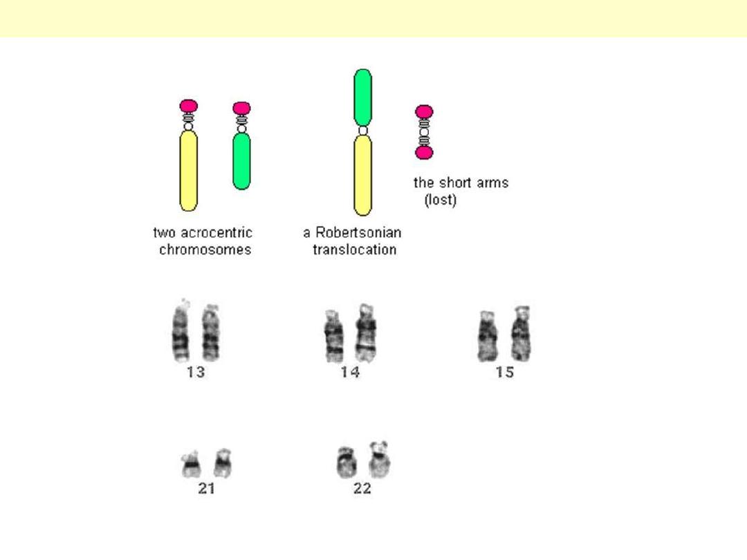
This special type of translocation occurs exclusively between two acrocentric chromsomes [Group D =
13, 14, 15 and Group G = 21, 22].
Robertsonian translocation
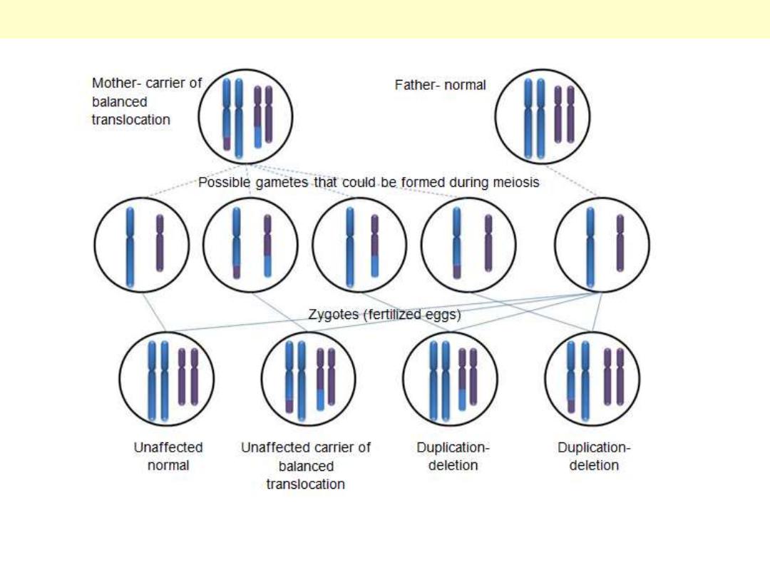
When a haploid cell (sperm or ova) carrying a Robertsonian translocation fuses with another normal
haploid cell, 4 possibilities arise, one is incompatible with life (monosomic) abortion.
Possible outcome of a Robertosnian translocation
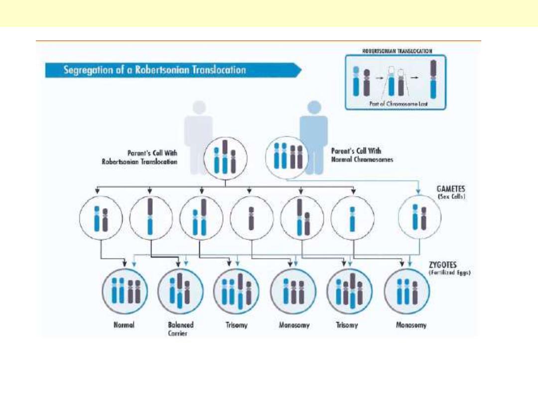
When a haploid cell (sperm or ova) carrying a Robertsonian translocation fuses with another normal
haploid cell, 4 possibilities arise, one is incompatible with life (monosomic) abortion.
Possible outcome of a Robertosnian translocation
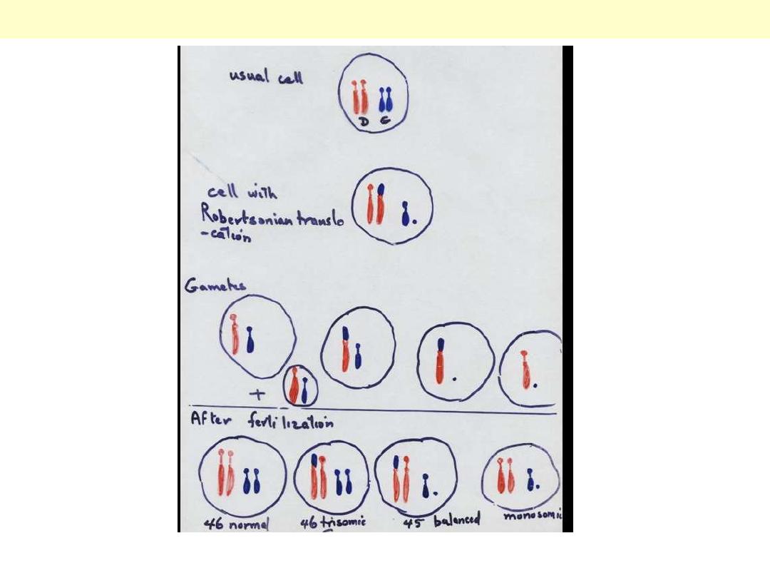
When a haploid cell (sperm or ova) carrying a Robertsonian translocation fuses with another normal
haploid cell, 4 possibilities arise, one is incompatible with life (monosomic) abortion.
Possible outcome of a Robertosnian translocation