The periodontal Ligament
cellsHistological structureThe periodontal ligament is formed of :
Fibers,
Intercellularsubstances
Synthetic
Resorptive
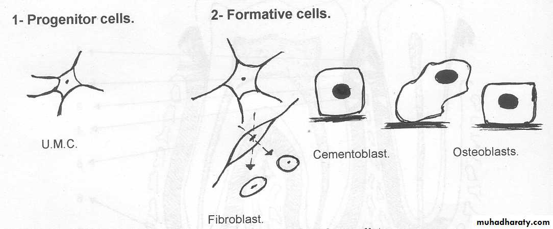
Progenitor
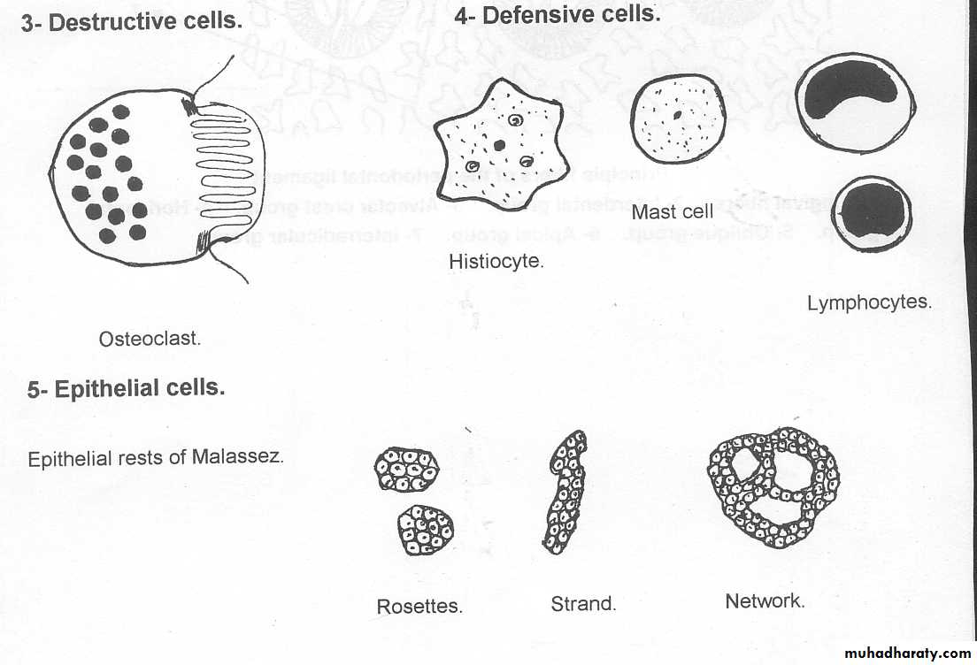
Defensive
ground substances
blood vessels,
nerves & lymphatics.&
epithelial cells
remnants of the epithelialroot sheath of Hertwig
The cells
Syntheticcells
Resorptive
cells
Progenitor
cells
Defensive
cells
fibroblasts, osteoblasts& cementoblasts.
cementoclasts , osteoclasts& fibroclasts.
undifferentiated mesenchymal cells
macrophage, lymphocytes &mast cells
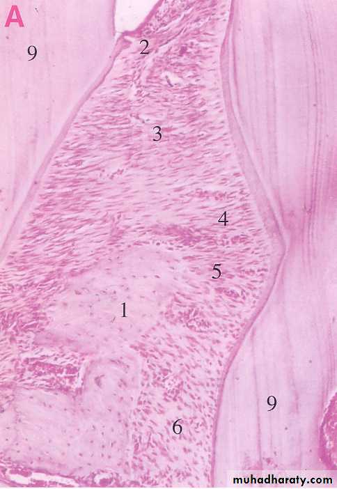
II- The fibers
*The fibers of the periodontal ligament are mainly collagen.They are divided into:
A) The principal fibers.
B) The accessory fibers.
C) The oxytalan fibers.
*Elastic fibers are restricted almost entirely to the walls of blood vessels.
• 1- The principal fibers: a- The gingival fibers:
It attaches the gingiva to the cementum.The fiber bundles pass from the cementum into the free and attached gingiva,
They break up into smaller bundles and interlace (bind intricately together) terminally with gingival fibrous tissue of lamina propria.
b- The transseptal ligament:
*It connects two adjacent teeth.*The ligament runs from the cementum of one tooth over the crest of the alveolus to the cementum of the adjacent tooth.
c- The alveolodental ligament:
1-Alveolar crest group:radiate from the crest of the alveolar process and attach themselves to the cervical part of the cementum.
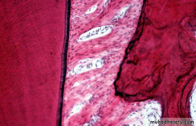
2-Horizontal group:
The fiber bundles run from the cementum to the bone at right angle to the long axis of the tooth.
1
2
dentin
bone
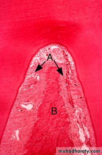
3- Oblique group:
The fiber bundles run obliquely.Their attachment in the bone is somewhat higher than the attachment in the cementum.
It is the greatest number of fiber bundlesfound in this group.
They perform the main support of the tooth against masticatory force.
bone
dentin
4- Apical group:
The bundles radiate from the apical region of the root to the surrounding bone.5- Interradicular group:
The bundles radiate from the interradicular septum to the furcation (branching)of the multirooted tooth.
dentin
bonedentin
bone
B- Accessory fibers:
It is collagenous in nature and run from bone to cementum in different planes, more tangentially to prevent rotation of the tooth and found in the region of the horizontal group.C- Oxytalan fibers
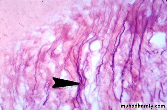
These are immature elastic (pre-elastic) fibers.They need special stains to be demonstrated.
They tend to run in an axial direction, one end being embedded in bone or cementum and the other in the wall of blood vessels.
At the apical region they form a complex network.
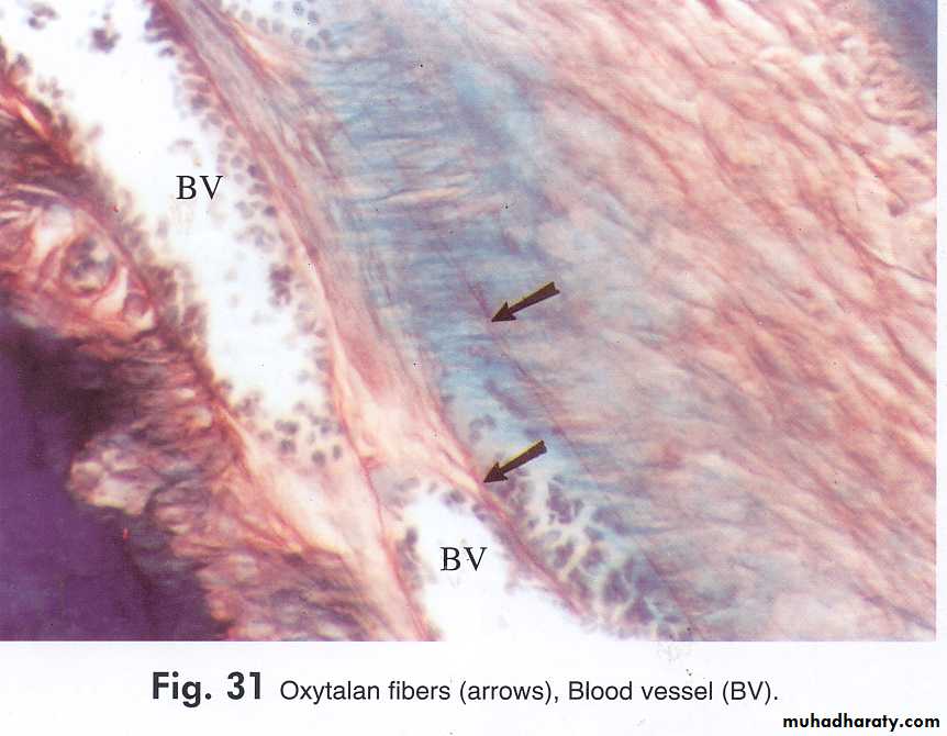
The function of the oxytalan fibers has been suggested that they support the blood vessels of the periodontal ligament during mastication i.e., it prevents the sudden closure of the blood vessels under masticatory forces.
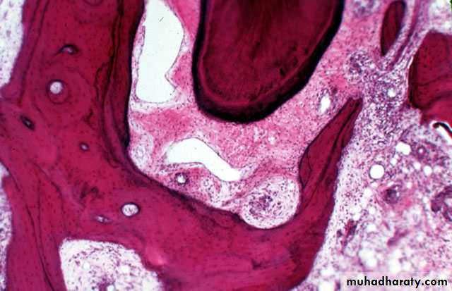
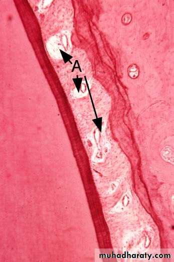
Interstitial tissue
It is found between the fibers of the periodontal ligament.They are areas containing some of the blood vessels, lymphatics and nervs and surrounded by loose connective tissue.
The Age Changes of periodontal ligament
*The periodontal ligament through aging shows:vascularity
cells
thickness
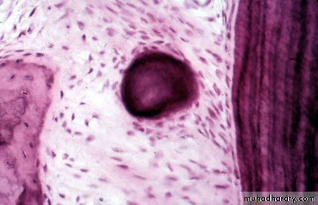
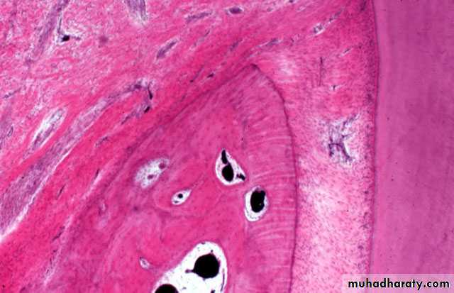
*They may contain cementicles.
The cementicles appear near the surface of cementum may be free , attached or embedded in the cementum.They have nidus favoring the deposition of concentric layers of calcospherite as degenerated cells, area of hemorrhage and epithelial rest's of Malassez.
Cementicles are usually seen in periodontal ligament by aging but in some cases they may be seen in a younger person after local trauma.