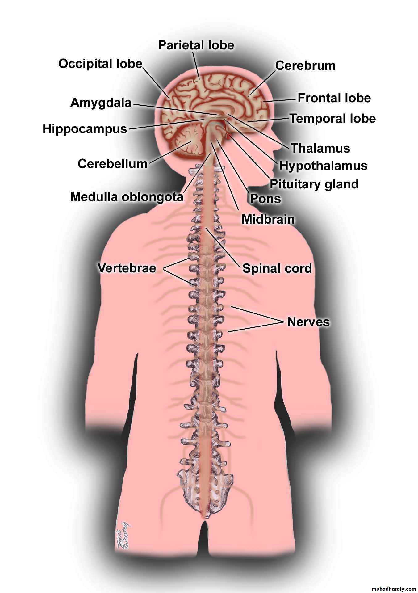
• Cerebral hemisphere
• Diencephalon• Brain stem
• Spinal cord
Neurology
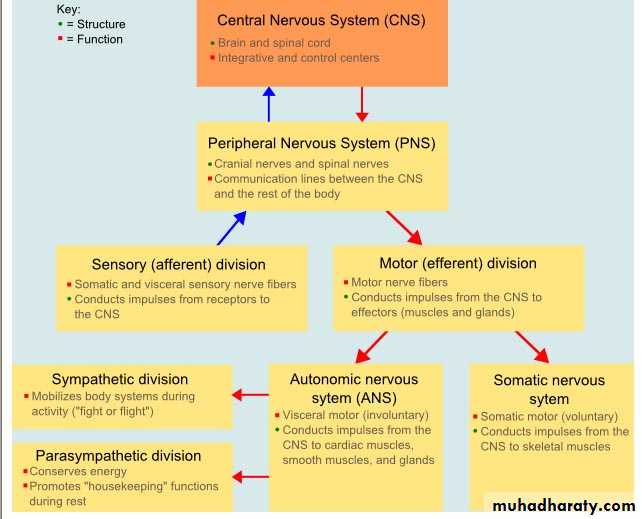
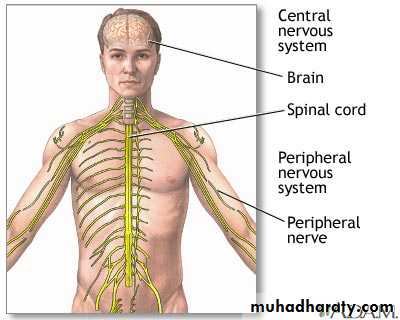
deals with the diagnosis and treatment of all categories of disease involving the nervous systemwhich comprises 3 parts :
• Central nervous systems
• peripheral nervous systems
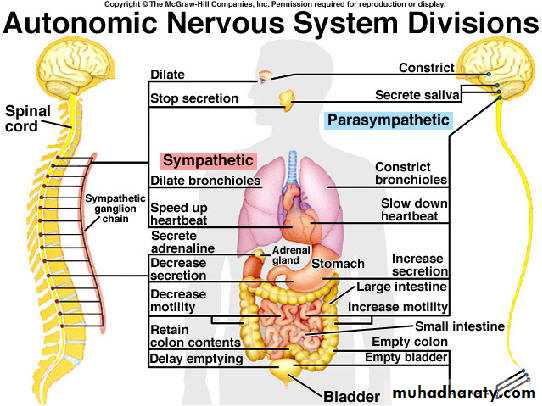
3) autonomic nervous systems,
Anatomy
What are the basic anatomical components of the nervous system ?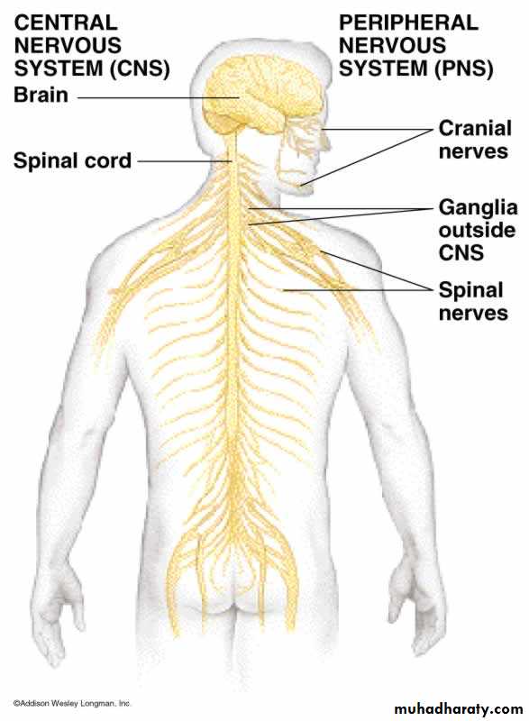
Upper motor neuron sign
== above ant. horn and cranial nuclei• No wasting
• Weakness [UMN]
• No Fasculation
• Hypereflexia
• Hypertonia [clasp knife spasticity ]
• Wasting
• Weakness [LMN]
• Fasculation
• Hyporeflexia
• Hypotonia
lower motor neuron sign
== anterior horn or cranial nerve nuclei and below
Clinical features differences between UMN and LMN lesion ?
FUNCTIONAL ANATOMY
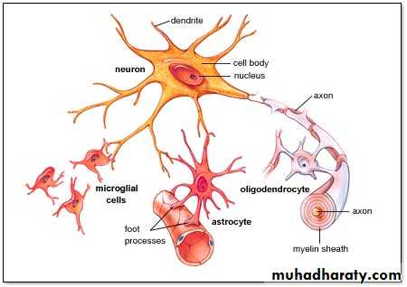
Types of cells includes:
• Neurons
• glial cells--- of 3 types
• Astrocytes
• structural framework
• control the biochemical environment around the neuron
• with the blood vessels forms the blood-brain barrier
• Oligodendrocytes
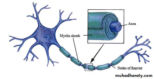
• formation and maintenance of the myelin sheath] inside the CNS
• Microglia[blood-derived mononuclear macrophages]
• ependymal cells lining the cerebral ventricles
Schwann cells : Peripheral neurons have axons invested in myelin made by Schwann cells which line the nerve axon [ OUTSIDE PNS
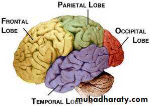
Cerebral hemispheres
has four functionally specialised lobes• Frontal
• parietal
• Temporal
• Occipital
• The brain stem
• midbrain
• pons
• medulla oblengata
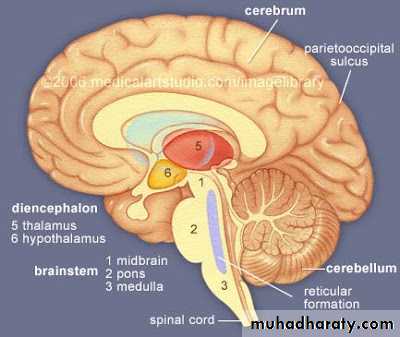
Brain stem
1-An important link between spinal cord and higher brain levels
2-relays motor and sensory impulses between other “higher” parts of the brain and spinal cord3-Midbrain – eye movement control
4-Pons/Medulla Signal relay
Involuntary functions
Many cranial nerves
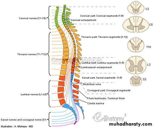
• Spinal cord : 31 different segments
• cervical• Thoracic [dorsal]
• lumber
• Sacral
• coccygeal
The terminal portion of the spinal cord is called the conus medullaris
The cauda equina (“horse’s tail”) the collection of nerves root at the end of the spinal cord
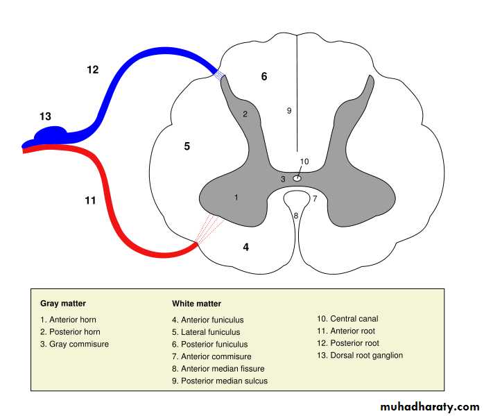
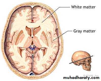
Gray Matter –masses of neurons + Absence of myelin accounts for the gray matter of the brain – Cerebral Cortex
White Matter - Myelinated neurons gives neurons a white appearance – inner layer of cerebrum
Physiology
Who does the nerve impulse take place ?THE GENERATION AND TRANSMISSION OF THE NERVOUS IMPULSE
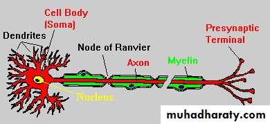
Synapse and
synaptic terminalNerve terminal
AXON
CELL BODY
MYLEINE
AXON
• conduction through the nerve followed by
• synaptic transmission
• Transmissions of information between different part of nervous system take place in 2 physiological steps
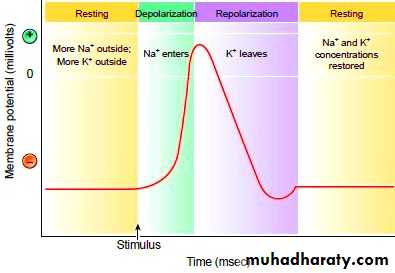
conduction [Nerve impulse] : Electrical wave conducted along the nerve leading to transmission of information between neurons through generation and propagation of an action potential
this is initiated by series of chemical transport of Na+ and K+ across the neuronal cell
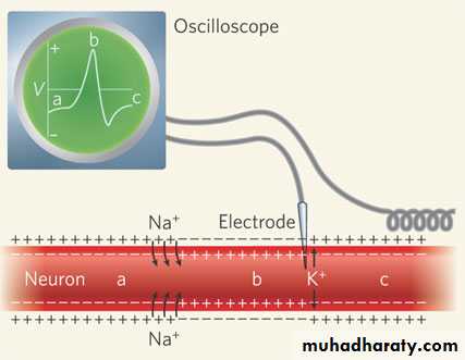
Synaptic transmission : Entry of calcium causes release of the neurotransmitter across the synaptic cleft which binds to receptors on the post-synaptic membrane which depolarize the membrane and initiate an action potential in the postsynaptic structure.
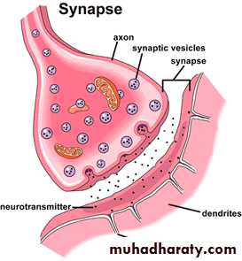
Neurotransmitter :are of 2 types
Excitatory1-Acetylcholine
2-Noradrenaline/
3- adrenaline
4-Glutamate
5-Aspartate
6- 5-hydroxytryptamine
Inhibitory
• Gamma-aminobutyric acid (GABA• Glycine
Terms
• Agnosia: faulty identification of recognition of an object which cannot be explained by primary sensory deficit .
• Finger agnosia: :inability to identify and differentiate between his fingers
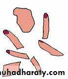
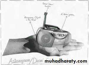
• Astereognosia :inability to identify an object by palpation
• Bilateral astereognosia = tactile agnosia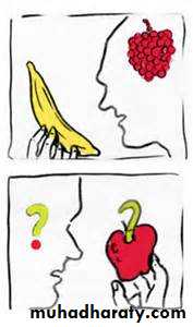
• Visual agnosia :inability to identify an object by vision
• Apraxia :• loss of ability to execute previously learned skills; in patients with normal sensory , motor ,cerebellar and extra pyramidal systems
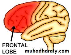
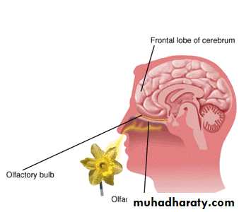
Frontal lobe
PersonalityDisinhibition
Emotional control
Lack of initiation
Social behavior
Antisocial behaviour
Contra lateral motor control
hemiplagia
Language[dominant lobe]
• Expressive dysphasia[dominant lobe]
• Micturition
IncontinenceOlfaction
Impaired smell[anosmia]
Apraxia of the left hand
Dominant


Parietal lobe :non dominantSpatial orientation and Constructional skills
• Apraxia[bilateral]• Tactile agnosia
• Agraphaesthesia
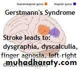
• Gerstman syndrom
• [Acalculia + agraphia+ Rt-Lt disorintation + finger agnosia ]
• Constructional apraxia
• Dressing apraxia
• Neglect of contra lateral side [anasognosia]
• Spatial disorientation
• Focal sensory seizures
• Contra lateral hemi sensory loss
• Contra lateral homonymous
• lower quadrantanopia
• Asymmetry of OKN

Parietal: dominant: - Language - Calculation
DysphasiaContralateral hemisensory loss
Focal sensory seizures
Dyscalculia
Astereognosis
Dyslexia
Agraphaesthesia
Apraxia
Contralateral homonymous
Agnosia
lower quadrantanopia
Asymmetry of optokinetic nystagmus
Temporal: dominant = Auditory perception =Language = Verbal memory = Smell = Balance
Receptive aphasia
Dyslexia
Amusia :non recognition of music
Impaired verbal memory
Contralateral homonymous upper quadrantanopia
Temporal: non-dominant Auditory perception= Melody/pitch perception = Non-verbal memory= Smell= Balance
Lesion leads to
Lesion leads to
Impaired non-verbal memory
Impaired musical skills (tonal perception)
Contralateral homonymous upper quadrantanopia
Occipital Visual processing
Visual inattentionVisual loss
Visual agnosia [dominant]
Homonymous hemianopia (macular sparing)
Investigations :
ElectrophysiologicalEEG[electroencephalogram]
EMG [electromyography ]
NCS [nerve conduction study]
evoked potential [visual , somatosensory ,brainstem audatory ]
IMAGING
CT [computerized tomography]
MRI [magmatic resonant tomography]
SPECT or PET
ultrasound (Doppler or duplex scanning)
MR angiography MR venography
CT angiography
Laboratory
CSF
Oligoclonal band
IgG index
antibodies