1
Third stage
Surgery
Lec-1-2
.د
زيد موفق
1/1/2014
Surgical Infections
Definition:
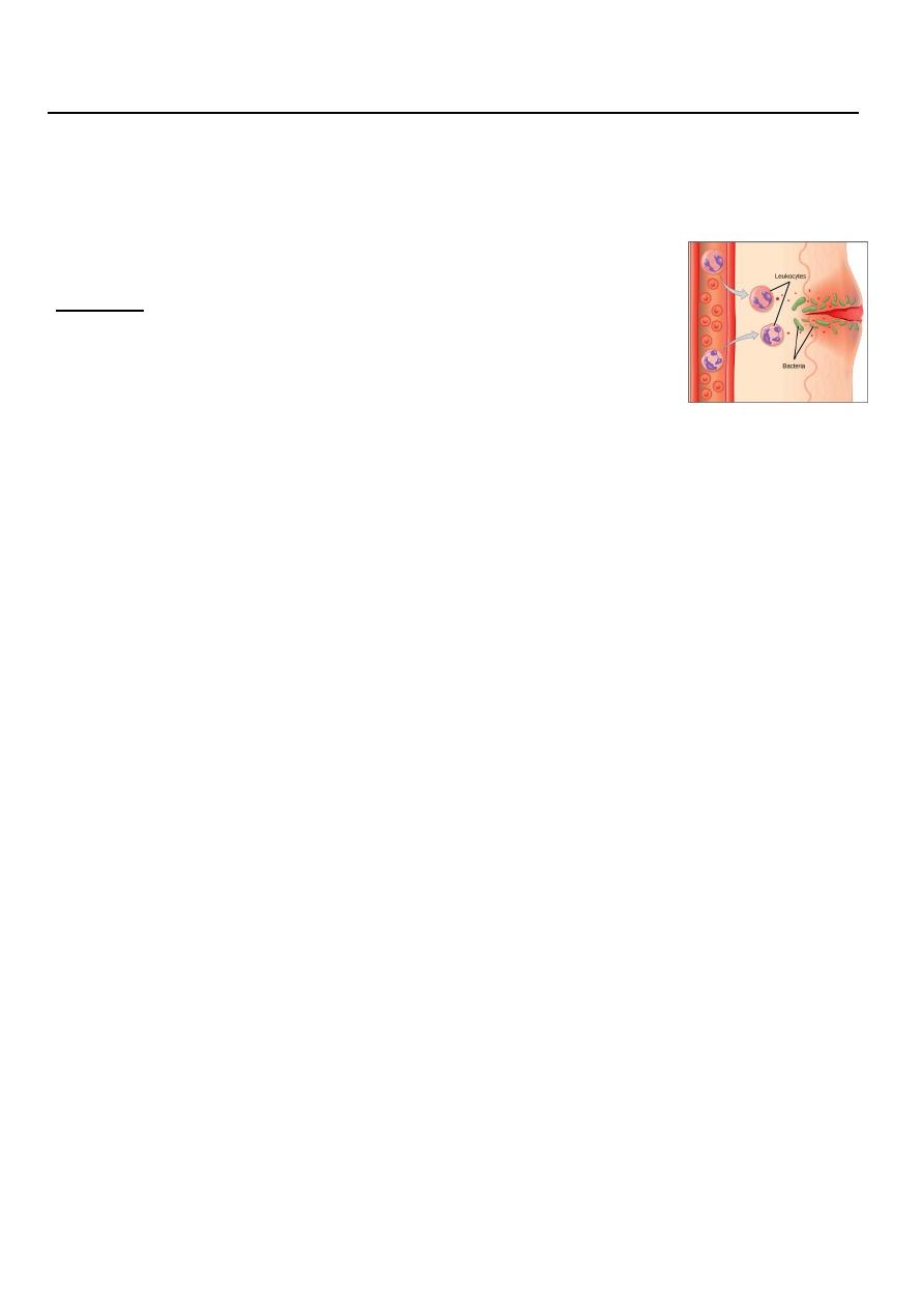
Infection :
Invasion of microorganism to the healthy tissue producing
inflammatory reaction
Pathogenesis and bacteriology:
Microorganisms usually prevented from causing infection in tissues by intact epithelial
surface, mainly the skin.
Protective mechanism includes:
1- Chemicals: low PH gastric juice.
2- Humoral: antibodies, complements and opsonin.
3- Cellular: phagocytic cells, macrophages, polymorphnuclear cells and killer lymphocytes.
Host response can be weakened by several factors:
1-Metabolic: malnutrition (including obesity), diabetes, uremia and jaundice.
2- Disseminated disease: cancer and acquired immune deficiency syndrome (AIDS).
3- Iatrogenic: radiotherapy, chemotherapy and steroids.
The chance of developing infection is also determined by:
1-the pathogenicity of the organisms present and the size of bacterial inoculum.
2- Devitalized tissues, excessive dead space or hematoma, all the result of poor surgical
technique increases the chance of infection.
Diagnosis:
1-History

2
2-Clinical examination
Clinical features of acute inflammation calor(heat) +rubber (redness), dolour (pain)+
tumour (swelling) + function laesa (loss/impairment of function)
3-Laboratory investigation / Radiology imaging.
Complication:
An infection may resolve spontaneously, or it may
1- destruct tissues.
2- abscess, which may rupture spontaneously.
3- sinus or fistula formation.
4- spreading infection death of tissue(gangrene), with it systemic effect.
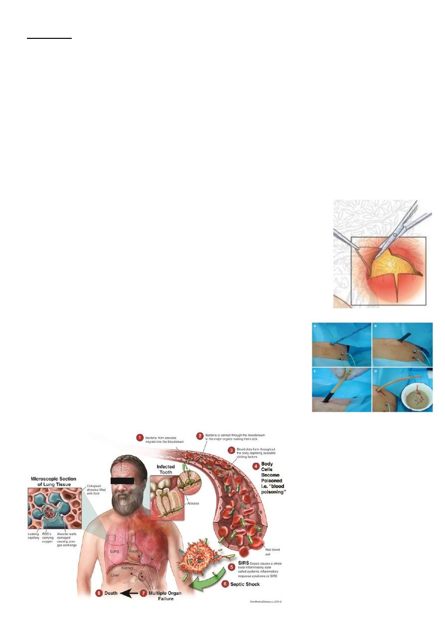
5- bacteremia, septicaemia, septic shoch, multiorgan failure ,disseminated intravascular
coagulopathy and death.
Treatment:
Source Control:
- draining an abscess
- resecting or débriding dead tissue
- diverting bowel, relieving obstruction, and closing a perforation.
Antibiotic treatment of a surgical infection without this mechanical solution will not resolve
the infection. (adjunctive therapies).
A/ Acute nonspecific surgical infection
1-Post operative wound infection, Surgical Site Infection(SSI)
#wound infection invasion of organisms through tissues following breakdown of local or
systemic host defense.
#SSI Are infection of the tissues, organs, or spaces exposed by surgeons during
performance of an invasive procedures.

3
Classification:
Superficial surgical site infection: infection of surgical wounds.
Deep surgical site infection: infection in the deeper musculofascial layer or organ spaces
infection.
Major surgical site infection: discharge significant quantities of pus either spontaneously or
need surgical drainage
+local signs of infection patient + systemic signs; tachycardia, pyrexia and raise WBC count.
Minor surgical site infection: may discharge pus or infected serous fluid (-ve systemic signs).
Treatment of surgical site infection:
Minor may resolve spontaneously by frequent dressing change ,may require drainage.
Major: drainage of pus, May require debridement (removal of dead devitalized tissue)
+Systemic antibiotic.
2-Cellulitis and Lymphangitis
- Is non suppurative invasive infection of tissues with poor locatization + cardinal signs of
inflammation.
-It is spreading infection (B-haemolytic streptococci , staphylococcus , C. perfringens).
-Tissue destruction, gangrene and ulcer may follow which are caused by release of
proteases and allow spread of infection.
Clinical features:
• well demarcated area of inflammation (hot ,red ,tenderness ,swelling and impairment of
function)
• malaise, chills,fever,rigors and a raised white cell count
• If not rapidly treated it can progress to lymphangitis and lymphadenitis
• Localized areas of skin necrosis may occur
Predisposing factors include:
o Lymphoedema

4
o Venous stasis
o Diabetes mellitus
o Surgical wounds
Management
• Rest and elevation of the affected limb
• Antibiotics (orally/ intravenous)
(Benzylpenicillin and flucloxacillin)
Lymphangitis is part of a similar process and presents as painful red streaks in affected
lymphatics.It is associated with painful lymph node groups in the related drainage area.
3-Erysipelas
Erysipelas is a sharply demarcated streptococcal infection of skin, usually associated
with broken skin on the face .
The area affected is erythematous and oedematous.
Fever+leukocytosis.
Broad-spectrum antibiotics
4-Boils
A boil, also called a furuncle, is a deep folliculitis, infection of the hair follicle.
-Staphylococcus aureus
Boils are red lumps around a hair follicle that are tender, warm, and very painful
(signs of inflammation).
- pea-sized to golf ball-sized.
- yellow or white point at the center of the lump / discharge pus.
severe fever, swollen lymph nodes, and fatigue.
chronic furunculosis is recurring boil + Systemic factors that lower resistance :
diabetes, obesity, and hematologic disorders.
Treatment:
Antibiotics against Staphylococcus aureus .
small boil burst and drain spontaneously
recurrent boils a systemic cause should be looked for and treated.

5
5-Carbuncle (A carbuncle is made up of several skin boils)
A carbuncle is a deeper skin infection that involves a group of infected hair follicles in one
skin location.
-back of the neck, shoulders, hips and thighs.
-diabetes
-Staphylococcus aureus, or Streptococcus pyogenes.
swelling + signs of inflammation + one or more openings draining pus. fatigue, fever and a
general discomfort.
Treatment
Antibiotic against gram positive bacteria, incision and drainage of pus collection followed by
frequent dressing.
6-Hidradenitis suppurativa
This is a chronic inflammatory disease of the apocrine gland containing skin(axillary and
groin ).
Less common sites: scalp, breast, chest and perineum.
Hidradenitis suppurativa +obesity and smoking.
Women are 4x.a
The pathophysiology involves follicular occlusion followed by folliculitis and secondary
infection with skin flora (usually Staphylococcus aureus and Propionibacterium acnes).
Clinically, patients develop tender, subcutaneous nodules which may not point and
discharge, but usually progress to cause chronic inflammation , suppurative skin abscesses,
sinus tracts .
Management
Patients should be
-advised to stop smoking and lose weight
-Symptoms can be reduced by the use of antiseptic soaps, tea tree oil, non-compressive
and aerated underwear.
-Medical treatments include topical and oral antibiotics and anti-androgen drugs.
-if abscess developed ,need drainage.
In selected cases, patients may require radical excision of the affected skin and
subcutaneous tissue+ skin grafting or flap transposition.

6
7-Abscesses
• An abscess is collection of pus within soft tissues
Pathology
• An abscess contains bacteria, acute inflammatory cells, protein exudate and necrotic
tissue,It is surrounded by granulation tissue (the 'pyogenic membrane')
The pus is composed of dead and dying white blood cells
• In superficial abscesses
o Staph. Aureus
o Strep. pyogenes
• In deep abscesses
o Gram negative species (e.g. E. coli)
o Anaerobes (e.g. Bacteroides)
Clinical features:
1- Superficial abscesses include infected sebaceous cysts, breast and pilonidal abscesses.
superficial abscess shows cardinal features of inflammation - calor, rubor, dolor,
tumor(Heat,Redness,Pain,Swelling)
After few days superficial abscess usually 'point' and are fluctuant
2-Deep abscesses like; diverticular abscess, subphrenic abscess and anastomotic
leaks(inside the abdomen)
• Patients shows signs of inflammation
o Swinging pyrexia
o Tachycardia
o Tachypnoea
• Physical signs are otherwise difficult to demonstrate
• Site of abscess may not be clinically apparent
Radiological imaging(Ultrasound and/or CT scan) often required to make the diagnosis

7
Treatment
(adequate drainage)
• Should be performed under general anaesthesia
• Antibiotics have little to offer as tissue penetration is usually poor
• Prolonged antibiotic treatment can result in a chronic inflammatory mass (an 'antibioma')
• Superficial abscesses open drainage
• For deep abscesses closed drainage
Open Technique
• Superficial abscesses can usually be drained through a cruciate
incision
• Position of incision may allow depended drainage
• Pus should be sent for microbiology
• Loculi should be broken down and necrotic tissue excised
• A dressing should be inserted into the wound
Closed Technique
• Deep abscess can be treated by ultrasound /CT guided
drainage

8
Definitions of infected states:
_ SSI(surgical site infection) is an infected wound or deep organ space
_ SIRS(systemic inflammatory response syndrome) is the body’s systemic response to
severe infection
SIRS when Two or more of:
hyperthermia (>38°C) or hypothermia (<36°C)
tachycardia (>90/min, no b-blockers)
tachypnoea (>20/min) or Paco2<32 mmHg or mechanical ventilation
white cell count >12 × 109/l or <4 × 10⁹/l
Sepsis is SIRS with a documented infection
Severe sepsis or sepsis syndrome is sepsis with new onset organ failures (respiratory (acute
respiratory distress syndrome), cardiovascular ,renal (acute tubular necrosis), hepatic,
blood coagulation systems or central nervous system)
septic shock = sever sepsis + hypotension (follows compromise of cardiac function and fall
in peripheral vascular resistance)
multiorgan dysfunction syndrome= acute potentially reversible dysfunction of 2 or more
organs
8-Bacteraemia and sepsis
Bacteraemia : presence of bacteria in the blood stream.
Sepsis(septicaemia): proliferation of bacteria in blood stream producing systemic
inflammatory response syndrome.
Bacteraemia occur :
1-anastomotic breakdown (deep space SSI).
2-procedures undertaken through infected tissues (particularly instrumentation in infected
bile or urine).
3- bacterial colonisation of indwelling intravenous cannulae.
Sepsis accompanied by MODS may follow anastomotic breakdown. Aerobic Gram-negative
bacilli are mainly responsible, but S. aureus and fungi may be involved, particularly after the
use of broad-spectrum antibiotics .
Bacteraemia is important when a prosthesis has been implanted, as infection of the
prosthesis can occur.

9
B/ Acute specific surgical infections
1-Gas gangrene
This is caused by C. perfringens, Gram-positive, anaerobic,
spore-bearing bacilli in soil and faeces.
-military , traumatic surgery and colorectal operations.
Risk factor for development of gas gangrene:
1- immunocompromised,diabetic
2- have malignant disease ,particularly if they have wounds containing necrotic or foreign
material, resulting in anaerobic conditions.
3- Military wounds
The cavitation which follows passage of a missile through the tissues causes a ‘sucking’
entry wound, leaving clothing and environmental soiling in the wound in addition to
devascularised tissue.
Clinical Picture
Infected wound with severe local wound pain and crepitus (gas in the tissues).
The wound produces a thin, brown, sweet-smelling exudate.
Oedema and spreading gangrene follow the release of collagenase, hyaluronidase, other
proteases and alpha toxin.
Early systemic complications with circulatory collapse and MSOF follow if prompt action is
not taken
Diagnosis
mainly clinical .
XR may show gas in the tissue, gram stain of the discharge show the bacteria.
Treatment
Aggressive debridement of affected tissues (this may be repeated as required)+ large doses
of intravenous penicillin

11
2-Tetanus
following deep or penetrating (civilian/military) wound and burn .
-developing countries.
In neonate (tetanus neonaturum) following the use of cow dung on the umbilicus
Microbiology
Due to Clostridium tetani,Gram-positive spore forming rod.
Widely found in the environment and soil.It is strict anaerobe that produces a powerful
exotoxins
Typically Infection produces few signs of local inflammation.
Pathogenesis
Germination of spores in wounds releases the exotoxin(tetanospasmin).
The toxin affects nervous system and reaches CNS via the peripheral nerves.
It acts on presynaptic terminals of nerves (myoneural junctions and the motor neurones of
the anterior horn of the spinal cord) and reduces the release of inhibitory
neurotransmitters (e.g. glycine)
resulting in excess activity of motor neurones produces muscle spasm.
Clinical features
The entry wound may show a localised small area of cellulitis+/- exudate
A short prodromal period, which has a poor prognosis, leads to spasms in the distribution of
the short motor nerves of the face followed by the development of severe generalised
motor spasms including :
Facial muscle spasm produces trismus
Typical facial appearance = 'risus sardonicus',
Back muscle spasm produces opisthotomous
Eventually exhaustion and respiratory failure leads to death
A longer prodromal period of 4–5 weeks is associated with a milder form of the disease.
Prevention
• Tetanus can be prevented by:
o Active immunisation with tetanus toxoid with booster every 5-10 years
o Adequate wound toilet of contaminated wounds

11
any patient with open traumatic wound (if not immunized) should receive toxoid with
benzylpenicillin.
If immunized should receive a booster of toxoid (if last immunisation >5 years).
The diagnosis is essentially clinical, Exudate from the wound can be stained to show the
presence of Gram-positive rods
Treatment
• In suspected cases:
o Passive immunisation with anti-tetanus immunoglobulin
o Adequate wound debridement
o Intravenous benzylpenicillin
o Relaxants may also be required, and the patient may require mechanical ventilation in
severe forms, which may be associated with a high mortality in Intensive care support
o Despite the use of ITU mortality is about 50%
3-Necrotising fasciitis
Necrotising fasciitis results from a polymicrobial, synergistic infection, most commonly a
streptococcal species (group A b haemolytic) in combination with Staphylococcus,
Escherichia coli, Pseudomonas, Proteus, Bacteroides or Clostridia.
80%have a history of previous trauma/infection and over 60 % commence in the lower
extremities.
Meleney’s synergistic gangrene(abdominal wall) and Fournier’s gangrene(perineum) are all
variants of a similar disease process .
Predisposing conditions include:
• diabetes;
• smoking;
• penetrating trauma;
• pressure sores;
• immunocompromised states;
• intravenous drug abuse;
• perineal infection (perianal abscess, Bartholin’s cysts);
• skin damage/infection (abrasions, bites, boils).

12
Classical clinical signs include: oedema extending beyond visible skin erythema; a woody
hard texture to the subcutaneous tissues; an inability to distinguish fascial planes and
muscle groups on palpation; disproportionate pain in relation to the affected area with
associated skin vesicles and soft tissue crepitus .
Early on, patients may be febrile and tachycardic, with a very rapid progression to septic
shock.
Radiographs should not delay treatment but if taken, they may demonstrate air in the
tissues.
Treatment
1-urgent fluid resuscitation, monitoring of haemodynamic status
2-administration of high-dose broad-spectrum intravenous antibiotics.
3- debridement as soon as possible until viable, healthy, bleeding tissue is reached.
Early re-look and further debridement is advisable together with the use of vacuum-
assisted dressings.
Early skin grafting in selected cases may minimise protein and fluid losses.
Mortality of between 30 and 50 per cent can be expected even with prompt operative
intervention.
C/ Chronic specific infection
1-Tuberculosis
Tuberculosis(Mycobacterium tuberculosis or Mycobacterium bovis) is common throughout
the world. It Causes significant morbidity and mortality particularly in Africa and Asia .
Primary tuberculosis
• Usually a respiratory infection that occurs in childhood, results in sub-pleural Ghon focus
and mediastinal lymphadenopathy (primary complex).Symptoms are often few,resolution
of infection usually occurs.
• Complications include:
o Haematogenous spread causing miliary TB affecting lungs, bones, joints, meninges.
o Direct pulmonary spread resulting in TB bronchopneumonia

13
Post-primary tuberculosis
Occurs in adolescence or adult life, due to reactivation of infection or repeat exposure.
Reactivation may be associated with immunosuppression (e.g. drugs or HIV infection)
It Results in more significant symptoms. Pulmonary infection accounts for 70% of cases of
post-primary TB.
Clinical features include cough, haemoptysis, malaise, weight loss and night sweats.
Infection of lymph glands results in discrete, firm and painless lymphadenopathy.
Confluence of infected glands can result in a 'cold' abscess.
Infection of the urinary tract can cause haematuria and 'sterile pyuria'
Investigations
If discharging sinus,discharge sent for ZN stain and culture.
• Repeated samples may be required
Microscopy
stained by Ziehl-Neelsen stain
• Negative microscopy does not exclude tuberculosis
Culture
Collect adequate specimens (e.g. early morning urine x3),Concentration of specimen (e.g.
centrifugation).
o Culture on Lowenstein-Jensen method at 35-37° for at least 6 weeks
Histology
• caeseating necrosis
• Tuberculous follicle consists of central caseaous necrosis surrounded by lymphocytes,
multi-nucleate giant cells and epitheloid macrophages Multinucleated (Langhan’s) giant
cell.
Skin tests
• Delayed hypersensitivity reaction used to diagnose tuberculosis.The two commonest tests
are the Mantoux and Heaf test.
• Positive skin test are indicative of active infection or previous BCG vaccination
Treatment
• First line chemotherapeutic agents are rifampicin, isoniazid and ethambutol
• Given as 'triple therapy' for first 2 months until sensitivities available

14
• Rifampicin and isoniazid are the usually continued for further 7 months
• Less than 5% of organisms are resistant to first-line agents
• Second line treatment includes pyrazinamide
2-Syphilis
Syphilis is a systemic sexually transmitted disease (STD) caused by the spirochete,
Treponema pallidum.
Stages of syphilis:
1-Primary Stage
One or more chancres (usually firm, round,small, and painless) appear at the site of
infection (most often the genital area) 10 to 90 days after infection
The chancres heal on their own in 3-6 weeks.Patient is highly infectious in the primary
stage.
2- Secondary Stage
Rashes occur as the chancre(s) fades or a few weeks after the chancre heals.
Rashes typically appear on the palms of the hands, the soles of the feet, or on the face,but
also may appear on other areas of the body.
Sometimes wart-like “growths” may appear in the genital area.
Rashes and syphilitic warts tends to clear up on their own within 2-6 weeks,but may take as
long as 12 weeks.Patient is highly infectious in the secondary stage.
Latent stage:
patient is serosensitive ,has no symptoms.
within one year of onset of infection,(Early latent stage )the patient is potentially
infectious.
more than one year after onset of infection(Late latent stage), patient is not infectious at
this stage.
3-Tertiary stage(late stage)
Lesion in the skin and bone(gummas), internal organs, central nervous system, and
cardiovascular system.
Signs and symptoms of late stage of syphilis include difficulty coordinating muscle
movements, paralysis,numbness,gradual blindness and dementia. This damage may be
serious enough to cause death.the patient is not infectious at the late stage.

15
Syphilitic ulcers are typically painless, rubbery , indurated, punched out ulcer.
Diagnosis:
-Dark-field examinations or direct fluorescent antibody tests of chancre tissue are the
definitive methods for diagnosing primary and secondary syphilis.
-sequential serologic tests (e.g. VDRL, RPR).
Treatment
is by use of a long-acting penicillin.
3-Actinomycosis
Chronic infectious inflammatory condition originating in the tissues adjacent to mucosal
surfaces caused mostly by infection with A. israelii ,but others, A. naeslundii, A.
odontolyticus, A. viscosus, A. meyeri also can cause human actinomycosis.
most common recognized infections: oral and cervicofacial regions.
The thoracic region, abdominopelvic region, and the CNS also frequently can be involved.
Actinomyces is normal inhabitants of some areas of the GI tract of humans and animals
from the oropharynx to the lower Bowel.
Lesions:
induration and sinuses , abscess , tissue fibrosis.
In pus, the most characteristic form is the sulfur granule .
The firm, fibrous masses are often initially mistaken for a malignancy
Diagnosis:
(Identification of microorganism is difficult)
-History : slowly progressive lesion with history of trauma predispose to mucosal invasion
by actinomyces.
-Biopsies for culture and histopathology (multiple sections )
Treatment:
Treatment of choice: Penicillin G. Other antimicrobics (tetracycline, erythromycin,
clindamycin) are active in vitro and have shown some clinical effectiveness
High doses of penicillin must be used and therapy prolonged for 4 to 6 weeks or longer
before any response is seen.
