
Detailed Review of Cranial
Nerves
Charlie Goldberg, M.D.
Professor of Medicine, UCSD SOM
CN 1- Olfactory: Sense of
Smell
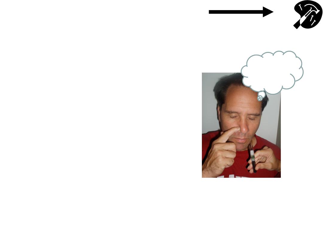
• Check air movement thru ea
nostril separately.
• Smell not usually assessed
(unless sx)
– use coffee grounds or other
w/distinctive odor
(e.g. mint, wintergreen, etc)
- check ea nostril independently
- detect odor when presented @
10cm.
Hmmm..
Coffee!
Hammer & Nails icon indicates A Slide
Describing Skills You Should Perform In Lab
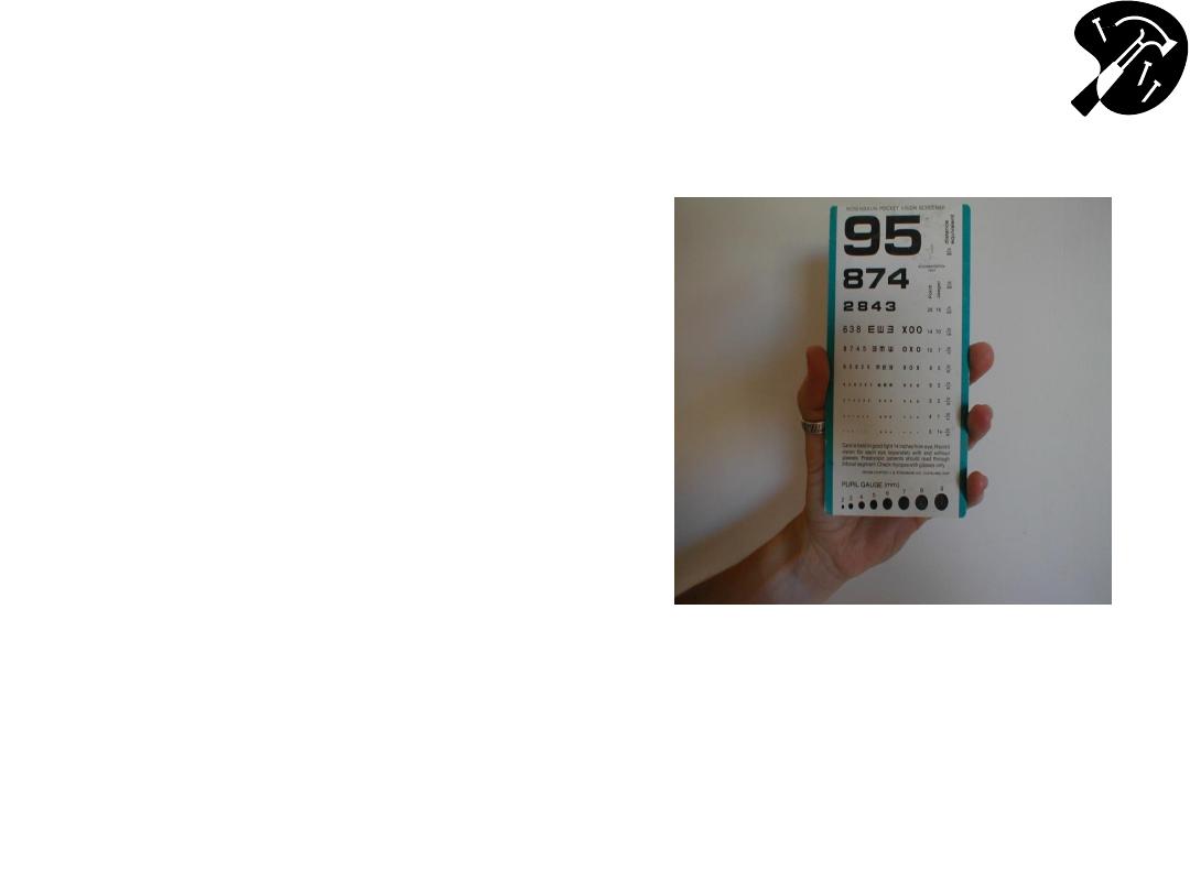
Functional Assessment
– Acuity (Cranial
Nerve 2
– Optic)
• Using hand held card
(held @ 14 inches) or
Snellen wall chart,
assess ea eye
separately. Allow
patient to wear
glasses.
• Direct patient to read
aloud line w/smallest
lettering that they’re
able to see.
Hand Held Acuity Card
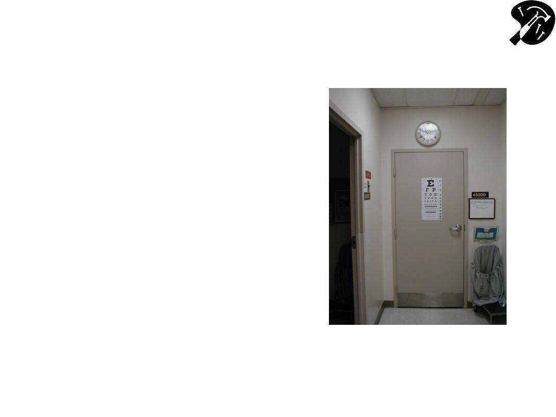
Functional Assessment
– Acuity (cont)
• 20/20 =s patient can read
at 20` with same accuracy
as person with normal
vision.
• 20/400 =s patient can read
@ 20` what normal person
can read from 400` (i.e.
very poor acuity).
• If patient can’t identify all
items correctly, number
missed is listed after a ‘-’
sign (e.g. 20/80 -2, for 2
missed on 20/80 line).
Snellen Chart For Acuity Testing
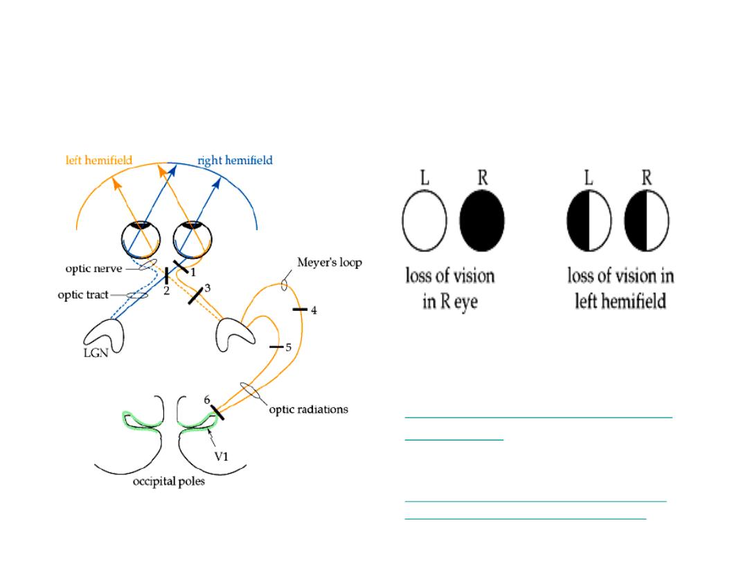
Functional Assessment - Visual
Fields (Cranial Nerve 2 - Optic)
Lesion #1
Lesion #3
Images from: Wash Univ. School
of Medicine, Dept Neuroscience
http://thalamus.wustl.edu/course
/basvis.html
NEJM Interactive case
– w/demo of visual
field losses:
http://www.nejm.org/doi/full/10.1056/NEJ
Mimc1306176?query=featured_home
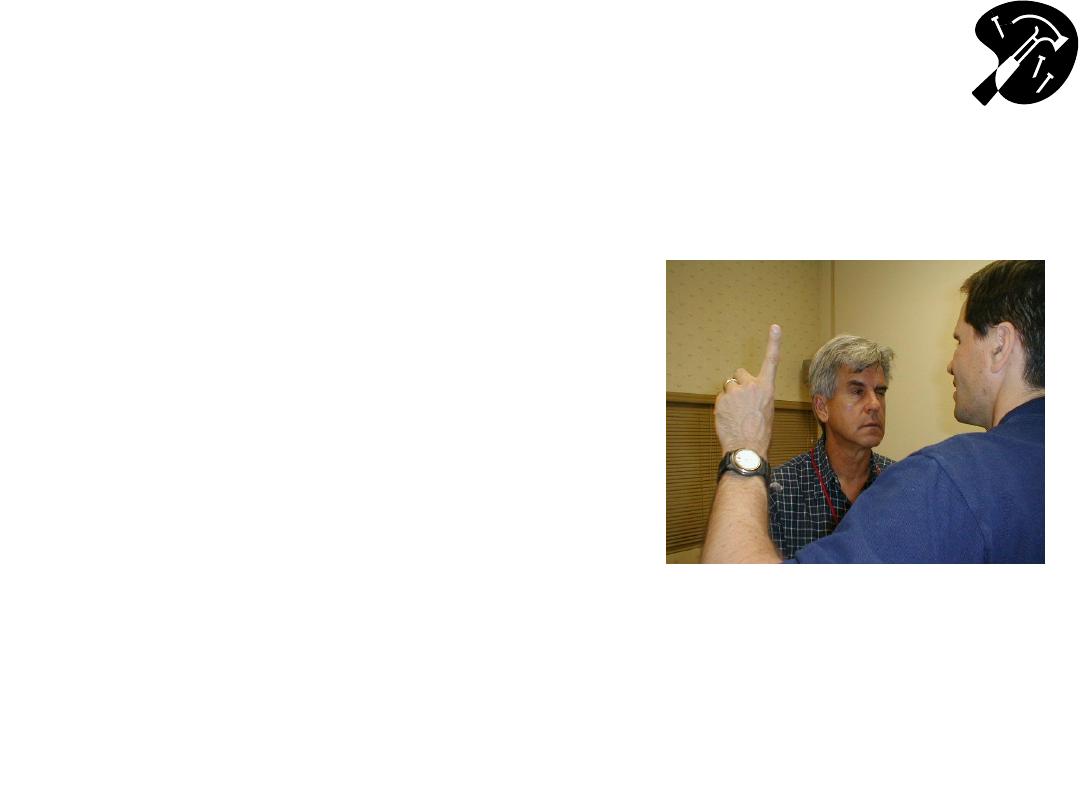
CN 2 - Checking Visual Fields By
Confrontation
• Face patient, roughly 1-2 ft
apart, noses @ same level.
• Close your R eye, while
patient closes their L. Keep
other eyes open & look directly
@ one another.
• Move your L arm out & away,
keeping it ~ equidistant from
the 2 of you. A raised index
finger should be just outside
your field of vision.
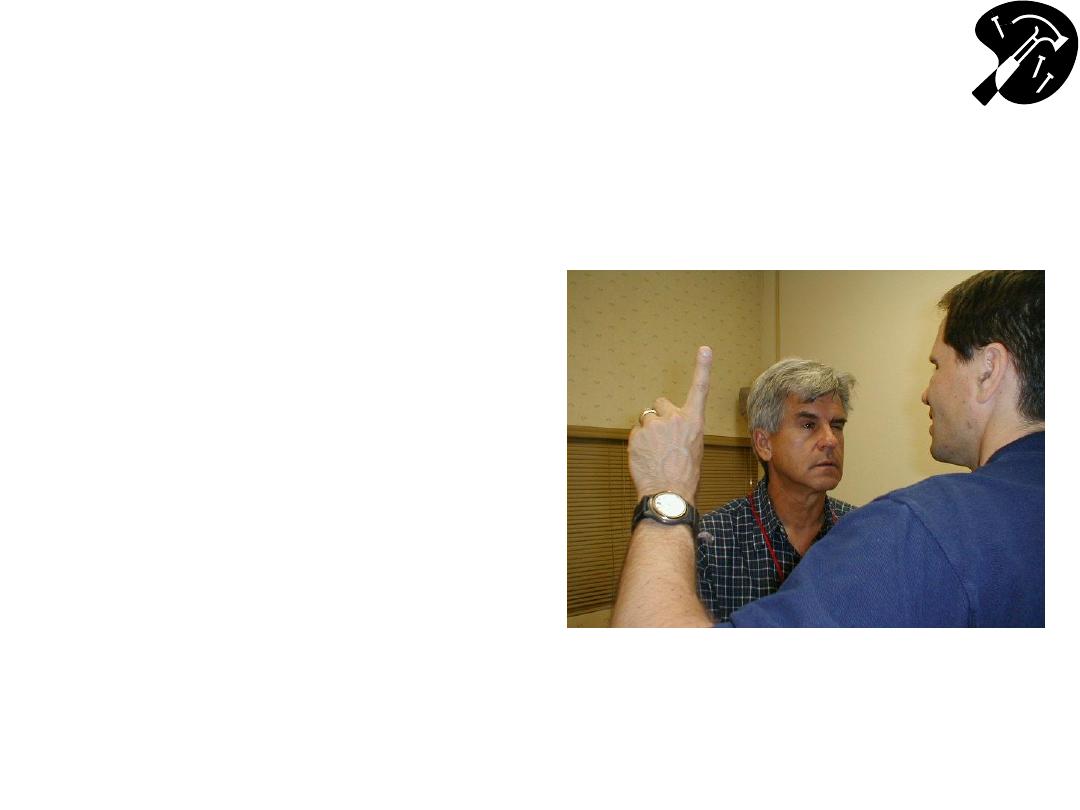
CN 2 - Checking Visual Fields By
Confrontation (cont)
• Wiggle finger & bring it in
towards your noses. You
should both be able to
detect it @ same time.
• Repeat, moving finger in
from each direction. Use
other hand to check
medial field (i.e. starting
in front of the closed eye).
• Then repeat for other
eye.

Pupillary Response
• Pupils modulate amount of light entering eye (like
shutter on camera)
• Dark conditionsdilate; Brightconstrict
• Pupils respond symmetrically to input from either
eye
– Direct response =s constriction in response to direct
light
– Consensual response =s constriction in response to
light shined in opposite eye
• Light impulses travel away (afferents) from pupil
via CN 2 & back (efferents) to cilliary muscles
that control dilatation via CN 3

Pupillary Response Testing
Technique
• Make sure room is darkpupils a little dilated,
yet not so dark that cant observe response
– can
use your hand to provide “shade” over eyes
• Shine light in R eye:
– R pupil constricts
– Again shine light in R eye, but this time watch L pupil
(should also constrict)
• Shine light in L eye:
– L pupil constricts
– Again shine light in L eye, but this time watch R pupil
(should also constrict)

Pupillary Response Testing
Technique
• Swinging Flashlight Test
– Looks for afferent pupil defect (CN II)
– After observing each eye individually, move
the flashlight between the left and right eye at
a steady rate
– See an example at Neuroexam.com:
•

Describing Pupilary Response
• Normal recorded as: PERRLA (Pupils Equal,
Round, Reactive to Light and Accommodation)
–
w/accommodation = to constriction occurring
when eyes follow finger brought in towards
them, directly in middle (i.e. when looking “cross
eyed”).
• Abnormal responses can be secondary to:
– direct or indirect damage to either CN 2 or 3
• Or parasympathetic injury to CN3 or damage to the
sympathetic neurons
– meds e.g. sympathomimetics (cocaine) dilate,
narcotics (heroin) constrict.

Pupil Response Simulator
University of California, Davis School of
Medicine
– Designed by Dr. Rick Lasslo,
M.D., M.S.
http://cim.ucdavis.edu/EyeRelease/Interface/
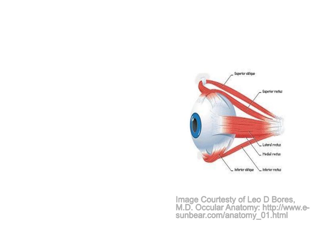
CNs 3, 4 & 6
Extra Ocular Movements
• Eye movement
dependent on Cranial
Nerves 3, 4, and 6 &
muscles they innervate.
• Allows smooth,
coordinated movement in
all directions of both eyes
simultaneously
• There’s some overlap
between actions of
muscles/nerves
Image Courtesty of Leo D Bores,
M.D. Occular Anatomy: http://www.e-
sunbear.com/anatomy_01.html

Cranial Nerves (CNs) 3, 4 & 6
Extra Occular Movements (cont)
• CN 6 (Abducens)
– Lateral rectus musclemoves eye laterally
• CN 4 (Trochlear)
– Superior oblique musclemoves eye down
(depression) when looking towards nose; also
rotates internally.
• CN 3 (Oculomotor)
– All other muscles of eye movement – also
raises eye lid & mediates pupilary constriction.
CNs & Muscles That Control
Extra Occular Movements
CN 6-LR
CN 6-LR
CN 4-SO
SO ‘4’, LR ‘6’, All The Rest ‘3’
SR
IR
MR
IO
SR
IR
LR- Lateral Rectus
MR-Medial Rectus
SR-Superior Rectus
IR-Inferior Rectus
SO-Superior Oblique
IO-Inferior Oblique
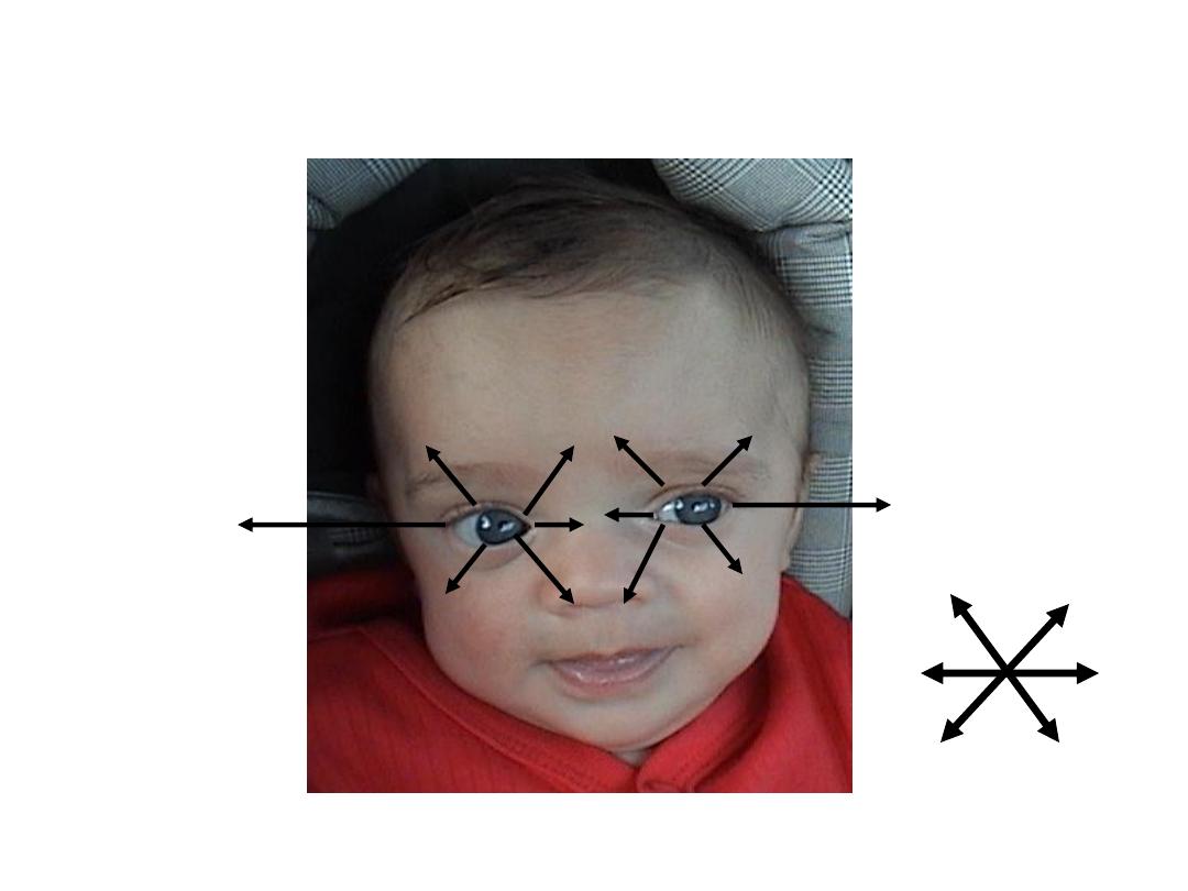
6 “Cardinal” Directions
Movement

Technique For Testing Extra-
Ocular Movements
• To Test:
– Patient keeps head immobile, following your
finger w/their eyes as you trace letter
“H”
– Alternatively, direct them to follow finger
w/their eyes as you trace large rectangle
• Eyes should move in all directions, in
coordinated, smooth, symmetric fashion.
• Hold the eyes in lateral gaze for a second
to look for nystagmus

Extra Occular Eye Movement
Simulator
University of California, Davis School of
Medicine
– Rick Lasslo, M.D., M.S.

Function CN 5 - Trigeminal
• Sensation:
– 3 regions of face: Ophthalmic, Maxillary &
Mandibular
• Motor:
– Temporalis & Masseter muscles
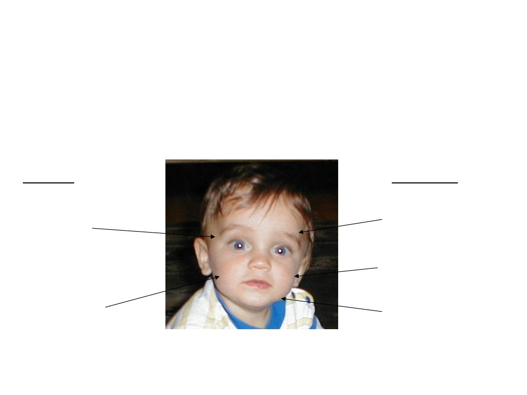
Function CN 5
– Trigeminal
(cont)
Ophthalmic(V1)
Maxillary (V2)
Mandibular (V3)
Temporalis
(clench teeth)
Masseter (move
jaw side-side)
Sensory
Motor
* Corneal Reflex: Blink when cornea touched - Sensory CN
5, Motor CN 7
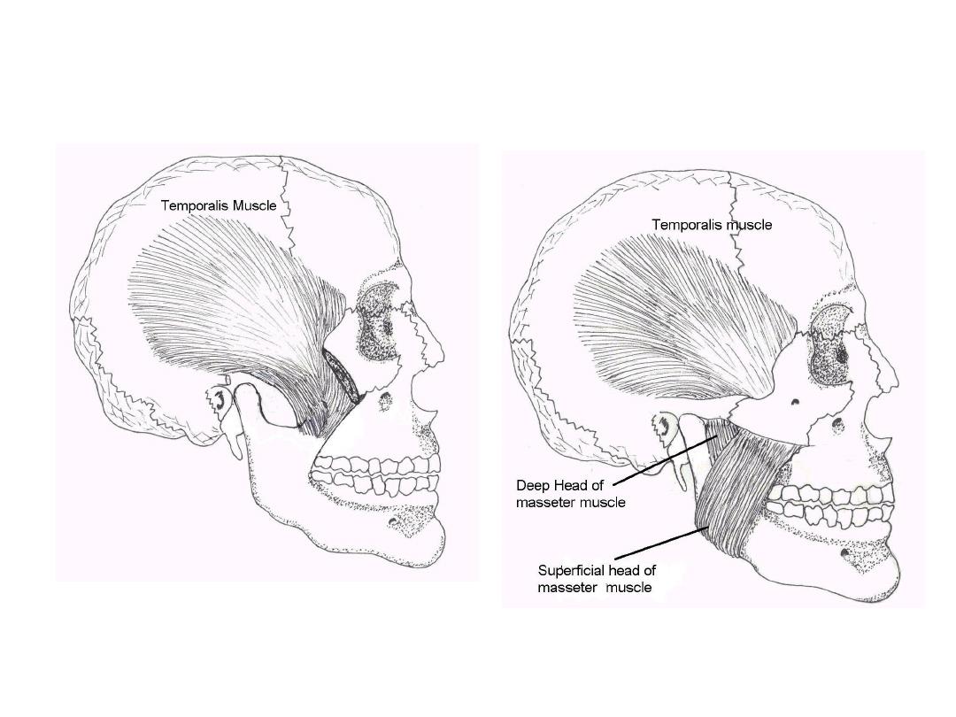
Temporalis & Masseter Muscles
Oregon Health Sciences University:
http://home.teleport.com/~bobh/

Testing CN 5 - Trigeminal
• Sensory:
– Ask pt to close eyes
– Touch ea of 3 areas (ophthalmic, maxillary, &
mandibular) lightly, noting whether patient detects
stimulus.
• Motor:
– Palpate temporalis & mandibular areas as patient
clenches & grinds teeth
• Corneal Reflex:
– Tease out bit of cotton from q-tip - Sensory CN 5,
Motor CN 7
– Blink when touch cornea w/cotton wisp
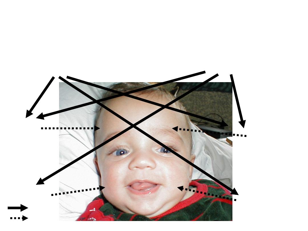
Function CN 7
– Facial Nerve
Facial Symmetry & Expression -
Precise Pattern of Inervation
L UMN
R UMN
R LMN -
Forehead
R LMN – Face
L LMN -
Forehead
L LMN -Face
Thick arrow =s UMN
Dashed arrow =s LMN
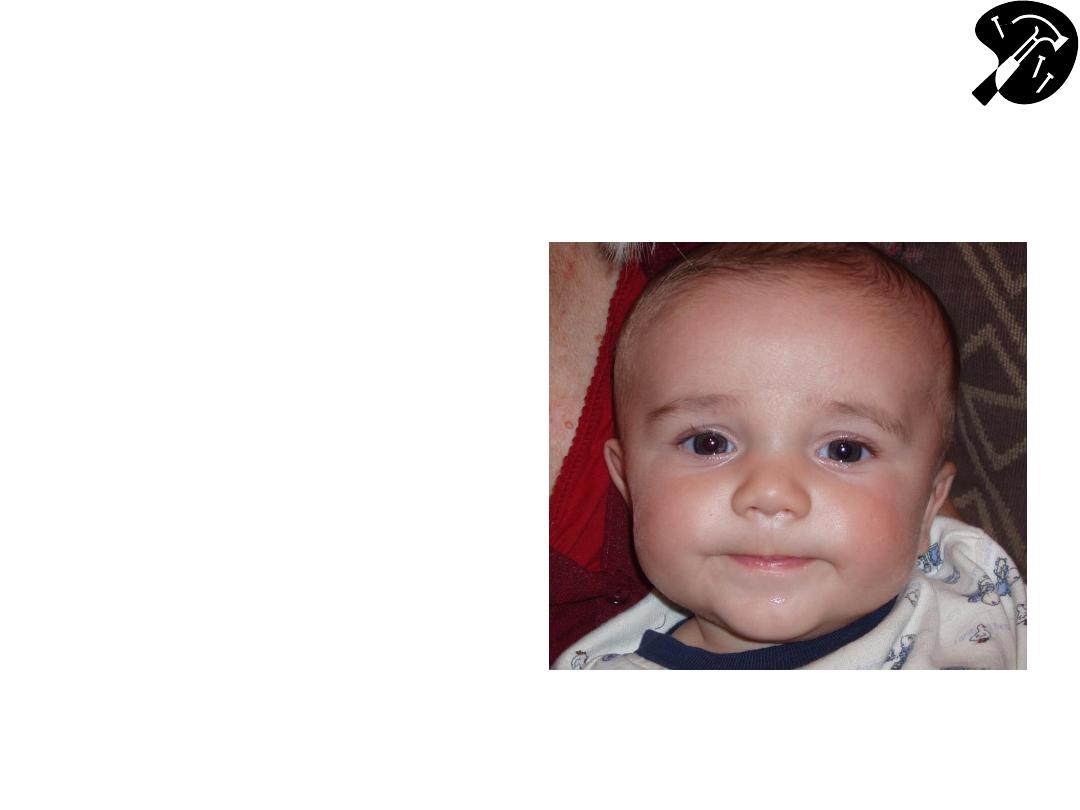
CN 7
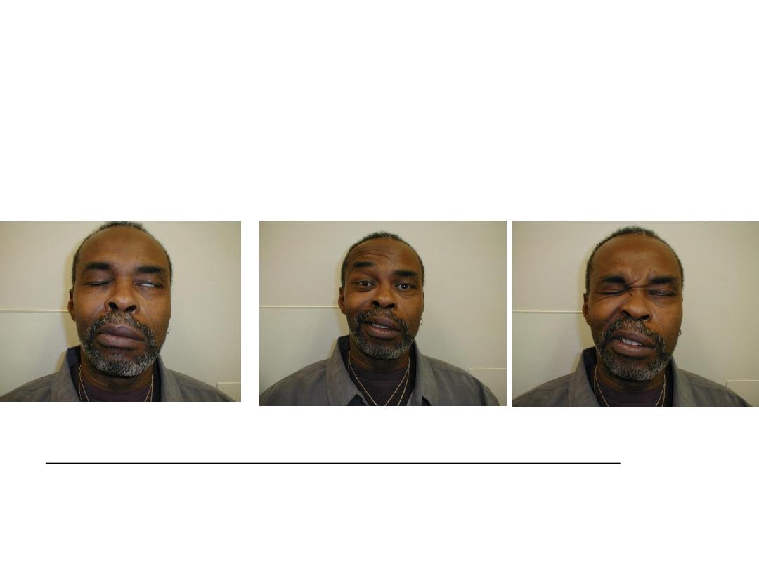
– Exam
• Observe facial
symmetry
• Wrinkle Forehead
• Keep eyes closed
against resistance
• Smile, puff out
cheeks
• Rarely you may need to
check taste to the anterior
2/3 of the tongue
Cute.. and symmetric!
Pathology: Peripheral CN 7 (Bell’s)
Palsy
Central (i.e. UMN) CN 7 dysfunction (e.g. stroke) - not shown: Can
wrinkle forehead bilaterally; will demonstrate loss of lower facial
movement on side opposite stroke.
Patient can’t close L eye, wrinkle L forehead or
raise L corner mouthL CN 7 Peripheral (i.e. LMN)
Dysfunction
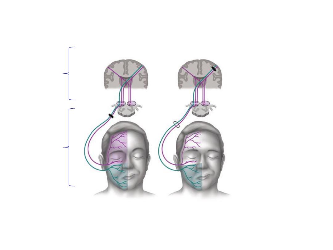
Comparison of a patient with (A)
a facial nerve (Bell’s Type - LMN) lesion
and (B) a supra-nuclear (UMN) lesion w/forehead sparing
Tiemstra J et al. Bell’s Palsy: Diagnosis and Management, Amer J Fam Practice, 2007;76(7):997-1002.
http://www.aafp.org/afp/2007/1001/p997.pdf
Note forehead
and lower face are affected on the
right, which is same side of the LMN lesion
Note forehead sparing on right side,
opposite the UMN lesion
Upper
Motor
Neuron
(UMN)
Lower
Motor
Neuron
(LMN)
A
B
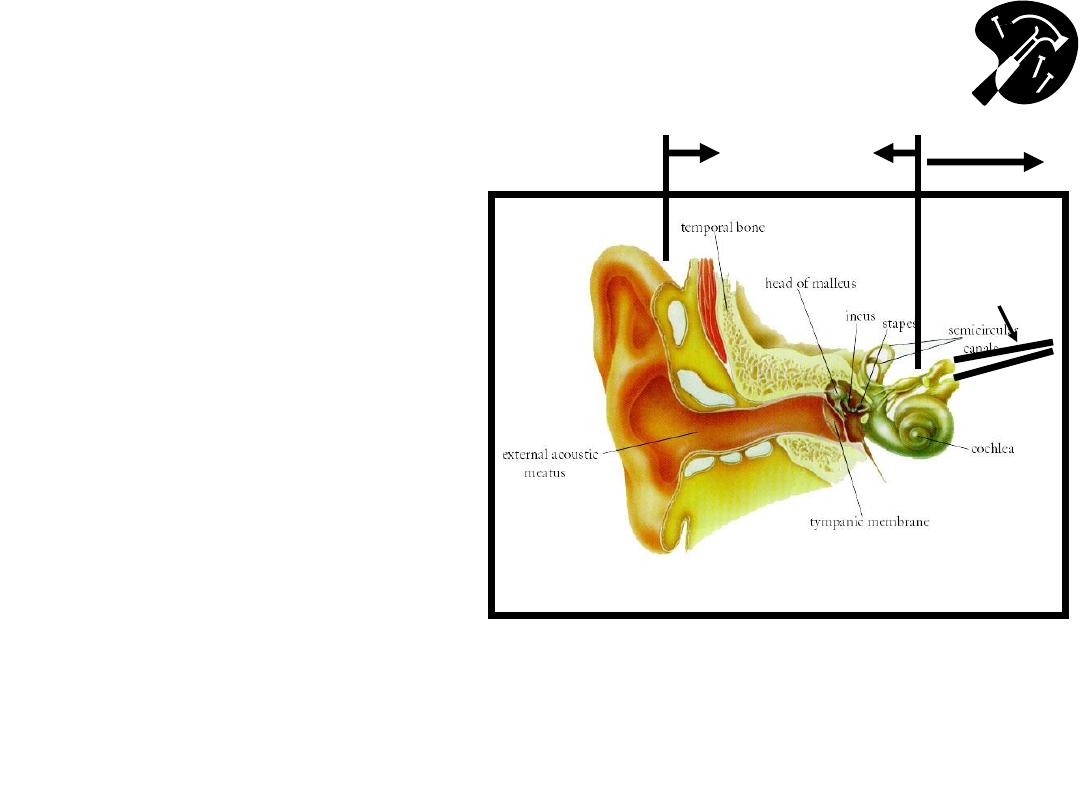
The Ear
– Functional Anatomy & Testing
(CN 8
– Acoustic)
• Crude tests hearing –
rub fingers next to
either ear; whisper &
ask pt repeat words
• If sig hearing loss,
determine Conductive
(external canal up to
but not including CN
8) v Sensorineural
(CN 8)
Image Courtesy: Online Otoscopy Tutorial
http://www.uwcm.ac.uk:9080/otoscopy/index.htm
Vestibular
CN8
Auditory
CN8
Conduction
Sensorineural
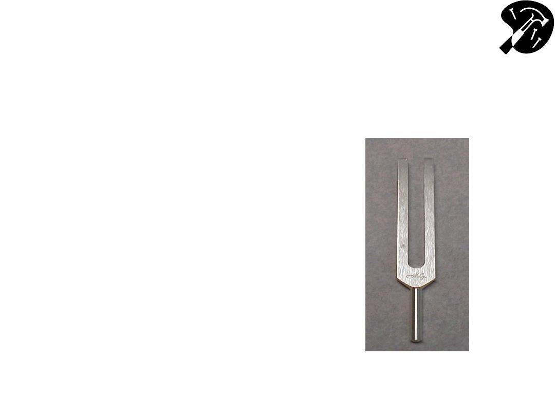
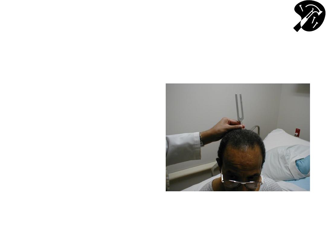
CN 8 - Defining Cause of
Hearing Loss - Weber Test
• 512 Hz tuning fork - this
(& not 128Hz) is well
w/in range normal
hearing & used for
testing
– Get turning fork vibrate
striking ends against heel
of hand or
Squeeze tips between
thumb & 1
st
finger
• Place vibrating fork mid
line skull
• Sound should be heard
=ly R and L bone
conducts to both sides.
CN 8 - Weber Test (cont)
• If conductive hearing
loss (e.g. obstructing
wax in canal on
L)louder on L as
less competing noise.
• If sensorineural on
Llouder on R
• Finger in ear mimics
conductive loss
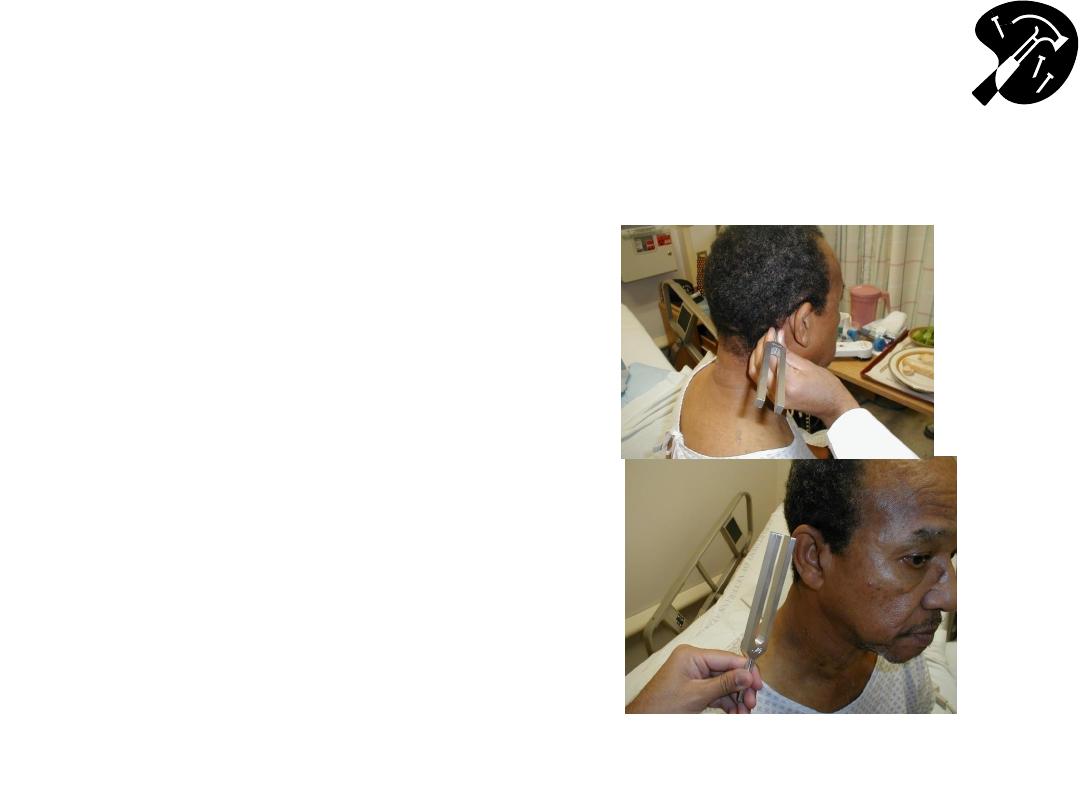
CN 8 - Defining Cause of Hearing
Loss - Rinne Test
• Place vibrating 512 hz
tuning fork on mastoid
bone (behind ear).
• Patient states when can’t
hear sound.
• Place tines of fork next to
ear should hear it again
– as air conducts better
then bone.
• If BC better then AC,
suggests conductive
hearing loss.
• If sensorineural loss,
then AC still > BC
Note: Weber & Rinne difficult to perform in Anatomy lab due to competing
noise
– repeat @ home in quiet room!
CN 8 Vestibular Division
• You will not routinely test; only w/patients who
present w/new onset “dizziness”
• If the patient has vertigo you will need to perform
a Dix-Hallpike maneuver
• You can seen an example of it here:
http://www.neuroexam.com/neuroexam/content.
php?p=23
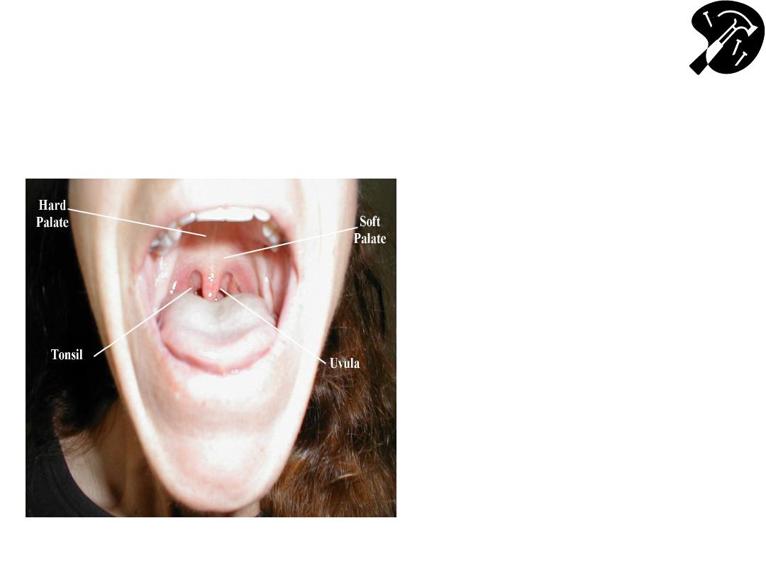
Oropharynx: Anatomy & Function CNs 9
(Glossopharyngeal), 10 (Vagus)
• CN 9 &10 are tested together
• Check to see uvula is midline
• Stick out tongue, say “Ahh” –
use tongue depressor if can
’t
see
– Nl response: palate/uvula rise
– We assume 9 is intact if the palate
rises symmetrically thus we test 9
and 10 indirectly here
• Gag Reflex – provoked with
tongue blade or q tip - CN 9
(afferent limb), 10 (efferent
limb)
– test this bilaterally
– This directly tests 9 and 10

Hypoglossal CN 12
• Tongue midline when patient sticks it outCN 12
– check strength by directing patient push tip into inside of either
cheek while you push from outside
– Observe for atrophy or fasciculations
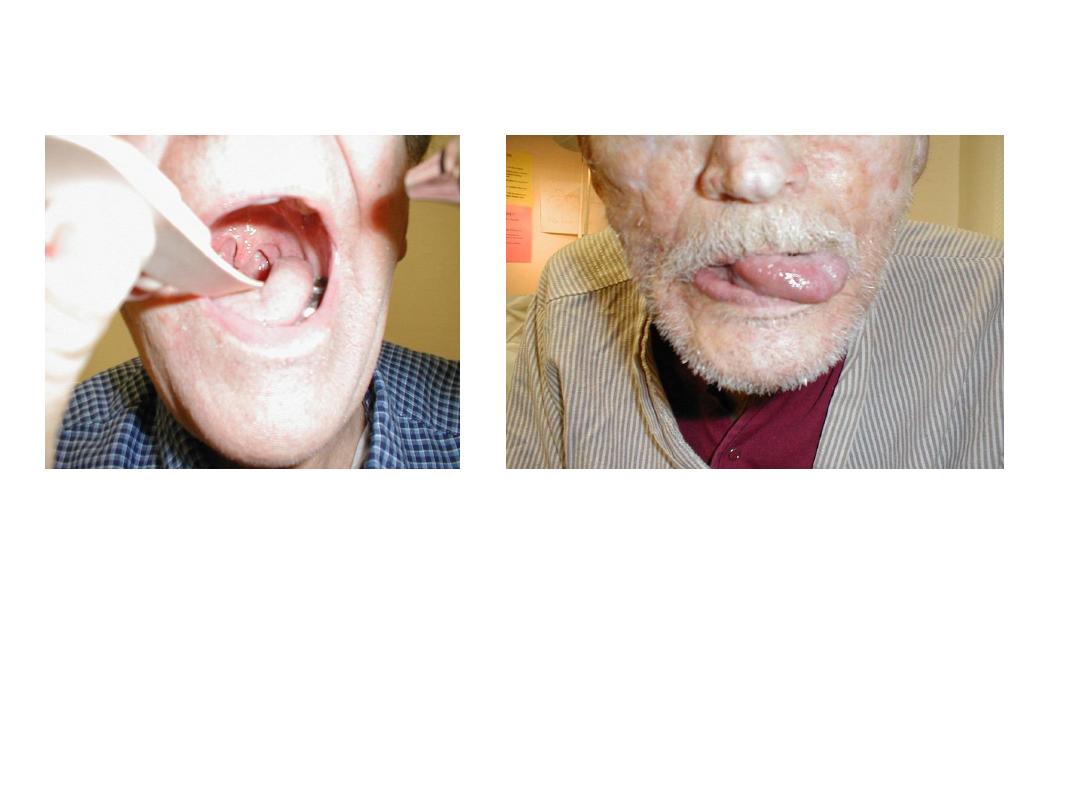
CN 9 & 12 Pathology
L CN 9 palsy: uvula
pulled to R
L CN 12 palsy: tongue
deviates L
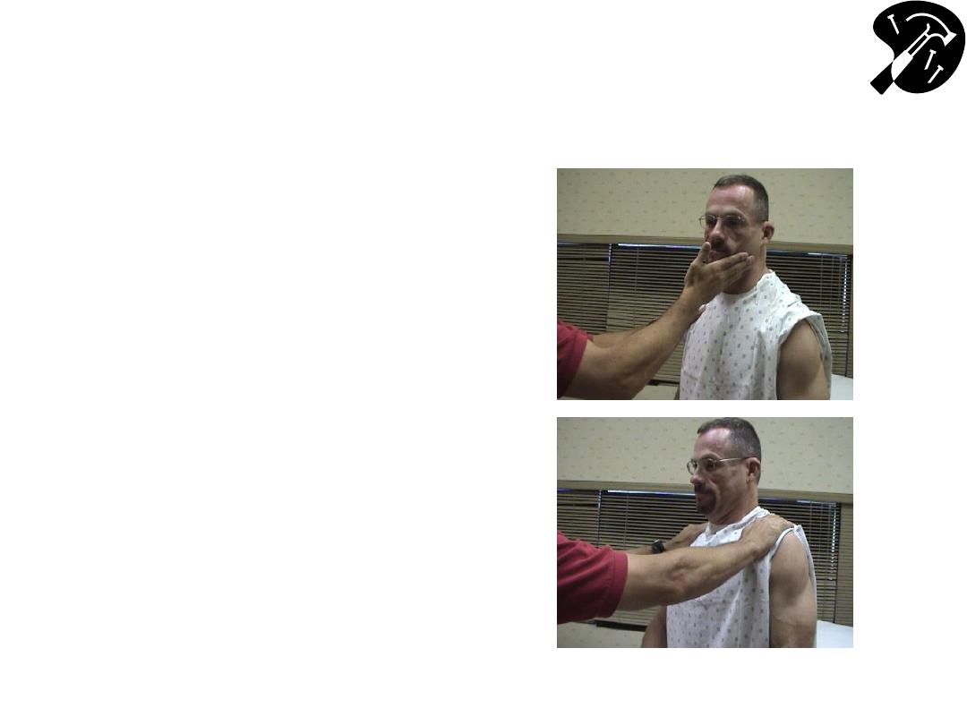
Neck Movement
(CN 11
– Spinal Accessory)
• Turn head to L into R
hand function of R
Sternocleidomastoid
(SCM)
• Turn head to R into L
hand (L SCM)
• Shrug shoulders into
your hands
Summary of Skills
□ Wash Hands
□
CN1 (Olfactory) Smell
□
CN2 (Optic) Visual acuity; Visual fields
□
CNs 2&3 (Optic, Occulomotor) Pupilary Response to light
□
CNs 3, 4 & 6 (Occulomotor, Trochlear, Abduscens) Extra-Occular
Movements
□
CN 5 (Trigeminal) Facial sensation; Muscles Mastication (clench jaw, chew);
Corneal reflex (w/CN 7)
□
CN 7 (Facial) Facial expression
□
CN 8 (Auditory) Hearing
□
CN 9, 10 (Glosopharyngeal, Vagus
) Raise palate (“ahh”), gag
□
CN 12 (Hypoglossal) Tongue
□
CN 11 (Spinal Accessory) Turn head against resistance, shrug shoulders
Time Target: < 15 minutes