The Trigeminal Nerve
ObjectivesList the trigeminal nerve nuclei and their location
Follow up the course of trigeminal nerve from its point of central connections to exit and down to its target areas.
Describe the sensory and motor components of the trigeminal nerve.
Trigeminal Nerve (Cranial Nerve V):
It is the largest cranial nerve and contains both sensory and motor fibers.It is sensory to greater part of head and motor to several muscles including muscles of mastication.
Course of the Trigeminal Nerve
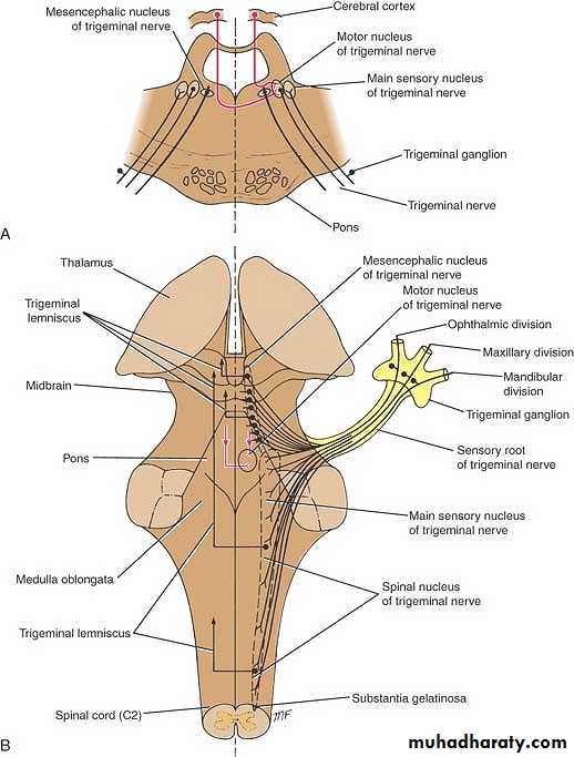
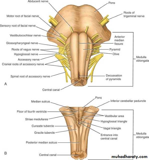
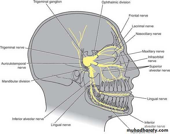
Trigeminal nerve leaves anterior aspect of pons as a small motor root and a large sensory root.It passes forward out of posterior cranial fossa and rests on apex of petrous bone in middle cranial fossa here sensory root expands to form trigeminal ganglion.
Ophthalmic, maxillary, and mandibular nerves arise from the anterior border of ganglion.
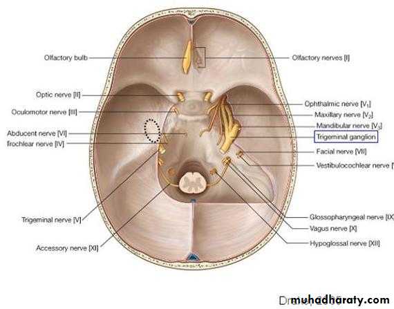
Ophthalmic nerve (V1) contains only sensory fibers leaves skull through superior orbital fissure to orbital cavity.
Maxillary nerve (V2) also contains only sensory fibers leaves the skull through foramen rotundum.
Mandibular nerve (V3) contains both sensory and motor fibers and leaves skull through foramen ovale.
The sensory fibers to skin of face from each division supply a distinct zone with little or no overlap of dermatomes .
Sensory Components of the Trigeminal Nerve
Pain, temperature, touch, and pressure from skin of face and mucous membranes travel along axons whose cell bodies are situated in the trigeminal ganglion.The central processes of these cells form sensory root of trigeminal nerve.
About half the fibers divide into ascending and descending branches when they enter the pons.
The remainder ascend or descend without division.