Ophthalmology
For
5
th
stage
http://goo.gl/rjRf4F
I
LOKA
©
http://www.muhadharaty.com/Ophthalmology
I
Content
Topics:
Page:
Ophthalmologic History
3
Physical examination
8
Ophthalmological instruments
18
Ophthalmological investigations
23
Notes
26
From Mosul medical college
29
Part1
: Ophthalmologic History
Personal data:
Name.
Age (congenital diseases, senile diseases).
Sex (sex related diseases).
Address (endemic diseases, social status).
Occupation (occupation related diseases, visual needs).
Date of admission.
Date of examination.
Chief compliant:
In patient words.
Don’t forget duration of chief compliant.
Don’t use diagnostic or medical words.
Return and correct unrelated compliant.
If more than one, arrange them by chronological order and by importance.
Types of compliant:
1. Visual compliant
sudden loss of vision, diminution of vision, blurring, photophobia,
others.
2. Non-visual compliant
discharge, itching, swelling, pain, others.
History of present illness:
Describe each compliant in specific words.
Analyze each compliant as regards:
o Onset.
o Course.
o Side.
o Duration.
o Associated symptoms.
o Previous medical advice and
medications.
Most common symptoms are loss
of vision, red eye.
Past ocular history:
Poor vision in one eye since birth.
Recurrence of previous disease, particularly inflammatory.
History of lazy eye/amblyopia.
Recurrent ocular problems, particularly inflammatory (iritis) and herpes simplex
keratitis.
Problems associated with contact lens wear (e.g. bacterial keratitis).
Previous history of trauma to the eye (associated with cataract, glaucoma, retinal
detachment).
Past medical history:
Chronological order of the major illness and major
operations.
Ask if similar or related problems have existed before.
Diabetes mellitus which may cause retinopathy.
Hypertension which may be associated with some
vascular eye diseases such as central retinal vein
occlusion.
Systemic illnesses such as sarcoid which may also
cause ocular inflammation.
Previous eye surgery, laser treatment.
History of previous glasses.
Recent cataract surgery (to look for complications of
surgery such as endophthalmitis, wound infection,
intraocular lens displacement causing a sudden drop
in visual acuity)
Past or recent refractive/corrective eye surgery
Drug history:
Some drugs such as isoniazid and chloroquine may be
toxic to the eye.
Presence of allergy.

Family history:
Ask if any other member of the family have similar condition or other eye related
problem.
Example of ocular diseases known to be inherited, such as retinitis pigmentosa, or of
disease where family history may be a risk factor, such as glaucoma.
Family history of corneal diseases (keratoconus corneal dystrophies), cataract, squint,
retinal diseases.
Social history:
Apart from asking the usual questions such as about occupation, smoking, alcohol and
the patient's everyday circumstances.
A sexual history can help in diagnosing Reiter's syndrome, where the patient can have
ocular problems such as uveitis or conjunctivitis.
Here are some of the possible questions that you might want to ask:
Is/are the eyelid(s) affected? (e.g. blepharitis, entropion, ectropion, trichiasis)
Is there any periorbital swelling? (may suggest orbital cellulitis if other features are
present like pain, reduced eye movements and systemic upset/pyrexia. Preseptal
cellulitis presents with perorbital swelling but eye movements are not impaired.)
Is it worse with eye movements? (scleritis)
Is the eye dry or gritty? (e.g. keratoconjunctivitis, blepharitis)
Is the eye 'sticky'? (e.g. bacterial conjunctivitis)
Is there an exudate? (ask about presence, amount, color)
Is the eye watering? (keratitis, iritis)
Is there any photophobia? (iritis, keratitis, glaucoma)
Is there a glare in sunlight or difficulty driving at night due to the glare from
headlights? (cataracts)
Is the vision impaired? (multiple causes including glaucoma, cataract, uveitis, etc)
Are there any floaters/flashes/haloes? (symptoms of retinal disease)
Is there a headache with it? (pituitary tumors causing bitemporal hemianopia)
Is there any urethral discharge? (Reiter's syndrome)

Some examples of chief compliant:
Burning:
o Blepharitis, dry eye syndrome, conjunctivitis corneal defect, episcleritis.
o Ocular toxicity (medication, makeup, contact lens solutions).
o Contact lens-related problems.
Crossed eyes in children:
o Esodeviations in children (eyes turned in).
o Exodeviations in children (eyes turned out).
Decreasd vision.
Discharge.
Double vision (diplopia):
o Monocular (diplopia remains when the uninvolved eye is occluded) occur in
corneal opacity or irregularity, iris defects, cataract dislocated natural lens or lens
implant, macular disease, retinal detachment, CNS causes (rare), nonphysiologic.
o Binocular (diplopia eliminated when either eye is occluded) occur in
neuromuscular imbalance, myasthenia gravis, isolated sixth-third-fourth nerve
palsy, orbital disease, internuclear ophthalmoplegia, vertebrobasilar artery
insufficiency, other CNS lesions.
Eyelid crusting:
o More Common Blepharitis, meibomitis, conjunctivitis.
o Less Common Canaliculitis, nasolacrimal duct obstruction, dacryocystitis.
Eyelids drooping (ptosis).
Eyelid twitch:
o Orbicularis myokymia (related to fatigue, excess caffeine, medication, or stress).
o Cornealor conjunctival irritation (especially from an eyelash, cyst, or conjunctival
foreign body).
o Dry eye.
o Blepharospasm (bilateral), hemifacial spasm.
Eyes bulging (proptosis):
o Inflammatory Thyroid eye disease (TED), idiopathic orbital inflammatory
syndrome (IOIS), sarcoidosis, Wegener granulomatosis.
o Infectious Orbital cellulitis, subperiosteal abscess.
o Neoplastic (benign and malignant) Dermoid cyst, capillary hemangioma,
rhabdomyosarcoma, metastasis, lymphangioma, optic nerve glioma, neurofibroma,
leukemia, lymphoproliferation (including lymphoma), neurilemmoma, mucocele.
o Trauma Orbital fracture, retrobulbar hemorrhage, orbital foreign body,
carotidcavernous fistula.
o Malformation Congenital, vascular.
Flashes of light:
o More Common Retinal break or detachment, posterior vitreous detachment,
migraine, rapid eye movements (particularly in darkness), oculodigital stimulation.
o Less Common CNS (particularly occipital lobe) disorders, vestibulobasilar artery
insufficiency, optic neuropathies, retinitis, entoptic phenomena, hallucinations.

Floaters.
Itchy eyes:
o Conjunctivitis (allergic, vernal, viral), blepharitis, dry-eye syndrome, topical, drug
allergy.
Light sensitivity (photophobia):
o 1. Abnormal eye examination:
More Common Corneal abnormality (abrasion or edema), anterior uveitis.
Less Common Conjunctivitis (mild photophobia), posterior uveitis, scleritis,
albinism total color blindness, aniridia, mydriasis of any etiology (pharmacologic,
traumatic), congenital glaucoma.
o 2. Normal eye examination: Migraine, meningitis, retrobulbar optic neuritis,
subarachnoid hemorrhage, trigeminal neuralgia, or a lightly pigmented eye.
Night blindness:
o More Common Refractive error (undercorrected myopia), advanced glaucoma
or optic atrophy, small pupil (from miotic drops), retinitis pigmentosa, congenital
stationary night blindness, status-post panretinal photocoagulation drugs
(phenothiazines, chloroquine, quinine).
o Less Common Vitamin A deficiency, gyrate atrophy, choroideremia.
Pain:
o Ocular dry-eye syndrome, blepharitis, infectious conjunctivitis, episcleritis,
scleritis inflamed pinguecula or pterygium, foreign body, corneal disorder, eye
strain from uncorrected refractive error, angle closure glaucoma.
o Periorbital inflammation (hordeolum, preseptal cellulitis, dacryocystitis,
dermatitis), contact, chemical, varicella zoster, or herpes simplex, referred pain
(dental, sinus).
o Orbital Sinusitis, orbital cellulitis, idiopathic orbital inflammatory syndrome,
orbital tumor or mass, optic neuritis, acute dacryoadenitis, migraine or cluster
headache, diabetic cranial nerve palsy.
o Asthenopia Uncorrected refractive error, phoria or tropia, convergence
insufficiency.
Red eye.
Spots in front of the eyes:
o Transient Migraine.
o Permanent or long-standing Posterior vitreous detachment, intermediate or
posterior uveitis, vitreous hemorrhage, vitreous condensations/debris.
Tearing:
o Adults Emotional states, corneal abnormality (abrasion, foreign body or rust
ring, recurrent erosion, edema), anterior uveitis, nasolacrimal duct obstruction,
punctal occlusion, lacrimal sac mass, pump failure (congenital or seventh nerve
palsy).
o Children ophthalmia neonatorum, nasolacrimal duct obstruction, congenital
glaucoma.

Part2
: Physical examination
1- Inspection:
Head position Congenital and longstanding paralytic squint often causes an
abnormal head posture with the head turned or tilted to minimize the diplopia.
Position of eyelids when looking straight ahead and on eye movement.
o A narrow palpebral fissure suggests ptosis or blepharospasm (tonic spasm in the
orbicularis oris muscle).
o Eyelid retraction or proptosis (forward bulging of the eyeball) makes the sclera
visible above the cornea.
o Ectropion makes the sclera visible below the cornea.
Lid lag:
o Examine the seated patient from the right.
o Hold your finger from a point 45° above the horizontal to a point below this plane.
o Watch how the upper eyelid moves with the downward movement of the eye.
o Normally, there is perfect coordination as the upper lid follows the downward
movement of the eye.
o In lid lag, as occurs in thyroid eye disease, sclera can be seen above the iris.
Periorbital appearance.
Lacrimal apparatus Causes of swelling of lacrimal gland: Inflammation (sarcoidosis),
Infection (mumps), Malignancy, lymphoma, cancer.
Eyelid margin Blepharitis (inflammation of the eyelid margin) may be associated
with systemic skin involvement.
Conjunctiva:
o Look for redness or chemosis (edema) of the white of the eye.
o Evert the eyelid to examine the upper subtarsal conjunctiva.
o Ask the patient to look down, hold the upper lid lashes, press gently on the upper
border of the tarsal plate with a cotton bud and gently pull the eyelashes up.
o Look for the giant papillae of allergic eye disease or a hidden foreign body.
o Redness around the corneal limbus uveitis.
o Diffuse redness scleritis or conjunctivitis.
o Look for injected (inflamed) conjunctiva, discharge, conjunctival hemorrhage.
Sclera Scleritis causes a dark-red color, tenderness and pain on eye movement.
Cornea:
o Is it clear? is there a bright reflection of light from the overlying tear film?
o To test for corneal ulceration, gently touch a fluorescein strip on to the conjunctiva,
where it will leave a yellow mark.
o Ask the patient to blink to distribute the dye on the cornea. The yellow dye reveals
epithelial defects which may be obvious to the naked eye.
o Use your ophthalmoscope with a +10 lens to visualize smaller defects. A light with a
cobalt blue filter highlights any defects.
o Resting appearance of the pupils.
o Signs in the cornea corneal arcus, Kayser–Fleischer rings, band keratopathy
(calcium deposition).
Anterior chamber is it intact (if penetrating injury is suspected), is a hypopyon
present?
Iris and pupil is the shape of the pupil normal?
Lens is there an opacity in the red reflex observed with the ophthalmoscope?
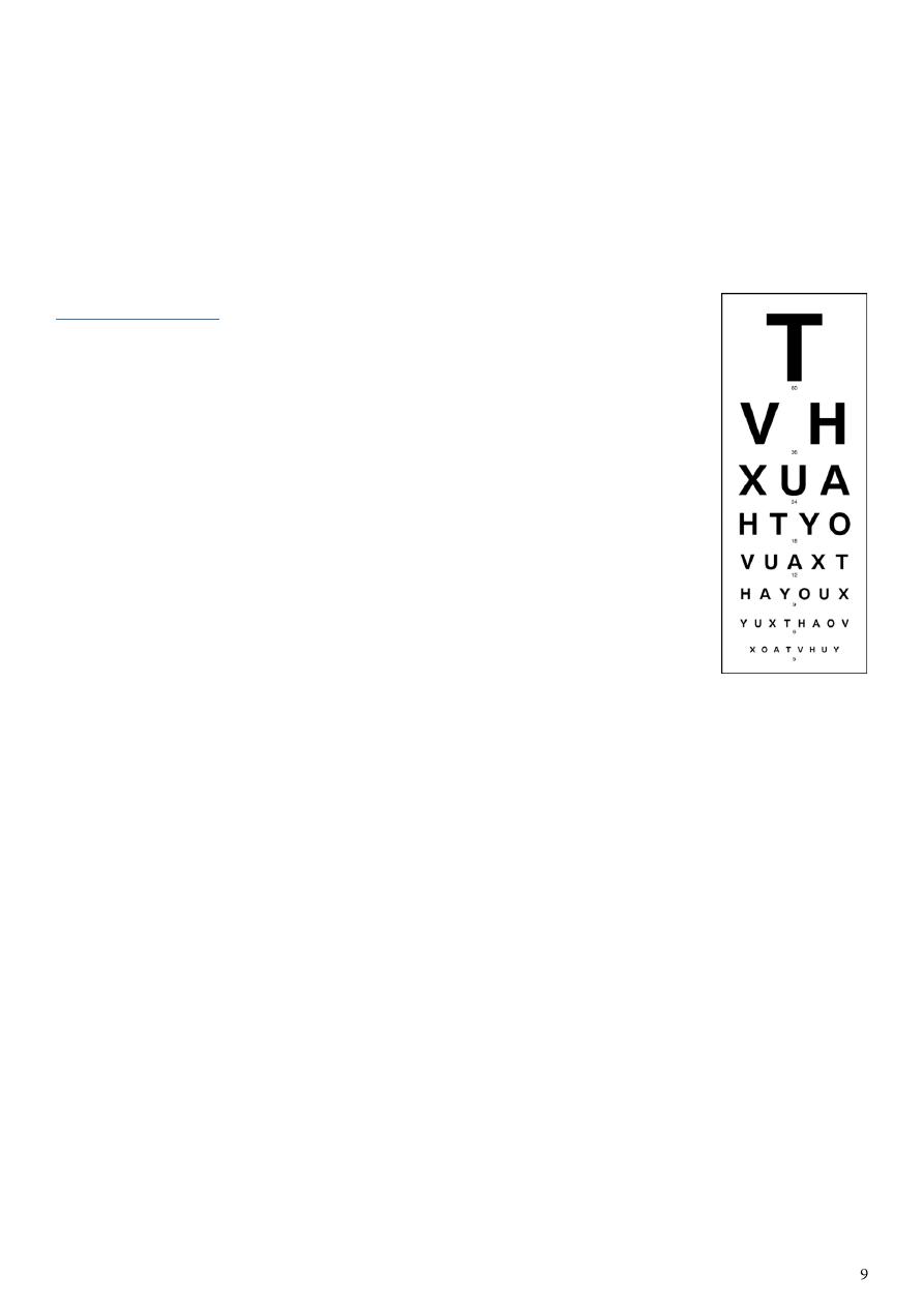
2- Visual acuity:
Visual acuity (VA) tests the resolving power of the eye.
The standard test is the Snellen chart, consisting of rows of letters of
decreasing size.
Procedure:
Ask patients to put on their distance glasses, if they use them.
Only use reading glasses when testing near vision.
Ensure good ambient lighting.
Place a Snellen chart 6 metres from the patient.
Cover one of the patient’s eyes with a card and ask him to read from
the top down until he can no longer distinguish the letters.
Repeat with the other eye.
Notes:
Snellen visual acuity is expressed as 6 (the distance at which the chart is read) over the
number corresponding to the lowest line read. This indicates the distance at which
someone with normal vision should be able to read that line, i.e. Snellen visual acuity
of 6/60 indicates that at 6 metres patients can only see letters they should be able to
read 60 metres away.
Normal vision is 6/6.
If the patient cannot read down to line 6 (6/6 vision), place a pinhole directly in front of
his glasses to correct refractive errors. This allows only central rays of light to enter the
eye and can correct for about 4 D of refractive error.
If patients cannot see the top line of the chart at 6 metres, bring them forward till they
can and record that vision, e.g. 1/60 – can see top letter at 1 metre.
If patients still cannot see the top letter at 1 metre, check whether they can count
fingers, see hand movements or just see light.
For children who can’t yet read, use different-sized objects instead of letters.
Repeat the process above for near vision, with the patient wearing any reading glasses.
Use a test card, held at a comfortable reading distance, to assess near vision. N5 is the
smallest size that most normal eyes can see (N8 is the size of normal newsprint).
In children:
Very young children are observed to see if they can follow objects or pick up ‘hundreds
and thousands’ cake decorations.
Infants up to 2 years: Cardiff cards
Toddlers 2-3 years: Kay's pictures
Young children 4-5 years: Sheridan-Gardiner test
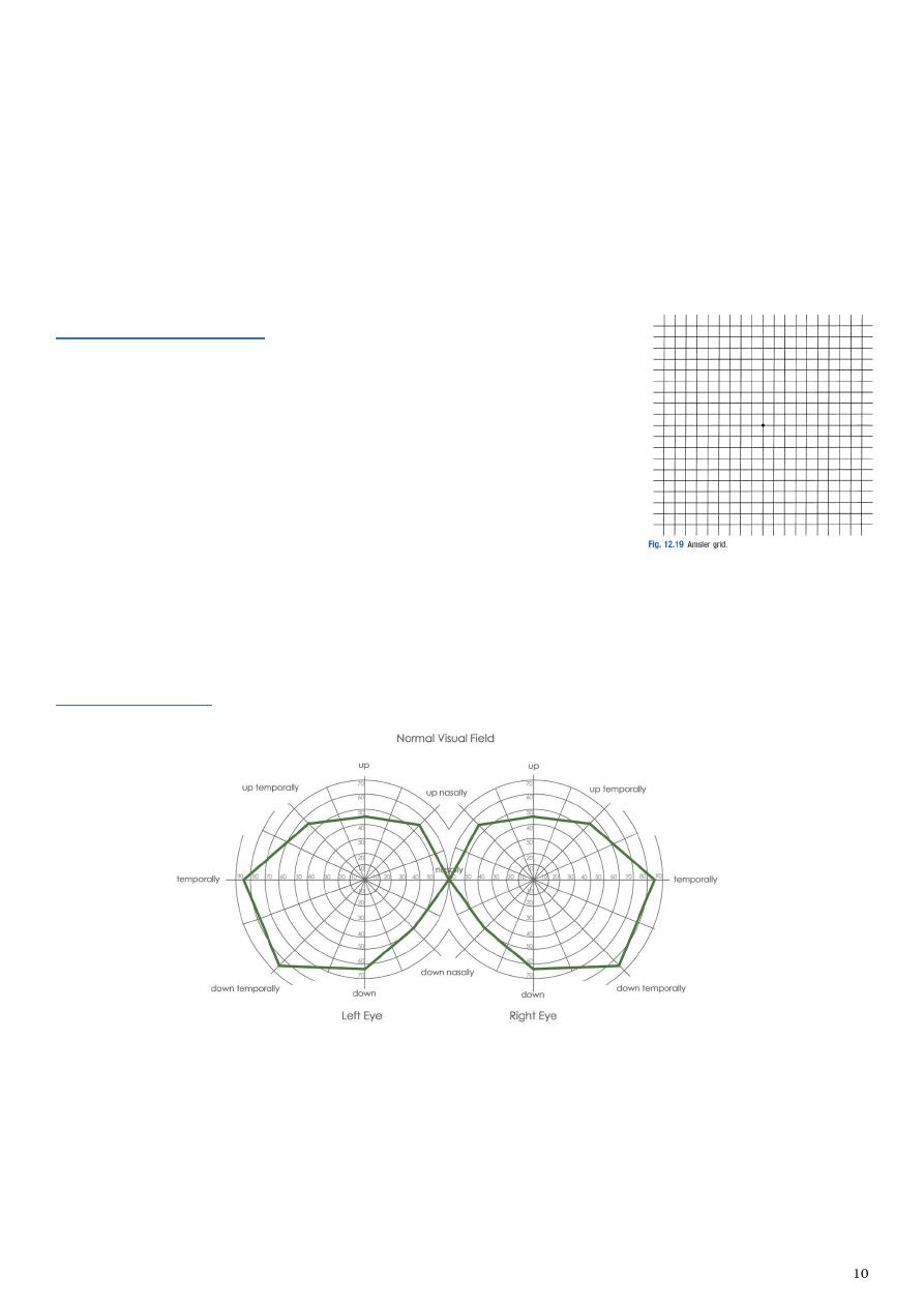
3- Macular function:
Use an Amsler grid to record visual defects, including central
scotomas, quadrantanopias and hemianopias, and distortion
of the central 10° of vision.
Ask the patient:
to cover one eye
to hold the grid at a comfortable reading distance
to fix on the central black spot with the eye being tested
to keep the eye still and look at the grid using the ‘sides of his vision’
to outline with a finger the areas where the lines are broken, distorted or missing.
4- Visual fields:
Information:
The normal visual field extends 160° horizontally and 130° vertically.
Fixation is the very center of a patient’s visual field.
The physiological blind spot is located 15° temporal to the point of visual fixation and
represents the optic nerve head.

The field is not flat; towards the center the eye is able to detect much smaller objects
than at the periphery. This produces a ‘hill of vision’ in which objects which are
resolved in finest detail are at the peak of the hill (at the fovea).
Confrontation tests:
Settings:
o Introduce yourself to the patient.
o Wash your hands.
o Sit directly facing the patient, about 1 meter away.
o Ask the patient to keep looking at your eyes.
o Remove the glasses from the patient as they could create artificial visual field
defect.
Homonymous defects:
o Keep your eyes open and ask the patient to do the same.
o Hold your hands out to their full extent. Wiggle a fingertip and ask the patient to
point to it as soon as he sees it move.
o Do this at 10 and 2 o’clock, and then 8 and 4 o’clock (to screen the four outer
quadrants of the patient’s visual field – superotemporal, superonasal,
inferotemporal, inferonasal).
Sensory inattention:
o Test both eyes together.
o Both you and the patient should keep your eyes open.
o Test both left and right fields at the same time.
o Note whether the patient reports seeing only one side move and which quadrant or
side is affected.
Peripheral visual fields:
o Test each eye separately.
o Ask the patient to cover one eye and look directly into your opposite eye.
o Shut your eye that is opposite the patient’s covered eye.
o Test each quadrant separately with a wiggling finger or white-tipped hatpin. Hold
the target equidistant between you and the patient.
o Start peripherally and move the target along the diagonal towards the centre of
vision until the patient detects it.
o Repeat for the other quadrants.
o Compare your visual field with the patient’s.
Central visual field:
o Test each eye separately using a red hatpin.
o Shut your eye that is opposite the patient’s covered eye.
o Ask the patient to cover one eye and look directly at your open eye.
o Hold the hatpin in the centre of the visual field, as close to fixation as possible.
o Ask the patient what colour the hatpin is. A ‘pale’ or ‘pink’ response implies colour
desaturation, usually because of a lesion affecting the optic nerve.
o Compare the four quadrants of the visual field centrally; each time ask about colour
desaturation. Note that the visual field for red may be smaller than for white.
See the video instead of this paragraph
www.muhadharaty.com/lecture/3520/

Blind spot:
o This is a physiological scotoma corresponding to absence of photoreceptors where
the optic nerve leaves the eye.
o Test one eye at a time
o Ask the patient to cover one eye and look directly at you.
o Shut your eye that is opposite the patient’s covered eye.
o Use red color hatpin because the central part of the visual field is more sensitive to
red light.
o Hold the hatpin at the fixation point; you and the patient focus on each other’s eye.
o Move the hatpin temporally and horizontally until it disappears from your visual
field. Maintaining the same temporal horizontal position, move it anteriorly or
posteriorly until it also disappears from the patient’s visual field.
o Compare the size of the patient’s blind spot to yours.
Tubular visual fields:
o These are often asymptomatic.
o Test visual fields by confrontation at 1 meter and 2 meters from the patient.
Findings:
Scotoma is a focal area of decreased sensitivity within the visual field, surrounded by a
more sensitive area.
Intraocular causes of visual field defects include:
o Macular lesions (age-related macular degeneration) cause central scotomas
(cannot see a face clearly but the rest of the visual field is unaffected).
o Peripheral retinal lesions spare central vision causing localised scotomas (retinal
detachment or scarring) or constriction of peripheral field (retinitis pigmentosa).
o Optic disc lesions cause horizontal or arcuate scotomas.
Lesions of the optic nerve within the orbit cause central scotomas (here red colors
appear orange or pink).
Unilateral optic nerve lesions cause a relative afferent pupillary defect.
Distension of the nerve sheath around the optic nerve (papilloedema) causes an
enlarged blind spot.
Lesions at the optic chiasm cause bilateral temporal visual field defects (bitemporal
hemianopia).
Optic tract lesions (due to suprasellar lesions, e.g. pituitary tumours) cause
asymmetrical homonymous visual field defects.
The optic radiations and occipital cortex cause:
o Symmetrical visual field defects (homonymous hemianopia)
o Lack of awareness of visual field loss: this suggests parietal lobe involvement.
o Superior homonymous quadrantanopia: this suggests temporal lobe involvement.
o Visual field defects: these may affect only central vision or spare the macula
because of the dual blood supply of the occipital cortex
o Functional visual loss is common, particularly bilateral visual field constriction that
does not expand on testing further away. This tubular constriction differentiates it
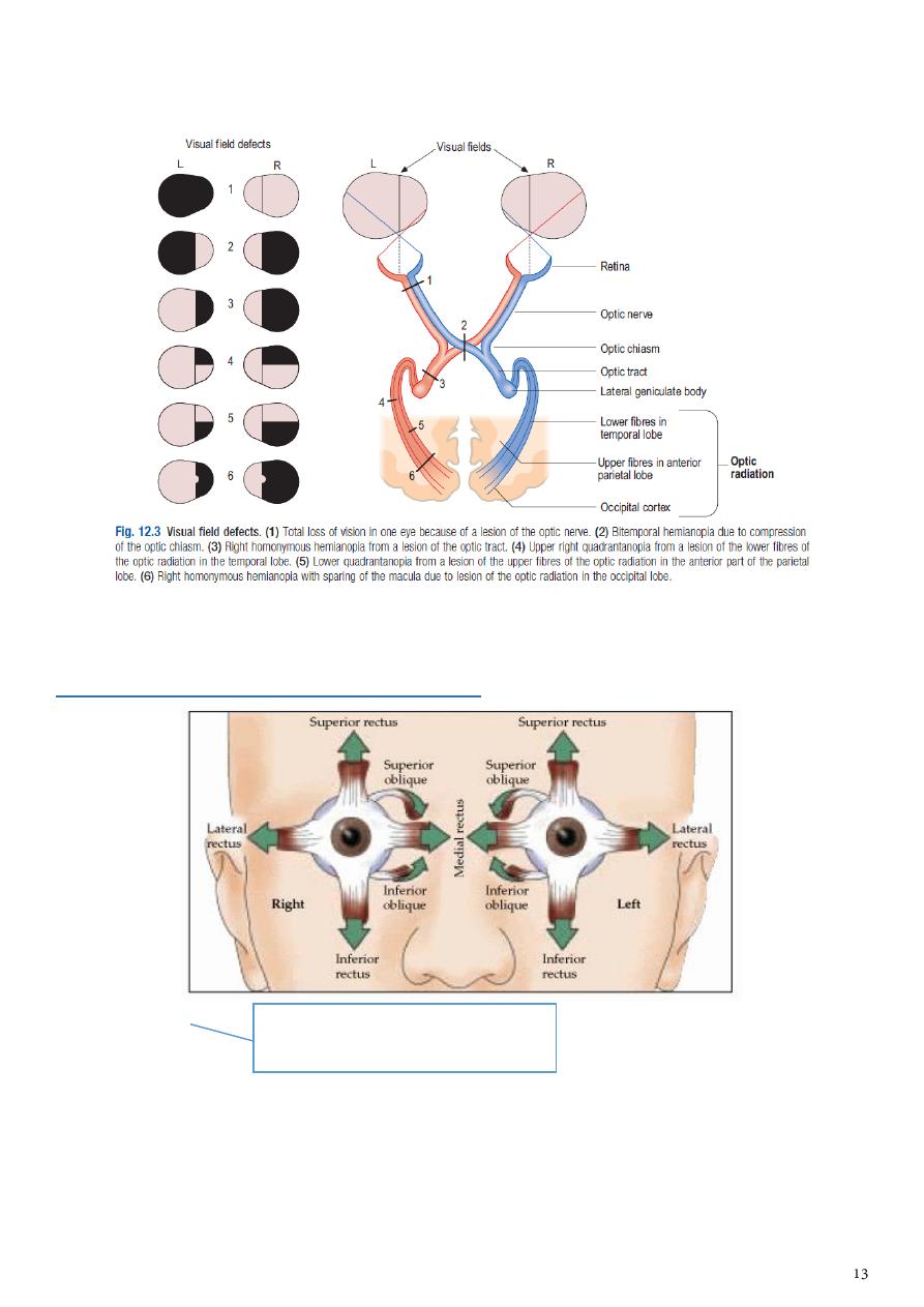
from the funnel constriction of bilateral retinal disorders, such as retinitis
pigmentosa or bilateral homonymous hemianopia (cortical visual impairment).
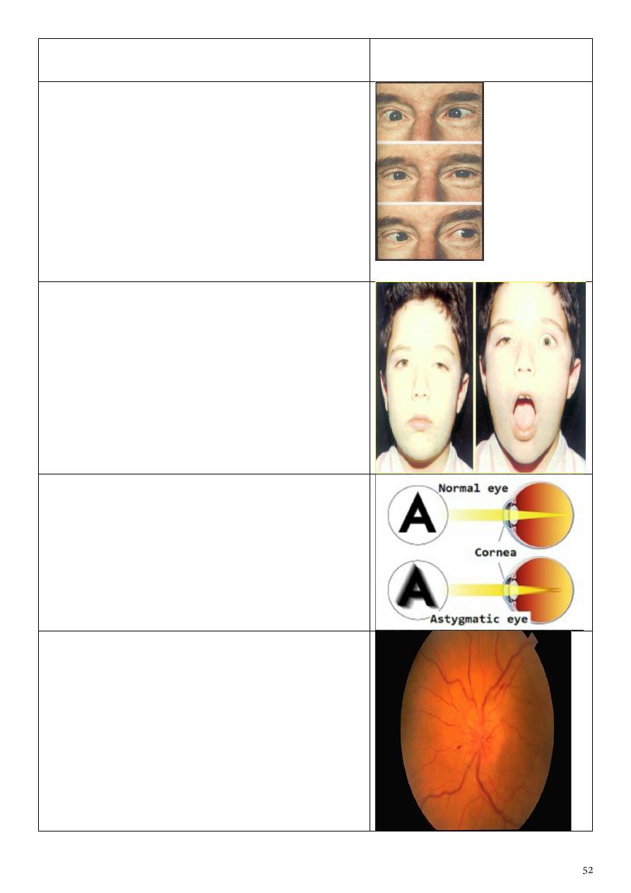
5- Ocular alignment and eye movements:
Examination:
Settings:
o Introduce yourself to the patient.
o Wash your hands.
o Sit directly facing the patient, about 1 meter away.
Inspection
o Look for head turns or tilts in the direction of underacting muscles.
o Hold a pen torch and ask the patient to look at the light.
See the video instead of this paragraph
www.muhadharaty.com/lecture/3528/

o Observe the position of the pen torch’s reflection on the cornea. The fixating (non-
deviating) eye has the pen torch’s reflection in the center; the deviating eye has an
off-center reflection. This test can be confusing if the paretic eye has better vision
than the non-paretic eye and the patient fixates with it. (Hirschberg test detects
any deviation in primary position.)
Ocular movements:
o Hold your finger vertically at least 50 cm away from the patient, and ask him to
follow it with his eyes, without moving his head.
o Move your finger steadily to one side, then up and down, then to the other and
repeat, describing the letter H in the air.
o Ask whether any diplopia is horizontal, vertical, tilted or a mixture of both.
o Cover one of the patient’s eyes to see if diplopia is monocular or binocular.
o If diplopia is binocular, ask which image disappears when you cover each eye; the
outer image corresponds to the affected eye.
o Note the direction of gaze in which diplopia is worst and work out which muscle is
affected.
o On horizontal movement, in the absence of proptosis, no ipsilateral sclera should
be seen on extreme gaze (‘burying the white’). Its presence suggests ipsilateral
muscle weakness.
o On down-gaze hold the eyelids open using two fingers from your free hand.
o For each eye, look for nystagmus while examining eye.
Squint (cover test)
o Examine visual acuity and the visual fields as above.
o Cover one eye and ask the patient to look at the light of your pen torch.
o Closely observe the uncovered eye for any movements.
o If it moves to take up fixation, that eye was squinting.
o Repeat the sequence for the other eye.
Oculocephalic (doll’s-eye) reflex
o With the patient supine, hold his head in both hands, with your thumbs holding his
eyes open; if the patient is conscious, ask him to focus on your eyes.
o Rock the head gently from side to side, noting the movement of the eyes as they
hold their gaze.
o An impaired reflex indicates brainstem abnormality.
Findings:
The eyes are normally parallel in all positions of gaze, except near convergence. If not,
a squint (strabismus) is present.
Squints are associated with:
o Paresis of one or more extraocular muscles (paralytic or incomitant squint).
o Defective binocular vision (non-paralytic or concomitant squint).
Acquired paralytic squints cause diplopia, in which the images are maximally separated
and squint is greatest in the direction of action of the paretic muscle.
Concomitant squints are the same in all positions of gaze.
In children, the visual acuity of the squinting eye falls, causing amblyopia (a ‘lazy’ eye).
Monocular diplopia is created by ‘ghosting’ from structural abnormality anywhere
between the cornea and fovea.
Pure horizontal diplopia usually results from involvement of the VI cranial nerve. The
symptoms are worse looking to the affected side.
Unlike VI nerve palsy, diplopia is worse on looking to the contralateral side.
Thyroid eye disease is a common cause of vertical diplopia.
The oculocephalic test differentiates supranuclear lesions from cranial nerve lesions
and is impaired in brainstem stroke, metabolic dysfunction or drug intoxication.
Causes of impaired horizontal movement include pontine gaze palsies (nuclear VI
nerve palsy), convergence spasm, Impaired vertical gaze (orbital floor fractures,
brainstem stroke, demyelination and children with hydrocephalus).
The ability to direct gaze rapidly from one object to another (saccadic eye movements)
can be tested by asking the patient to look at targets (such as the finger) held at either
side of the head.
Types of squint(-tropia):
-- eye moves outward to refixate exotropia
-- eye moves inward to refixate esotropia
-- eye move up to refixate hypertropia
-- eye move down to refixate hypotropia
6- Nystagmus:
See this video: www.muhadharaty.com/lecture/3529
Examination:
Hold your finger an arm’s length from the patient, in front of him.
Ask the patient to look at your finger and follow it with his eyes without moving the
head.
Move your finger steadily to each side and up and down, describing an H.
Watch the patient’s eyes carefully for nystagmus and note:
o The position in which it occurs
o The direction in which it is most marked
o Whether it is horizontal, vertical, rotatory or multidirectional
o Whether there are fast and slow phases (jerk) or equal oscillations about a central
point (pendular).
Some types of nystagmus:
Biphasic or jerk nystagmus.
Acquired pendular nystagmus cerebellar or brainstem disease.
Congenital nystagmus a horizontal jerk nystagmus, can be pendular.
Peripheral vestibular nystagmus associated with vertigo.
Central vestibular nystagmus vertigo is less prominent.
Vertical nystagmus brainstem damage.
Upbeat nystagmus upper brainstem lesions in multiple sclerosis, infarction,
Wernicke’s encephalopathy.
Downbeat nystagmus lesions around the craniocervical junction.
Periodic alternating nystagmus congenital, lesions at the craniocervical junction,
drug intoxication.
Optico-kinetic nystagmus present in functional blindness.
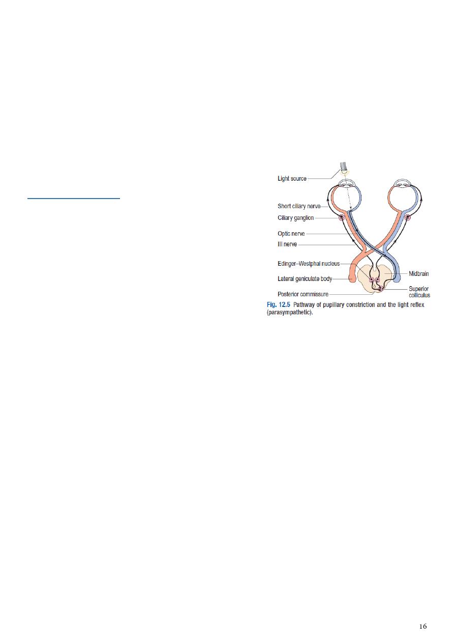
7- Pupillary reflex:
See:
www.muhadharaty.com/lecture/3530
www.muhadharaty.com/lecture/3531
Examination:
Assess the pupils’ shape and symmetry, taking
account of ambient lighting.
Ask the patient to fix his eyes on a distant point straight ahead.
Bring a bright torchlight from the side to shine on the pupil.
Look for constriction of that pupil (direct light reflex).
Repeat and look for constriction of the opposite pupil (consensual light reflex).
With his vision still fixed on a distant point, present an object about 15 cm in front of
the eyes and ask the patient to focus on it (convergence). Look for pupil constriction
(accommodation reflex)
Findings:
Simple, or essential, anisocoria, a >0.4 mm difference between the pupil diameters, is
common.
A pathologically small pupil (after damage to the sympathetic nervous system) will be
more apparent in dim illumination, since dilation of the normal pupil will be greater.
A pathologically large pupil (seen in disease of the parasympathetic nervous system)
will be more apparent in the light.
Small pupils that respond poorly to pharmacological dilatation diabetes mellitus,
autonomic neuropathy.
Pinpoint, irregular pupils that constrict only on convergence (Argyll Robertson pupil)
syphilis.
Adie pupil is usually mid-dilated and responds poorly to convergence
malfunction within the orbit.
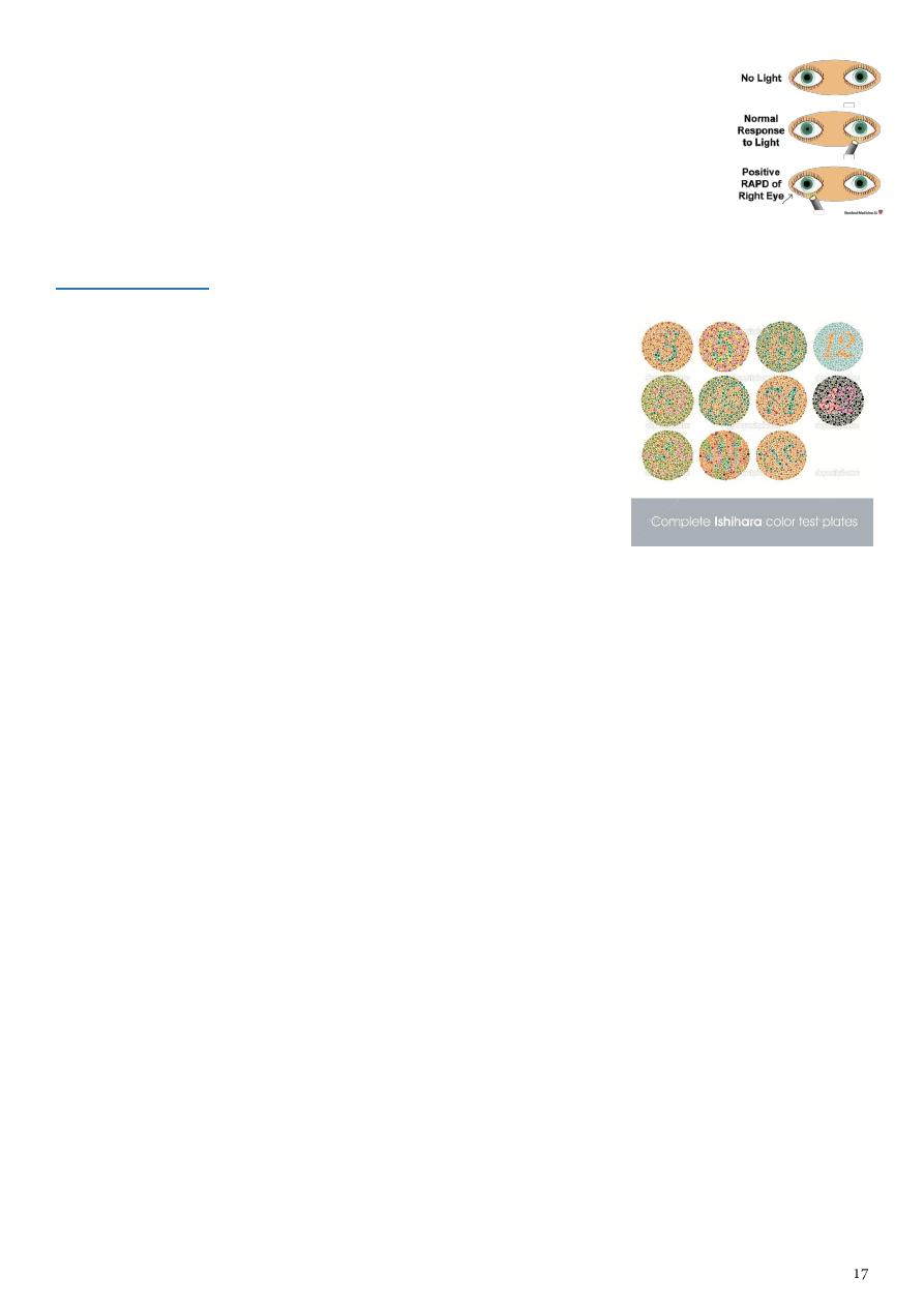
Marcus Gunn pupil Optic nerve damage.
A unilateral defect in optic nerve conduction is demonstrated as a
relative afferent pupil defect (RAPD).
8- Color vision:
See this video: www.muhadharaty.com/lecture/3535/
Examination:
Assess red–green color vision using Ishihara test plates.
These are colored spots forming numbers which the
patient reads out.
The first plate is a test plate; if the patient cannot see the
number, he has poor visual acuity or functional visual loss.
Findings:
Red desaturation is impaired ability to identify red objects and an early indicator of
optic nerve pathology.
Congenital red–green blindness is an X-linked recessive condition and affects 7% of the
male population.
Red–green color vision is impaired before loss of visual acuity with optic nerve damage
anywhere from the photoreceptors to the lateral geniculate nucleus of the thalamus.
Part3
: Ophthalmological instruments
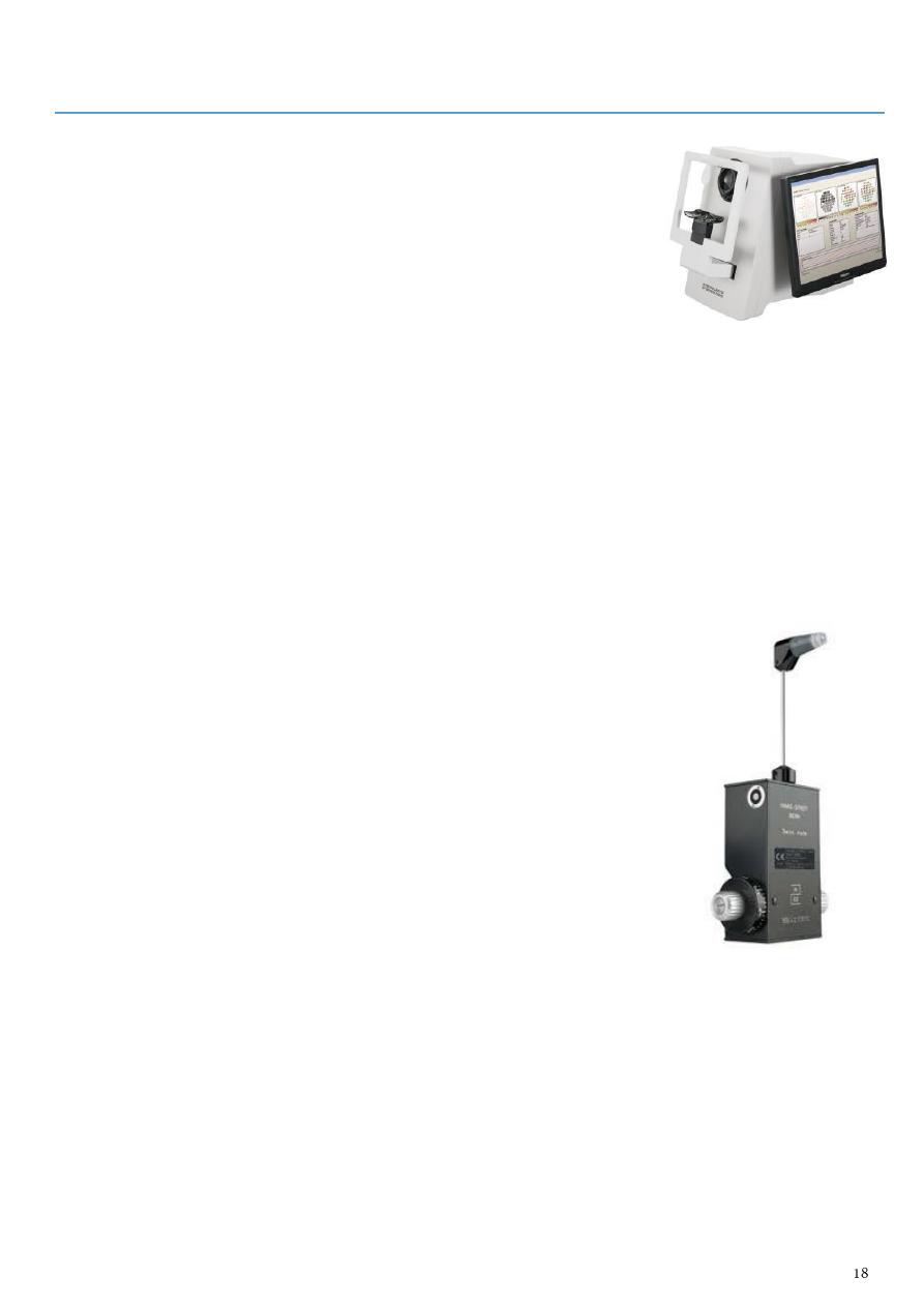
Perimeters:
These machines permit more accurate plotting of the visual
field.
They measure:
o The kinetic visual field in which the patient indicates when he first sees a light of
a specific size and brightness brought in from the periphery. This is rather like the
moving pinhead of the confrontation test.
o The static visual field in which the patient indicates when he first sees a
stationary light of increasing brightness.
These techniques are particularly useful in chronic ocular and neurological conditions
to monitor changes in the visual field (e.g. in glaucoma).
See this video www.muhadharaty.com/lecture/3521
Goldmann tonometer:
Intraocular pressure is measured with a Goldmann tonometer.
A clear plastic cylinder is pressed against the anaesthetized
cornea.
The ring of flattening, viewed through the cylinder, is made
visible by the presence of fluorescein in the tear film.
A horizontally disposed prism, within the cylinder, splits the ring
of contact into two hemicircles.
The force applied to the cylinder can be varied to alter the
amount of corneal flattening and thus the size of the ring.
It is adjusted so that the two hemicircles just interlock.
This is the endpoint of the test, and the force applied, converted into units of ocular
pressure (mmHg) can now be read from the tonometer.
Optometrists use a puff of air of varying intensity to produce corneal flattening rather
than the prism of the Goldmann tonometer.
Various other tonometers are also available including small hand held electronic devices.
See this video www.muhadharaty.com/lecture/3527
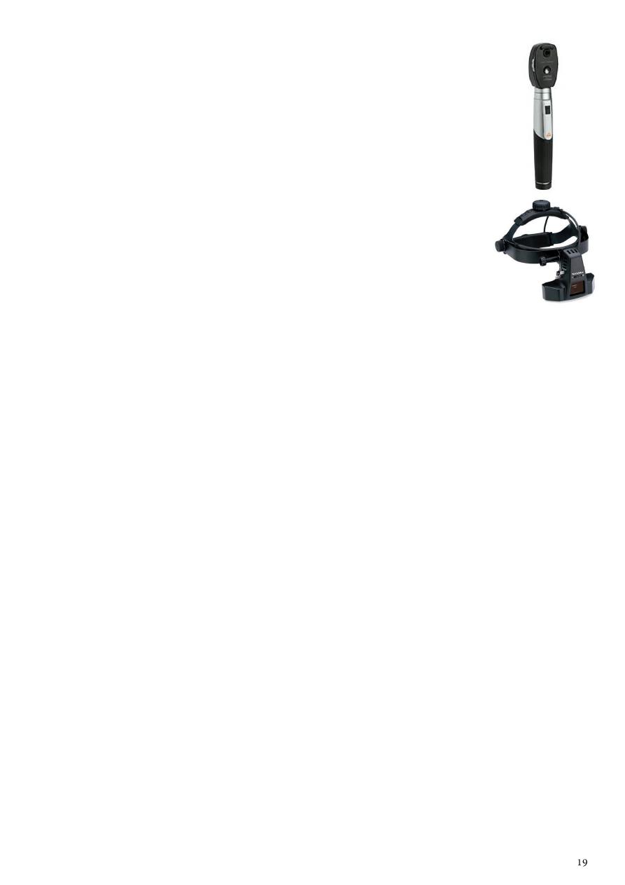
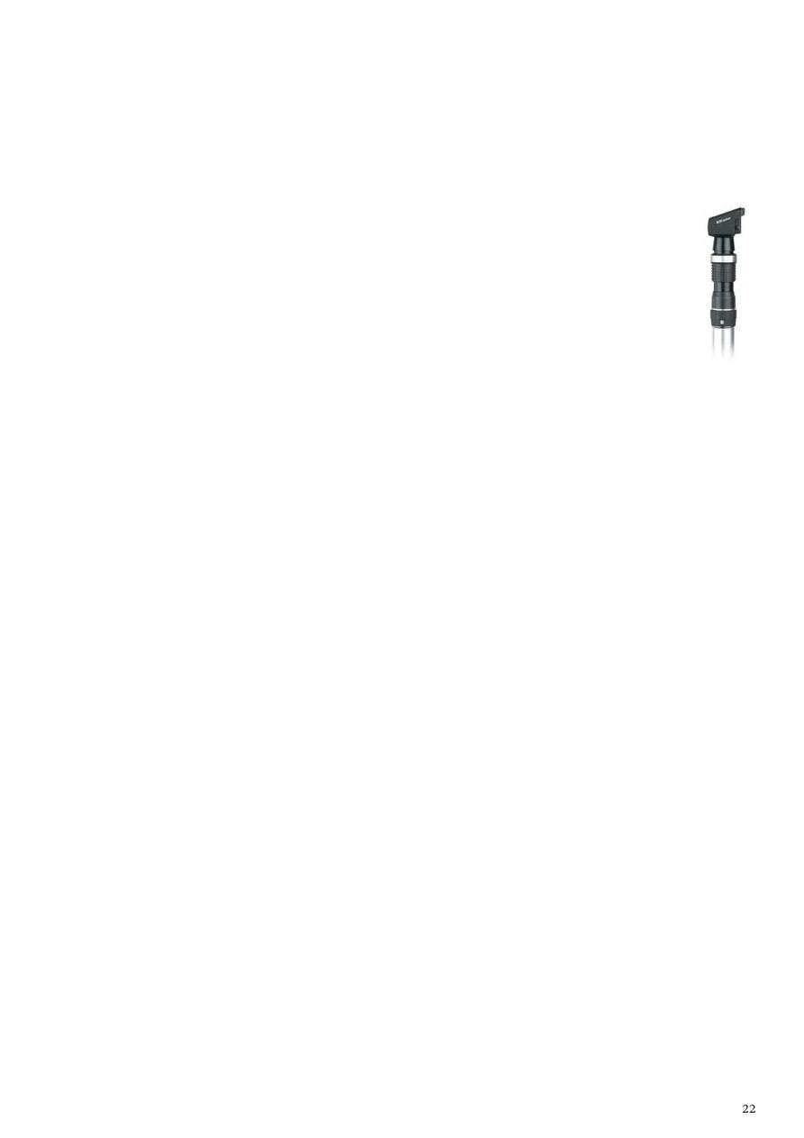
Ophthalmoscopy:
See this video: www.muhadharaty.com/lecture/3536/
Type:
Direct ophthalmoscopy (the conventional ophthalmoscope) provides an
image of the red reflex, a magnified view of the optic nerve head, macula,
retinal blood vessels and the retina to the equator.
Indirect ophthalmoscopy which allows the extreme retinal
periphery to be viewed. The examiner wears a head-mounted
binocular microscope with a light source. A lens placed between the
examiner and the eye of the subject is used to produce an inverted
image of the retina.
Settings:
Examine the eye undilated first to see the pupils and iris; then examine the eye dilated
using tropicamide drops, to visualize the lens, vitreous and retina.
Only the optic nerve can be reliably assessed without pupillary dilatation.
If patients have particularly thick lenses, examine their eyes with their glasses in place;
however, this reduces your field of view.
Advise the patient not to drive or use machinery until the effect of the mydriatic has
completely worn off. This may take several hours.
Examination:
Hold the ophthalmoscope in your right hand and use your right eye to examine the
patient’s right eye. Hold the ophthalmoscope in your left hand and examine the
patient’s left eye with your left eye.
Find ‘0’ and then rotate the ‘lenses’ clockwise until you obtain the number 10 (plus
‘10’). This should be the same color as the ‘1’ clockwise to ‘0’. If not, you have gone too
far.
Place your free hand on the patient’s forehead and ask the patient to look down. Catch
the upper eyelid and gently retract it against the orbital rim. Holding the eyelid against
the brow enables you to approach the patient’s head as closely as possible without
bumping into it, and prevents the upper eyelid from obscuring your view.
Ask the patient to fixate on a distant object straight ahead.
From a distance of about 10 cm bring the red reflex into focus. In this way the cornea,
iris and lens can be visualized and any opacity appears black against this red
background.
Now come close to the patient’s head so that you are touching the hand you are
resting on the patient’s forehead.
As you do so, rotate the lenses anticlockwise, progressively increasing the focal length.
Look for black opacities in the vitreous until the retina comes into focus.
Fundal examination:
If you approach at a slight angle above the horizontal from the temporal side you
should bring the optic disc into view.
The normal disc is pink with a pigmented temporal margin. Pallor can be difficult to
judge. Confirm it by checking the relative afferent pupil response, which will be
diminished with pallor.
Change the focus if you cannot see the disc clearly and assess its shape, color and
vessels.
Follow the blood vessels as they extend from the optic disc in four directions:
superotemporally, inferotemporally, superonasally and inferonasally.
Ask the patient to look superiorly (examine horizontally), temporally (examine
vertically), inferiorly (examine horizontally) and nasally (examine vertically).
Ask the patient to look directly at the light to locate the center of the macula. Ask her
to keep her eye still while you look around the macula.
Finding:
Optic disc margins abnormality, color of the disc (pale), optic cup abnormalities.
Macular region normal foveal reflex, any abnormal lesions such as hemorrhages,
exudates or cotton wool spots.
Major vessel branch diameter, A/V nipping, emboli in the arterioles.
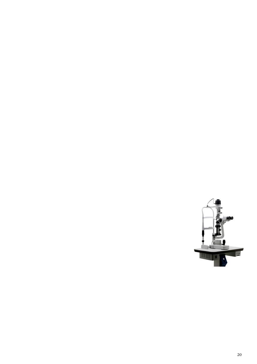
Slit lamp:
See this video: www.muhadharaty.com/lecture/3537/
Ophthalmologists use a biomicroscope (slit lamp) to examine the
eye and lids.
This allows the examiner to obtain a magnified stereoscopic view.
The slit of light permits a cross-section of the transparent media
of the eye to be viewed.
By adjusting the angle between this beam and the viewing
microscope the light can be used to highlight different structures and pathological
processes within the eye.
Each structure is carefully examined, starting with the lids and working inwards.
Examine the following:
o Lid and lashes.
o Conjunctiva conjunctival reactions, chemosis, membrane, pterygium,
pinguecula, concretions.
o Sclera redness, nodule, discoloration, episcleritis, scleritis.
o Cornea: (can use special stain)
Epithelial signs punctate epithelial erosions (PEE), punctate epithelial
keratitis (PEK), filaments.
Stromal signs stromal infiltration, stromal edema, vascularization.
Descemet's membrane signs breaks, folds (Striate keratopathy).
o Anterior chamber blood (hyphema), pus (hypopyon), cells (AC reaction), depth.
o Lens opacity (Cataract), ectopia lentis, intraocular lens.
Fluorescein:
Fluorescein has the property of absorbing light in the blue
wavelength and emitting a green fluorescence.
The application of fluorescein to the eye can identify
corneal abrasions (where the surface epithelial cells have
been lost) and leakage of aqueous humour from the eye.
To examine an abrasion:
o A weak solution of the dye is applied to the eye.
o The eye is examined with a blue light.
o The area of the abrasion will fluoresce bright green.
To determine if fluid is leaking from the eye (e.g. after penetrating corneal injury):
o Non-fluorescent, 2% solution of fluorescein is applied to the eye.
o The eye is examined with a blue light.
o The dye, diluted by the leaking aqueous, becomes bright green at its junction with
the dark fluorescein.
Cotton bud:
The underside of the upper lid is examined by everting it over a small blunt ended
object (e.g. a cotton bud) placed in the lid crease.
This is an important technique to master as foreign bodies may often lodge under the
upper lid causing considerable pain to the victim.
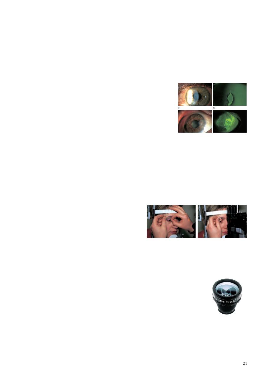
Diagnostic lenses:
See this video: www.muhadharaty.com/lecture/3538
Ophthalmologists employ special lenses that can be used in conjunction with the slit
lamp to examine particular ocular structures.
o Gonioscopy lens is a diagnostic contact lens, with a built-in mirror that permits
visualization of the iridocorneal angle.
o Larger lens with three mirrors allows the peripheral retina to be seen.
Both are applied to the anaesthetized cornea with a lubricating medium.
Other lenses can be used to obtain a stereoscopic view of the retina.
Other lenses: Hruby lens, Panfundoscopic lens.
Retinoscope:
See this video: www.muhadharaty.com/lecture/3539/
The technique of retinoscopy allows the refractive state of the eye to be
measured (the required strength of a corrective spectacle lens).
A streak of light from the retinoscope passes into the eye.
The reflection from the retina is observed through the retinoscope.
By gently moving the retinoscope from side to side the reflected image is seen to
move.
The direction in which this image moves depends on the refractive error of the eye.
By placing trial lenses of differing power in front of the eye the direction in which the
reflected image moves is seen to reverse.
When this point is reached the refractive error has been determined.

Part4
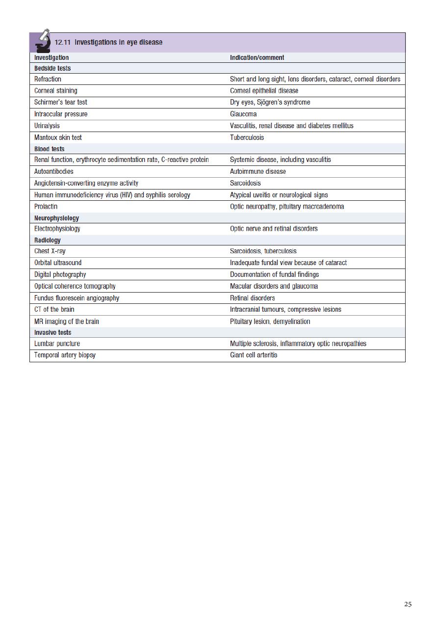
: Ophthalmological investigations
Ultrasound:
This is used extensively in ophthalmology to provide information about the vitreous,
retina and posterior coats of the eye, particularly when they cannot be clearly
visualized (if there is a dense cataract or vitreous hemorrhage).
Ultrasound is also used to measure the length of the eyeball prior to cataract surgery
to estimate the power of the artificial lens that is implanted into the eye.
Keratometry:
The shape of the cornea (the radius of curvature) can be measured from the image of a
target reflected from its surface.
This is important in contact lens assessment, refractive surgery, and in calculating the
power of an artificial lens implant in cataract surgery.
The technique of photokeratometry allows a very accurate contour map of the cornea
to be produced.
Synoptophore:
This machine permits the assessment of binocular single vision, the ability of the two
eyes to work together to produce a single image.
It is also able to test the range over which the eyes can move away from (diverge) or
towards each other (converge) whilst maintaining a single picture (to measure the
range of fusion).
Exophthalmometer:
This device measures ocular protrusion (proptosis).
Electrophysiological tests:
The electrical activity of the retina and visual cortex in response to specific visual
stimuli, for example a flashing light, can be used to assess the functioning of the retina

(electroretinogram), RPE (electro-oculogram) and the visual pathway (visually evoked
response or potential).
Radiological imaging techniques:
The CT and MRI scans have largely replaced skull and orbital X-rays in the imaging of
the orbit and visual pathway.
The newer diagnostic techniques have enhanced the diagnosis of orbital disease (e.g.
optic nerve sheath meningioma) and visual pathway lesions such as pituitary tumors.
They have also become the first line investigation in orbital trauma.
Fluorescein angiography:
This technique provides detailed information about the retinal circulation.
Fluorescein dye is injected into the antecubital vein.
A fundus camera is used to take photographs of the retina.
A blue light is shone into the eye to ‘excite’ the fluorescein in the retinal circulation.
The emitted green light is then photographed through a yellow barrier filter which
removes any reflected blue light.
In this way a fluorescent picture of the retinal circulation is obtained.
The dye leaks from abnormal blood vessels (e.g. the new vessels sometimes seen in
diabetic eye disease).
Areas of ischemia, due to retinal capillary closure, fail to demonstrate the normal
passage of dye (e.g. in a central retinal vein occlusion).
The technique is useful both in diagnosis and in planning treatment.
Digital imaging and laser scanning techniques:
New techniques of retinal imaging are being developed to improve the quality of
retinal and optic disc pictures and to permit quantitative assessment of features such
as the area of the optic disc and optic disc cup.
These will help in the assessment of patients with chronic diseases such as glaucoma
and diabetes where the management requires an accurate assessment of any change
in the disc or retina.

Part5
: Notes
Causes of sudden-onset, uniocular, permanent visual loss:
Retinal artery occlusion
Anterior ischemic optic neuropathy
Retinal vein occlusion
Traumatic optic neuropathy
Causes of acute anterior uveitis (iritis):
Idiopathic
HLA B27 association
Sarcoidosis
Herpes simplex keratitis
Post-cataract surgery
Tuberculosis
Neurosyphilis
Trauma
Causes of retinitis:
Cytomegalovirus
Herpes simplex
Herpes zoster
Varicella zoster
Causes of periorbital edema:
Allergic eye disease
Thyroid eye disease
Orbital cellulitis
Nephrotic syndrome
Heart failure
Angio-oedema

Causes of proptosis:
Thyroid eye disease (exophthalmos)
Caroticocavernous fistula
Orbital cellulitis
Orbital haematoma
Granulomatosis with polyangiitis
Orbital tumours, including lymphoma, metastasis, meningioma, glioma
Pseudoproptosis (pathological myopia, shallow orbits, contralateral enophthalmos)
Causes of ptosis:
Mechanical:
o Chronic orbital inflammation
o Degenerative
o Eyelid tumour
o Intraocular surgery
o Long-term use of contact lenses
o Trauma
Myogenic:
o Chronic progressive external ophthalmoplegia
o Congenital myogenic ptosis secondary to levator dysgenesis
o Myotonic dystrophy
o Oculopharyngeal dystrophy
Neuromuscular junction:
o Myasthenia gravis
Neurogenic: congenital:
o Congenital Horner’s syndrome
o Congenital III nerve palsy
Neurogenic: acquired:
o Acquired Horner’s syndrome
o III cranial nerve palsy
o Synkinetic neurogenic ptosis, e.g. Marcus Gunn
Common causes of arteriolar occlusion:
Accelerated hypertension
Diabetic retinopathy
Human immunodeficiency virus (HIV) retinopathy
Retinal vein occlusion
Systemic lupus erythematosus
Systemic vasculitis

Causes of oculomotor nerve palsy:
Medical (pupil sparing) Hypertension, Diabetes.
Surgical (pupillary involvement) Aneurysm, Trauma, Uncal herniation.
Causes of anisocoria:
Physiological anisocoria (20% of population)
Sympathetic tract lesion (Horner’s syndrome)
Parasympathetic tract lesion (oculomotor n. palsy, Adie’s pupil)
Argyl Robertson syndrome
Pharmacological
Iris damage (traumatic mydriasis, iritis, angle closure glaucoma, siderosis)
Anisocoria in light: parasympathetic problem
– Cranial nerve III defect
– intracranial pressure
– drug-induced mydriasis
– Adie’s pupil
– iris damage
– simple anisocoria
Anisocoria in dark: sympathetic problem
– Horner’s syndrome
– simple anisocoria

Part5
: From Mosul medical college
History taking:
Include:
Personal data.
Chief compliant.
History of present illness.
Previous ophthalmological history.
Previous medical and surgical history.
Drug history.
Loss of vision:
1- Gradual painless LOV:
Cataract.
Open angle glaucoma.
Diabetic retinopathy.
Tumor.
2- Sudden painless LOV:
Retinal detachment.
Optic neuritis.
Retinal venous occlusion.
Retinal arterial occlusion.
Vitrous hemorrhage.
Sudden discovery of preexisting unilat pinguecula.
3- Painful LOV:
Uveitis.
Iritis.
Close angle glaucoma.
Keratitis.
Trauma.
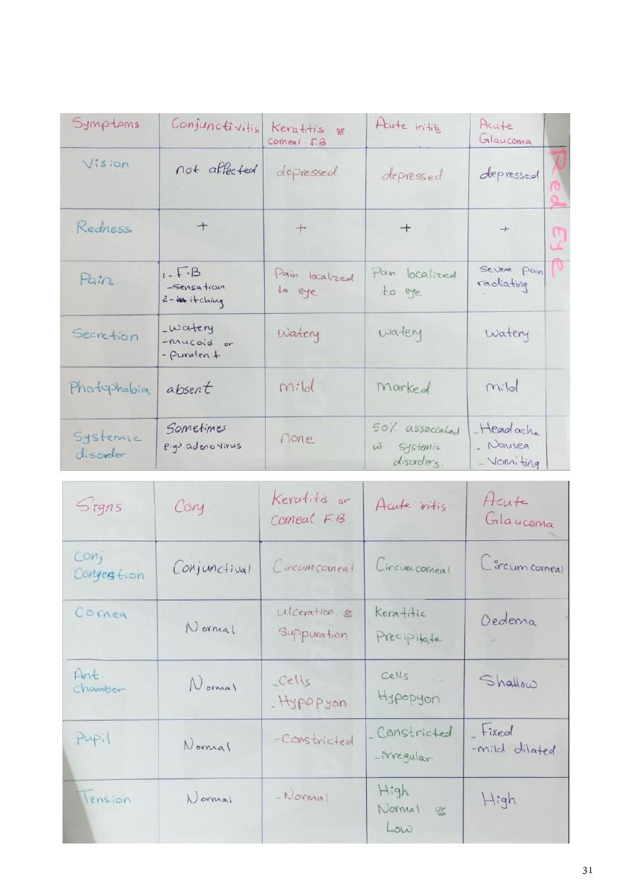
Red eye:
Conjunctivitis.
Keratitis.

Iritis.
Closed angle glaucoma.
Scleritis and epi-scleritis.
Blepharitis.
Allergy.
Trauma.
Side effects of corticosteroids eye drop:
Flare up of pre-existing eye infection.
Predispose for microbial keratitis (viral, bacterial, fungal).
Inhibit collagen synthesis of cornea and predispose to corneal thinning.
Cataract (chronic case).
Open angle glaucoma (chronic case).
Indications of topical steroid in eye disease:
Decrease inflammatory reaction in post ocular surgery (cataract operation).
Inflammatory noninfectious disorders (iritis).
Hypersensitivity to microbial antigens phlyctenular keratoconjunctivitis (under
cover of topical antibiotics) Disciform keratitis (under cover of antiviral).
Sometimes in non-severe microbial keratitis involving the axial part of the cornea to
decrease scaring under the follow precaution:
o Identify causative agent by lab investigations and giving appropriate antibiotic.
o There is clinical response or treatment.
o There is no thinning or impending perforation of cornea.
o Contraindicated in pseudomonas, fungal, epithelial viral keratitis.
o Patient must not immune compromised.
o Close follow up (in hospital).
Contraindications of steroids:
Active bacterial keratitis.
Pseudomonas, fungal, epithelial viral keratitis.
Immune compromised patient (D.M).
Thinning or impending perforation of cornea.
When we don’t know causative agent of disease.
Severely infected and complicated eye disease that need management with antibiotics
and follow up.
No clinical response on treatment.
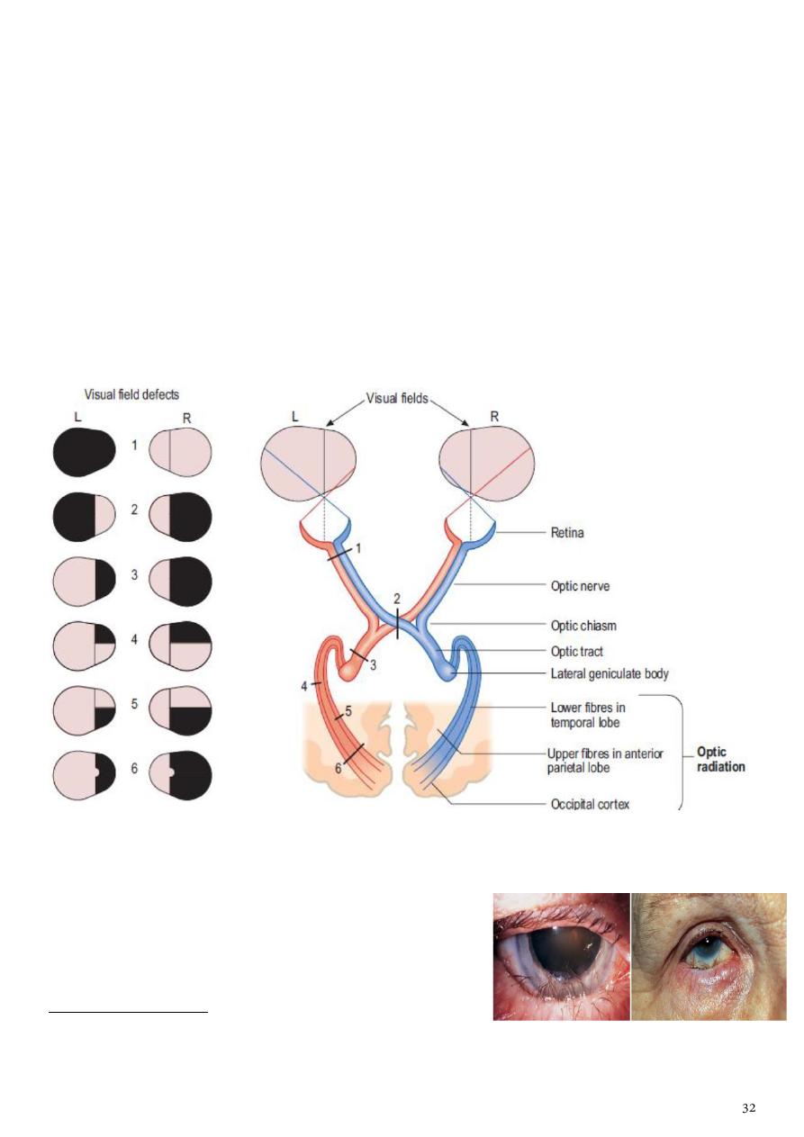
Effects of lesions in optic pathway on visual fields:
1- Blindness of the left eye.
2- Biltemporal hemianopia.
3- Right homonumus hemianopia.
4- Right upper quadrantic hemianopia due to lesion of lower fibers of optic radiation in the
temporal lobe.
5- Lower right quadrantic hemianopia due to lesion of the upper fibers of optic radiation in
the upper anterior parietal lobe.
6- Right homonymus hemianopia lesion in optic radiations in the posterior parietal lobe
here the light reflex is present.
Entropion:
Malposition (inward turning) of eyelid toward the
eyeball with trichiasis (misdirection of the eyelashes
toward the eyeball).
Causes of entropion:
1) Congenital (rare).
2) Acute spastic conditions such as infections, inflammatory & traumatic.
3) Cicatricial (scaring of the palpebral conjunctiva).
Causes of trichiasis:
1) Infections (trachoma, herpes).
2) Auto-immune (ocular cicatricial pemphigoid).
3) Inflammatory (Stevens-Johnson syndrome, vernal keratoconjunctivitis).
4) Trauma.
Complications (entropion & trichiasis):
1) Infections.
2) Corneal ulceration, corneal opacity.
3) Chronic conjunctivitis, conjunctival scar.
Treatment of entropion:
1) Eyelid hygiene.
2) Antibiotics to treat the causative microorganism.
3) Steroids.
4) Lubrication.
5) Small amount of botulinum toxin (BOTOX).
6) Surgical repair (Lateral canthotomy, lateral canthoplasty).
Treatment of trichiasis:
1) Treatment of the underlying disease (Stevens-Johnson syndrome & ocular cicatritial
pemphigoid).
2) Lubrication.
3) Treatment of infection.
4) Surgery (follicle destroying or lash/follicle repositioning).
Ectropion:
Abnormal eversion (outward turning) of the lower
eyelid margin away from the globe.
Patient complaint: Lacrimation.
Causes:
1) Congenital.
2) Acquired:
a) Senile.
b) Paralytic (facial nerve palsy).
c) Cicatricial (due to scar) such as burns, glaucoma drops and chronic dermatitis ….etc.
d) Mechanical (tumor) such as neurofibromas.
Complications:
1) Conjunctival keratinization.
2) Corneal breakdown (ulceration)
3) Epiphoria (watery eye: tears flow onto the cheek).
4) Pain.
Treatment:
1) Treatment of infection (Chlamydia, herpes).
2) Lubrication.
3) Steroid.
4) Surgical repair (V to Y plasty or Z plasty).
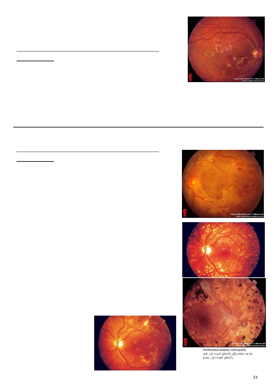
Diabetic retinopathy:
The problem is in the vessels.
First type leakage (non-proliferative diabetic
retinopathy)
Loose pericyte of vessels wall.
Features:
o Hemorrhage red dots.
o Transudate (edema) change in color.
o Exudate (lipoprotein) golden yellow lesion.
o Aneurysm red dots near blood vessels.
Site of leakage: close it by laser.
Second type occlusion (proliferative diabetic
retinopathy)
Because viscosity of blood increase and platelets
aggregation increase and vessels wall thick due to
hyperplasia of tunica media and RBC stick with each
other called roloux formation lead to ischemia in the
periphery.
Vascular endothelial growth factor:
o Produce new blood vessels that is fragile and lead to
hemorrhage.
o Produce fibrosis could lead to retinal detachment.
Features:
o Ischemia.
o Fibrosis.
o New vessels.
o Localized area of ischemia called cotton wall spots.
Site of ischemia: photocoagulation, this destroy new
vessels but the fibrosis not destroyed, and this
procedure not lead to any problem to the patient.
Description
Photo
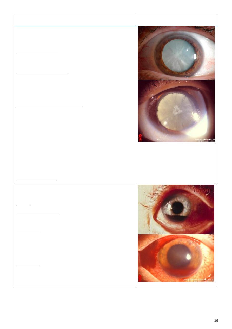
Cataract and dilated pupil
Any opacity in the lens is cataract (affect the vision
or not).
Causes of cataract:
1-age (commonest) 2-congenital
3-drugs like steroid 4-radiation 5-trauma
Causes of dilated pupil:
1- Drugs (sympathomimetic, anticholinergic)
2- Third CN palsy (parasympathetic damage)
3- Optic nerve damage
4- Traumatic mydriasis (damage of sphincter muscle)
Assess the state of fundus by:
1- Visual acuity.
2- Light perception (+ve other disease, -ve optic
damage).
3- Light perception (from which side the light
come?).
4-pupillary reflex
5- Color discrimination (done by put color filter in
the slit lamp, it is used to check the function of optic
nerve).
6- B scan US.
Treated by surgery (extraction of lens).
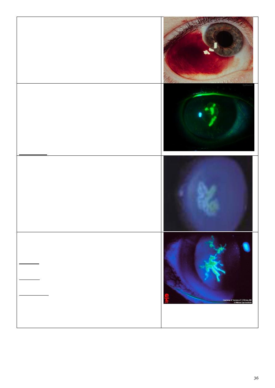
Hyphema
(blood in the anterior chamber)
+
subconjunctival hemorrhage + dilated pupil
Cause: trauma.
Cause of hyphema: Trauma is the major cause,
surgery (during & after), intraocular tumors,
anticoagulant treatment.
Assess IOP:
1- The blood will block the trabecular meshwork
space
no drainage
high IOP.
2- Wound on sclera or other parts
leak of eye
fluid
low IOP.
Treatment:
1-bed rest with head elevated
2-anti-glucoma drugs if increased IOP
3-drainage of blood if high amount
Subconjunctival hemorrhage
Ask the patient about trauma, straining (cough,
vomiting), bleeding tendency, drug.
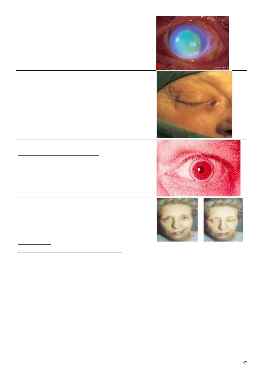
HSV-1 eye lesion
Branching greenish discoloration of the cornea =
dendritic ulceration due to HSV-1 infection.
The color of fluorescence is orange but here we see
it greenish because we use the slit lamp filter.
Fluorescence stain has affinity to collage fibers, so
areas of no epithelium or removed epithelium will
be stained by the florescence stain.
Treatment: Acyclovir, trifluridine, idoxuridine.
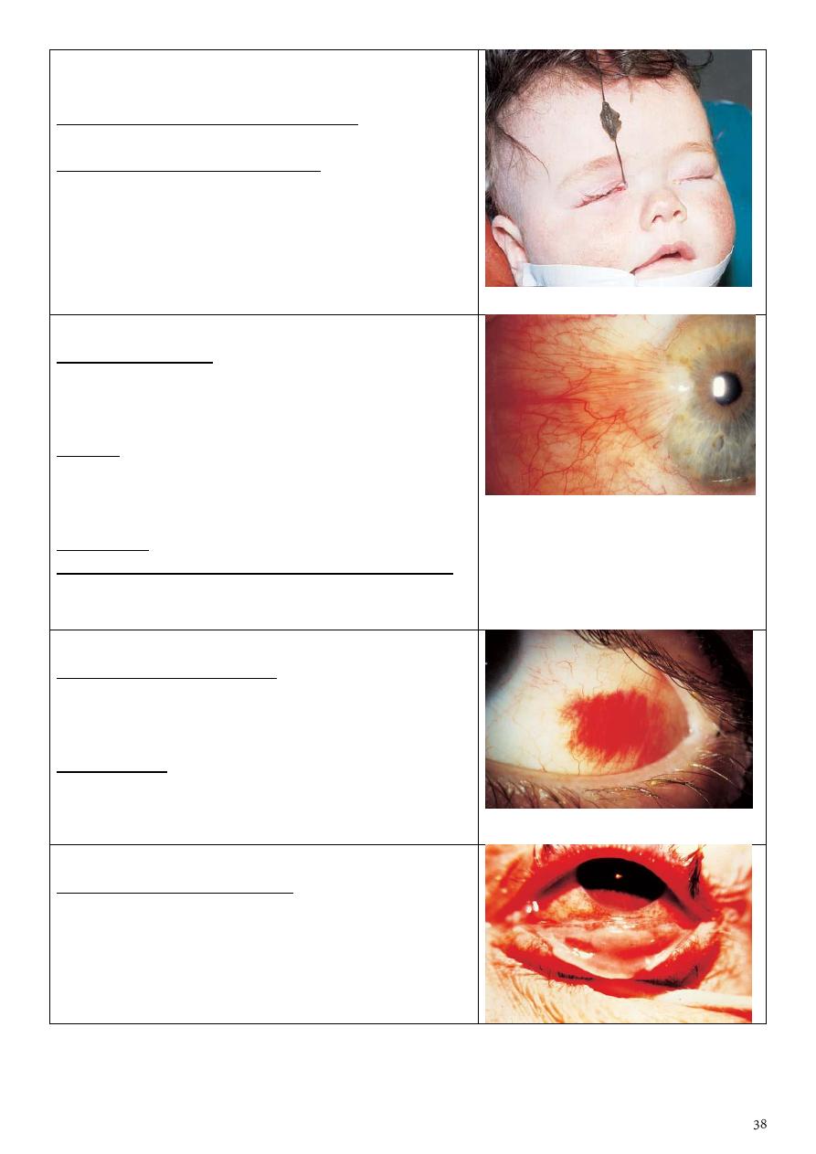
Ameboid ulcer
Cause:
Mistreatment of dendritic ulcer by steroid.
Geographical ulcer
Fluorescein stain showing ulcer in the cornea.
Cobalt blue filter in slit lamp.
Causes: 1-steroids 2-chemical substances 3-bacterial
infections 4-F.B. 5-trauma 6-contact lens users
History: patient with HSV-1 wrongly treated by
steroid
Treatment:
1- according to the cause
2- gradual decrease in steroids
3- antiviral (if dendritic)
4- patching and soft bandage (if traumatic)
Corneal ulceration
-Fluorescein stain showing corneal ulceration
-Same causes of any ulcers
-If we see pus in the anterior chamber (Hypopium)
the cause is bacterial.
Dacryocystitis
Cause: obstruction of the nasolacrimal system.
Most common organism is staphylococcus.
Presentation: painful swelling on the medial side of
the orbit, which is the enlarged, infected lacrimal
sac.
Treatment:
1- Systemic antibiotics 2- Dacryocystorrhinostomy
(DCR) to prevent recurrence.
Corneal arcus (lipidis) or arcus senilis
What is the clinical significance? It indicate
hyperlipidemia in young people which may causes
systemic diseases.
What is the visual prognosis? It never affect
vision(never reach cornea)
Facial palsy on R. side with lagophthalmos &
Bell's phenomenon
Presentation: Mouth deviated to the left side,
cannot close right eye, eye pulled upward (bell's
phenomina).
Significance? Exposure keratopathy
How do you treat the eye of this patient?
1-artificial tears in days
2-closure of eye at sleep (taping)
3-If chronic do surgery (lateral tarsorraphy)
4-shield glasses
Congenital failure of canalization of right
nasolacrimal duct
What is the presentation of this child? Lacrimation
on right side(epiphora) or conjunctivitis
What is the proper management?
1-reassurance of parents
2-topical AB with massage till the end of the first
year
3-if not opend (after the first year, most open
spontaneously during the first year) probing and
irrigation with antiseptic solution
Ptergyium
Describe this lesion: Localized redness located at
nasal corner of the eye, triangular in shape with the
apex extend to involve cornea due to fibrovascular
ingrowths
Causes?
1- reflected or direct UV light exposure 2- irritation
3- Exposure to irritant materials (dust, smoke…)
4- SCC (mass) 5- fibrosis due to injury
Treatment? Surgical removal with conjunctival graft
What are the indications of surgery in this patient?
1-affect visual axis. 2-severe discomfort. 3- cosmetic.
4- poor vision. 5- dryness of cornea. 6-astigmatism.
Subconjuctival hemorrhage
What will you ask in the Hx? 1-history of trauma,
straining conditions (vomiting, constipation, cough)
2-history of hypertension. 3-bleeding tendency.
4-history of drugs: anticoagulants and NSAIDs.
Management: assurance of patient, resolved after
10-14 days, it is simple condition put if the patient
want treatment you can give artificial tear or
lubricants.
Membranous conjunctivitis
What are the possible causes?
1-true membrane: diphtheria.
2-pseudomembrane: most bacteria like staph +
adenoviral infection.
What is this sign? Papillae on the inner surface of
upper lid (giant cobblestones papillae)
What are its causes? vernal keratoconjunctivitis,
allergic conjunctivitis: atopic, seasonal, giant
papillary conjunctivitis.
Treatment:
Avoiding exposure to the causative allergen, topical
steroids, mast cell stabilizers, antihistamine, dark
glasses & cold compresses.
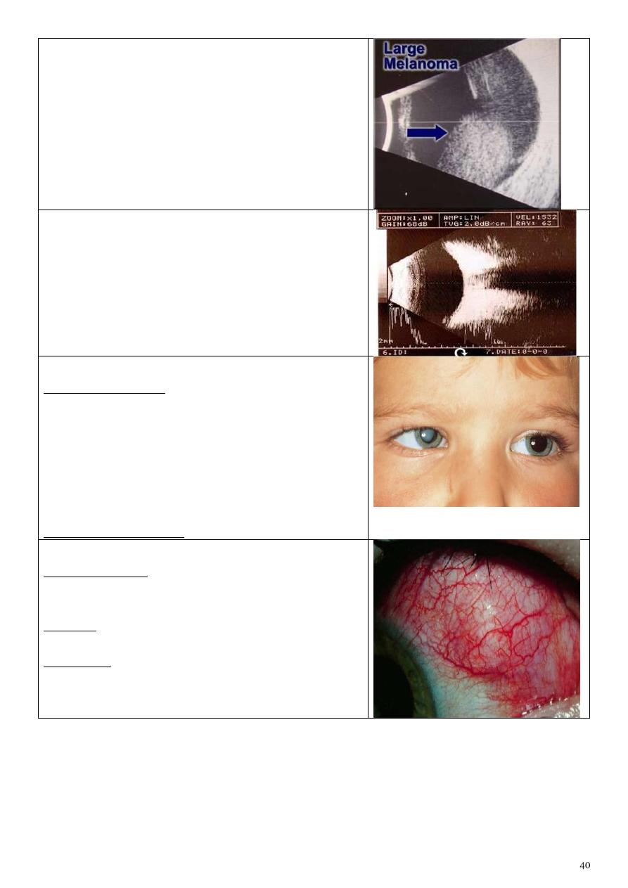
Corneal topography
Color coded map used for studying the shape,
curvature, refractive power of cornea in every point
with details.
Staphyloma
-Occur in high myopia as the globe is large.
-Thinning of retina, bulging, exposed sclera.
US in high myopia
US showing large melanoma
B scan sonography showing the vitreous
cavity
Leukocoria & pseudosquint
Describe the lesion? Right white pupil.
Enumerate the most common causes according to
their frequency?
1-congenital cataract.
2-retinoplastoma.
3-retinal fibrosis.
4-persistent hyperplastic primary vitreous.
5-retinopathy of prematurity.
Cause of pseudosquint: Epicanthal fold.
Scleritis
Symptoms/Signs: Pain, Significant ocular tenderness
to movement and palpation, Watering,
photophobia.
Etiology: immune, associated systemic disease,
connective tissue disease, rheumatoid arthritis.
Treatment: underlying condition, NSAIDs,
corticosteroids, immunosuppression.
Episcleritis
Symptoms/Signs: Often asymptomatic, Mild tearing/
irritation, Tender to touch, Self-limiting (may last for
months).
Treatment: Lubricants, NSAIDS, Rarely low dose
steroids.
Goldmans applanation tonometry
Put florescence stain to the tear film then press the
external surface of eye by the tonometer and when
it become flat it means equalization of the pressure
inside and outside the eye, and the semi-circles will
meet each other, so take this point as IOP.
Schiotz tonometry
There are 3 things here:
1- Weight (g).
2- Degree of corneal indentation (mm).
3- Table of pressure measurement.
Pressure reversely related with the indentation.
Snellen's test
For assessment of visual acuity
By using which test we can get this view?
1- Gonioscopy (visualization of the angle of the
anterior chamber = irido-corneal angle)
2- With goniolens (for detecting glaucoma).
Describe the aqueous circulation:
It is produced by the ciliary process of ciliary body
>posterior chamber >pupil> anterior chamber
>trabecular meshwork> Canal of
Schlemm>extraocular veins.
Measurement of AC depth
Comment on the AC depth: Shallow anterior
chamber depth.
If this patient is asymptomatic, how you treat him?
1-Medical Tx: mitotic agents ,alpha agonist, b-
blocker, carbone anhydrase inhibitor,
prostaglandin analogs
2-peripheral laser iridotomy (YAG laser)
*it consider pre-glaucoma so it is urgent.
3- hypermetropia: convex lenses.
4- Treatment of glaucoma: pylocarpin, iridotomy
Comment on the anterior chamber depth:
1) Eclipse sign: we shine a light on the temporal
aspect of the patient anterior chamber & watching
the iris, if the AC is deep the whole iris is illuminated,
if the AC is shallow, only the temporal part is
illuminated.
2) Important in angle closure glaucoma.
If this patient is asymptomatic, how would you treat
him?
Miotic agents, alpha agonist, carbonic anhydrase
inhibitors and prostaglandin analogs.
Abnormal cupping of the optic disk
(CD ratio >
0.3)
Normal cup:disk ratio 0.3 – 0.4
If CD ratio increase it means there is large cup.
Cause: primary open angle glaucoma
Criteria of cupping:
1-cup:disk ratio > 0.3.
2-progressive enlargement
3- asymmetrical between the two eyes.
Criteria to diagnose glaucoma:
1) Increased IOP. Normally 15±6 mmHg.
2) Abnormal cupping.
3) Changes in visual field.
Glaucomatous cupping of optic nerve
-Expected field of vision: Tubular field.
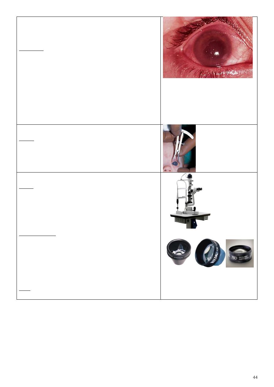
Automated static perimetry (visual filed)
perimetry graph showing paracentral scotoma in
open angle glaucoma
-No patient complain
-Diagnosed by chance
Peripheral laser iridotomy
Indication:
Shallow anterior chamber (angle closure glaucoma)
so we do it to make direct shunt between anterior
and posterior chambers.
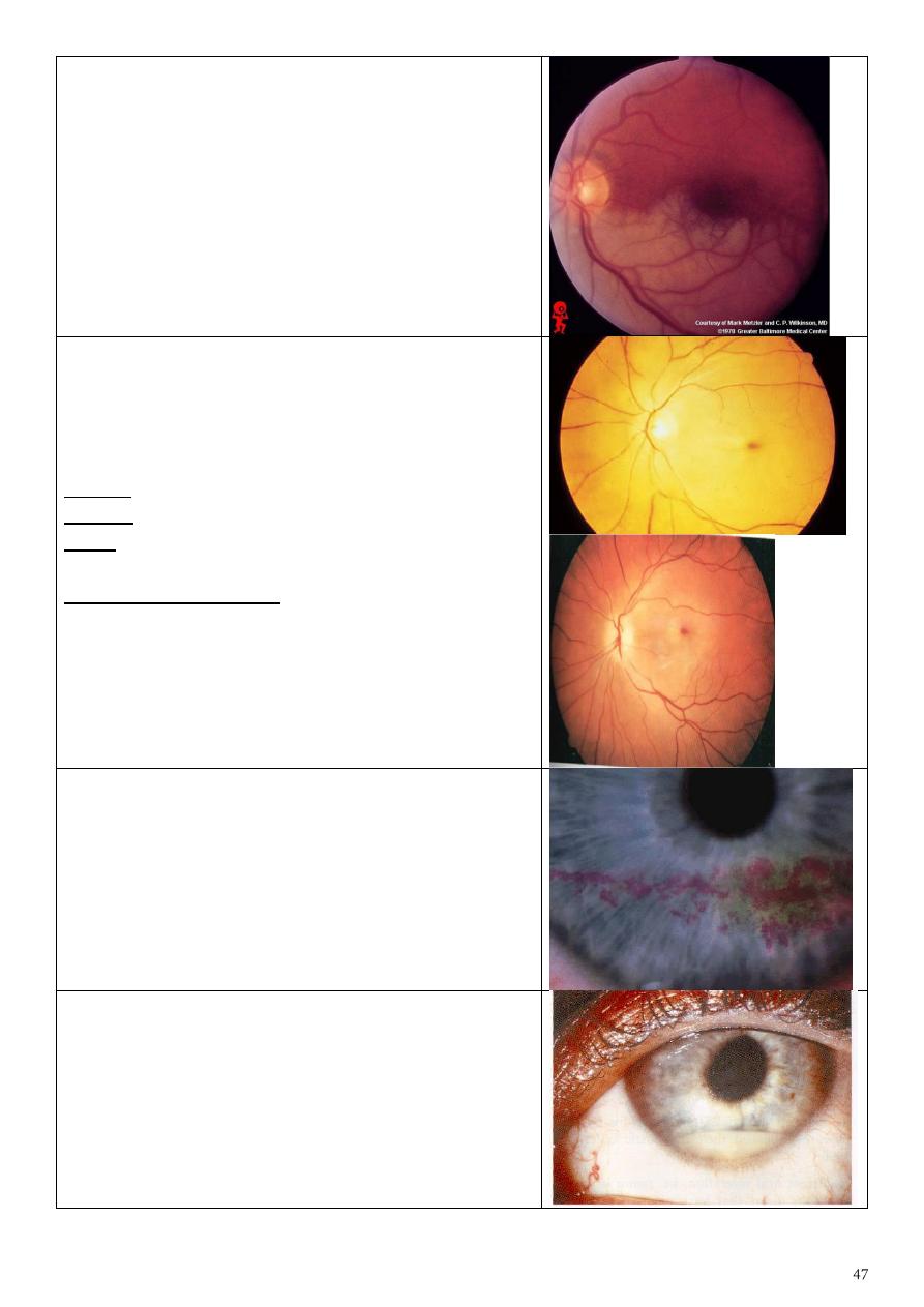
Posterior capsule opacification
Treatment: YAG laser capsulotomy to open
thickened posterior capsule after cataract surgery.
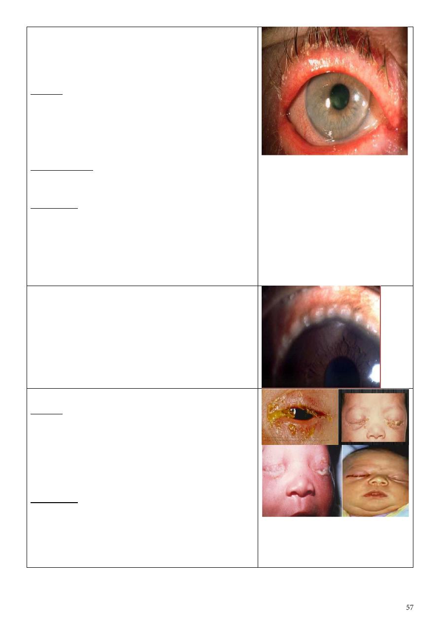
Dx: Red congested eye (acute angle closure
glaucoma) with corneal edema, ciliary flash and
dilated pupil.
Features:
Painful red eye.
Achy, abdominal pain.
Misty vision.
Go from light into dark.
Small eye, shallow anterior chamber.
Pupil mild dilated.
Iris lens contact.
Push the iris forward.
Eye feels hard.
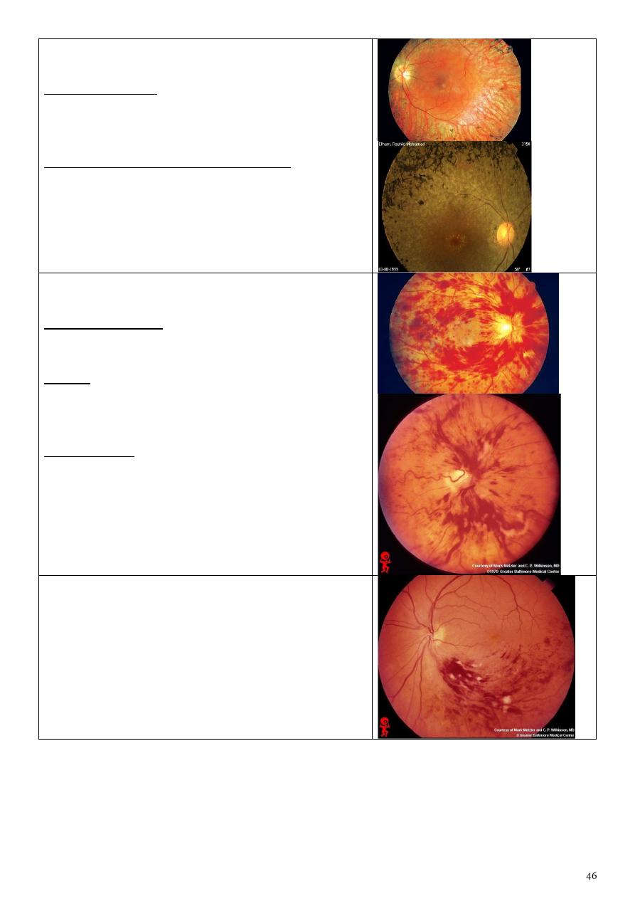
Measuring corneal diameter by caliber
Why?
To detect and follow up congenital glaucoma (
buphthalmos)
Slit lamp
Uses:
1- Examine the anterior segment of the eye.
2- Examine the posterior segment with special
lenses.
3- Measure the IOP with Goldman aplination
tonometer.
Added lenses:
1-Goldman 3 mirrors contact lens (use topical
anesthesia, vescoelastic substance).
2-Central lens.
3-Dome shape lens (goniolens): used to see
iridocorneal angle (the test called gonioscopy).
4-Plus lens 78D (convex lens).
See: upside down and laterally reversed image (real
image) by using this instrument.

Direct ophthalmoscope
1-Portable, light instrument.
2-Examine the patient in any position (lying, setting,
standing).
3-Not specialized instrument (used by general
physician).
4-Cause magnification X15 times so see narrow field.
5-We can see the fundus without dilatation of it.
6-The image seen here is erect (virtual image).
7-This examination is affected by refractive error of
the patient so use the lenses inside this instrument.
8-Right eye-right hand-right eye.
Indirect ophthalmoscope
1-It is specialized instrument.
2-Need special training, difficult to use.
3-Magnification 3-5 times depend on the lens we
use so not see the fundus directly.
4-The field here is larger than in direct method.
5-Examine the patient with your both eyes.
6-Should dilate the eye of patient.
7-Not affected by refractive error of patient.
8-The 3D image (with depth) because we use both
eyes.
9-During retinal surgery the indirect
ophthalmoscope is instrument of choice.
10-Use +20 or +30 lens.
Describe this corneal lesion: Dendritic ulcer
What is the most likely diagnosis? HSV type 1
How would you treat? Antiviral drugs (acyclovir ,
trifluoridine and idoxuridine) // avoid steroids.
Subhaloid hemorrhage
Hemorrhage between retina & vitreous
Retinitis pigmentosa
Bone spicules, waxy disc & attenuated blood vessels.
Patient complains:
1-night blindness (nyctalopia).
2-visual loss
3-seeing flashes of light (photopsia).
Mention 2 syndromes associated with it.
1-bardet biedl syndrome
2-refsum syndrome
3- Autosomal dominant / recessive
Central retinal venous occlusion (CRVO)
Congested dilated blood vessels.
History (ask about):
Hypertension, hyperlipidemia, DM, smoking,
obesity, life style, psychological problems, stress.
Causes:
1) Extraluminal causes: hypertension & tumor.
2) In the wall: vasculitis.
3) Intraluminal: thrombus.
Complications:
1) Neovascular glaucoma.
2) Chronic macular edema.
Branch retinal vein occlusion
Occlusion of the lower temporal branch of retinal
vein marked by hemorrhage and multiple spots
(cotton wool spots) of ischemia in the lower
temporal zone of retina
Occlusion of Lower temporal and lower nasal
branches of retinal artery a marked by complete
ischemia
Central retinal arterial occlusion (CRAO)
Attenuation & constriction of the arteries (cattle
track appearance), the whole retina is pale except
the fovea (cherry red spot) due to double blood
supply.
Causes: Usually embolic.
History: ask same questions in venous occlusion.
Could lead to neovascular glaucoma (due to
occlusion of iris root by new vessels).
DDx For cherry red spot:
-Commotio retinae
-Quinine poisoning
-Macular hole surrounded by RD
-Amauratic family idiocy
Filamentray keratitis
Stain: rose bengal stain
Causes : 1-dry eye 2-sjogren syndrome
3- facial palsy 4-blepharoptosis
Presentation: patient with dry eye and the mucin
become thick and adhere to the cornea and give
sensation of F.B.
Hypopium
It is pus in the anterior chamber
Causes:
1- Infected: endophthalmitis (pneumococcus).
2- Sterile: severe iritis.
3- keratitis.
4-any inflammation or infection.
Hypopium with cloudy cornea and red eye
Describe this lesion?
Nodulo-ulcerative lesion Oval, elevated,
ulcerated, congested, dirty lesion on the lower lid
Differential diagnosis?
1-basal cell carcinoma
2-sequamous cell carcinoma
3-sebaeous cell carcinoma
4-keratoacanthoma
5-malignant melanoma
Chalazion
Describe this lesion Localized swelling in the upper
lid with normal skin over it
Enumerate four causes
1-chalazion 2-stye 3-sebaceous cell carcinoma
4-haemangioma 5-blockage of tarsal glands
6-Seborrhea 7-Chronic blepharitis 8-Acne rosacea.
How would you treat it?
1-conservative: massage, hot compress, AB
ointment, local steroid.
2-surgery: surgical incision and curettage of the
gland.
Ectropion of the right lower lid
What is the complaint of this patient?
Lacrimation, congested red conjunctiva, repeated
infections.
What are the possible causes?
1-aging 2-congenital 3-lower lid scar
4-faiacl palsy 5-neurofibromas 6- mechanical cause
Entropion of the lower lid with trichiasis
What are the possible causes?
1-aging
2-congenital
3-palpebral conjunctival scaring
4- trachoma, trauma, infection, inflammation
Lens dislocation (ectopic lens)
What are the possible visual symptoms? Blurred
vision and diplopia and refractive errors.
Enumerate three possible systemic causes:
1-trauma
2-marfan syndrome
3-homocystinuria
4-high myopia (eye problem)
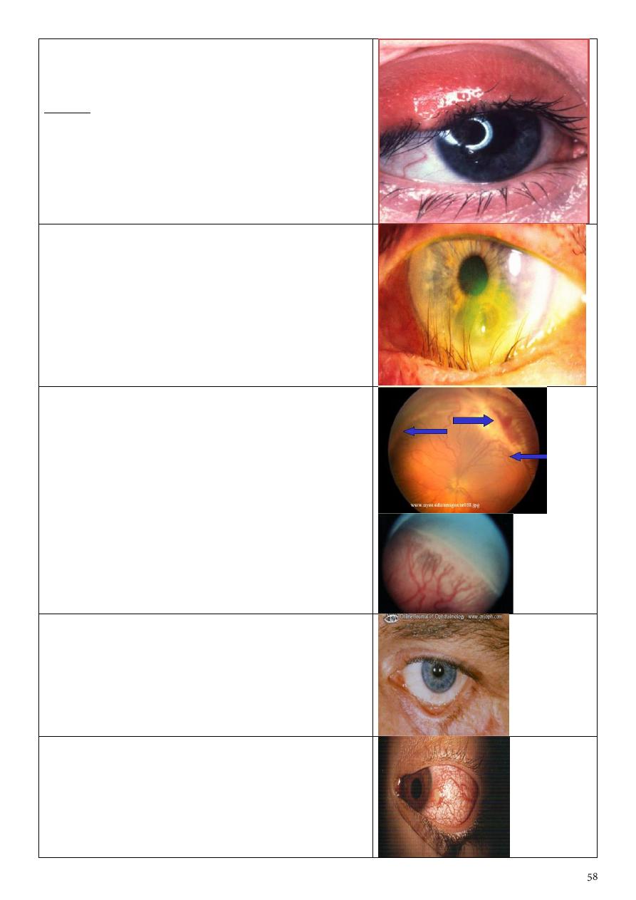
Keratic precipitate (white dots in cornea)
What is your diagnosis? It is a feature of iritis or
anterior uveitis
What do you think the presentation of this patient?
Blurred vision, photophobia, pain, redness, and
lacrimation.
Treatment:
1- topical: atropin sulphate and corticosteroid
2- systemic: steroids (severe cases) or AB (infected
cases)
Herpes zoster ophthalmicus
Describe the skin lesion:
Erythematous base lesion with crust, pastule and
some sloughing involving forehead and side of the
nose and upper + lower lid of lift eye.
Describe the ocular lesion:
FLOURESCEIN stain showing corneal ulcer of left
eye+ skin lesion involve upper + lower lid.
Thyrotoxicosis
Describe this facies:
Staring facies with periorbital edema
Enumerate four ocular features of this disease:
1-lid lag
2-lid retraction
3-exophthalmos
4-excessive lacrimation
5-Chemosis.
6-Periorbital soft tissue changes as chemosis & lid
swelling.
7-Exposure keratopathy.
8-Ophthalmoplegia.
Enumerate four systemic features of this disease:
1-sweating 2-palpitation 3-tremor 4-tachycardia
5- Goitre 6- Increased appetite with loss of weight
What is this procedure? Eversion of upper lid
What are its benefits?
1-very simple
2-F.B. detection
3-papillae detection
4-follicular reaction detection
5- Arlet line of trachoma.
6- Giant papillary conjunctivitis.
7- Adrenochrome deposits.
8- Melanoma.
9- Conjunctival concretion.
What is this procedure?
• Cover/uncover test
In which disease you want to use it in your
examination?
• Used to detect inapparent squint (phoria)
Retinal detachment
Left complete ptosis
What are the possible causes?
1- 3
rd
palsy 2-congenital 3-birth injury 4-horner
syndrome
Would you go for surgery for this child? Yes, to
avoid amblyopia or reduction of visual acuity and
here we do sling procedure.
Munson sign
Describe what you see: This patient asked to look
down, lower lid is seen pushed by cornea due to
increased corneal curvature with opacity.
What is the disease? Keratoconus with acute
hydrops
Significance: It is one of the common causes of
reduction in visual acuity (myopia & Astigmatism).
Diagnosis of keratoconus:
1-Ask the patient to look down.
2-Placido disc.
3-Corneal topography (green normal, blue
depression, red elevation).
4-Keratometry (give the reading in diopter).
What are the lines of treatment for this case?
1-contact lens or patching
2-hypertonic drops to cause withdrawal of hydrops
3-corneal graft
Lines of treatment of keratoconus in general:
1- Corneal Collagen Cross-linking, in early cases to
arrest the disease
2- Glasses
3- Contact lenses (Rigid)
4- Shaving the corneal surface
5- Intra-stromal corneal rings
6- Penetrating keratoplasty or Lamellar
keratoplasty..in advanced cases or corneal scarring
• Is it a concomitant or incomitant squint?
incomitant
• Which muscle is affected? Right lateral rectus
• What is the nerve supply? Abducent nerve
• concomitant: angle of squint not changed in
different visual directions
• incomitant: angle changed
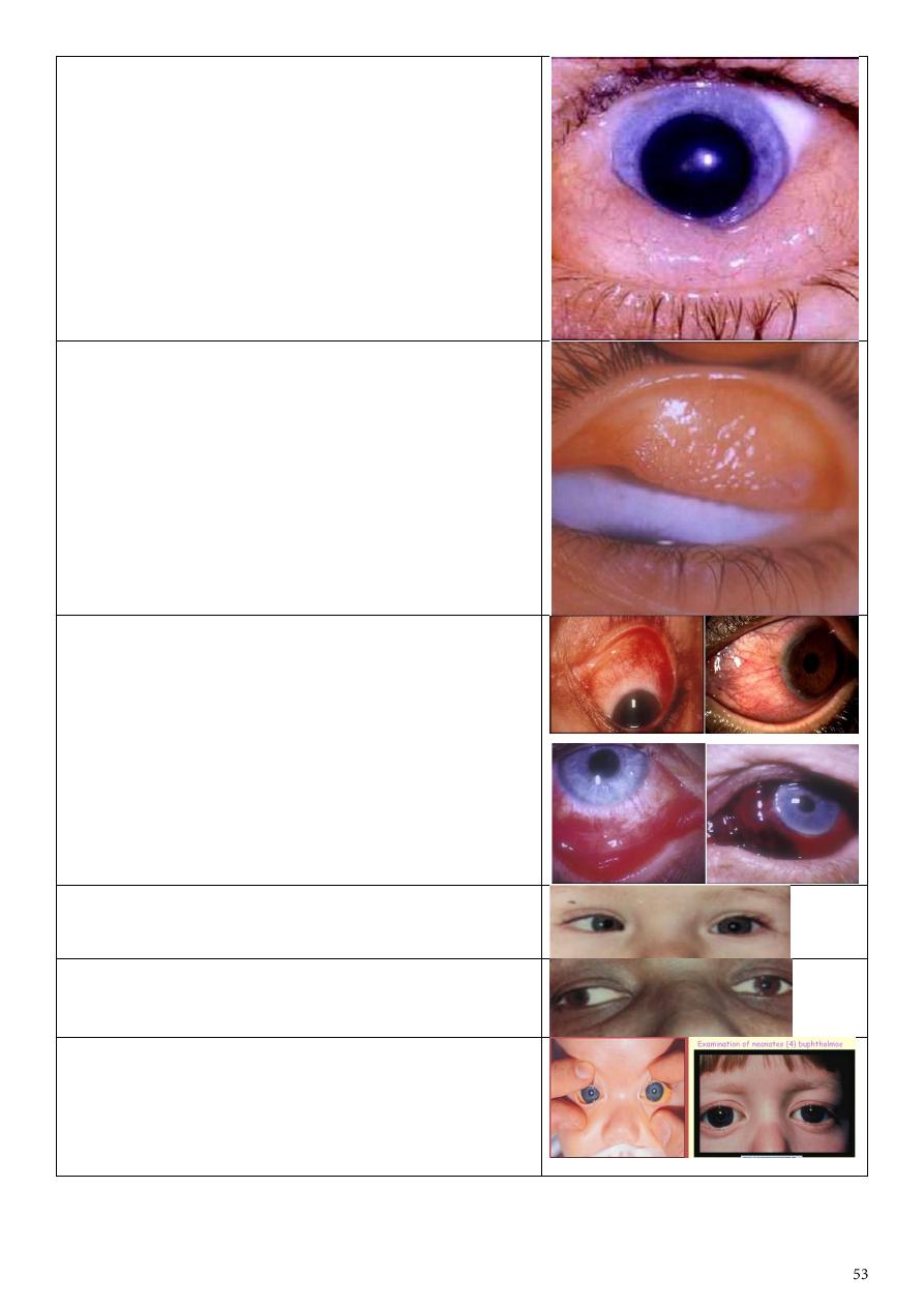
Marcus - Gunn phenomenon
Pathogenesis:
Faulty Innervation (motor fibers from 5th nerve
reach levator instead of the 3rd nerve).
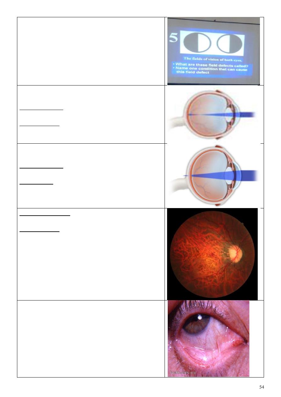
Astigmatism
Treatment:
Cylindrical lenses & refractive eye surgery
.
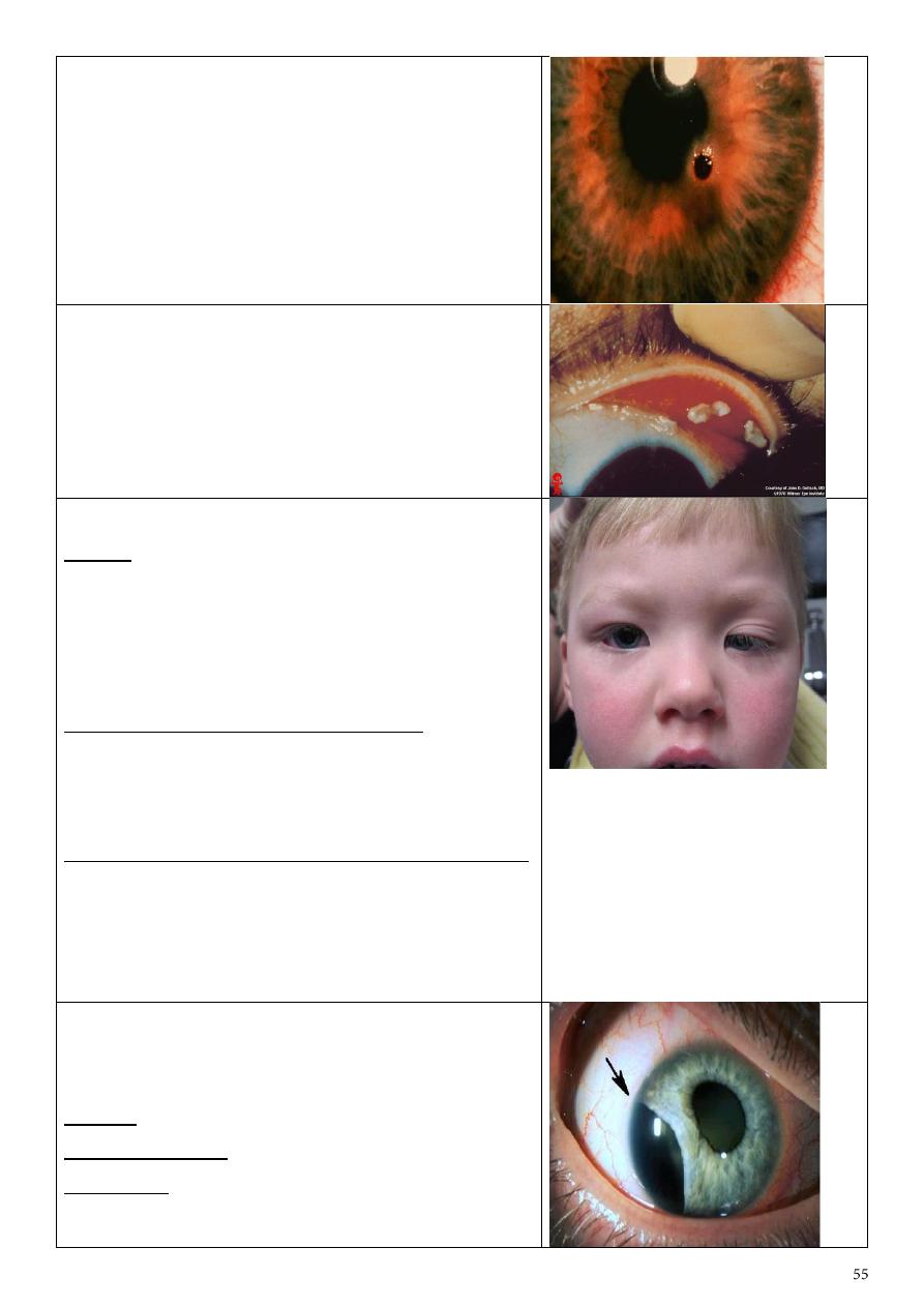
Papilleodema
Cause:
Elevated intracranial tension.
Chemosis
Edema of the conjunctiva.
Causes:
1) Conjunctivitis.
2) If drainage of blood & lymph from around the eye
is obstructed.
Describe:
Upper eyelid eversion, follicular formation.
Causes:
Trachoma & viral conjunctivitis.
Red congested eye
Common Causes:
1- Conjunctivitis.
2- Acute iritis (ant. Uveitis).
3- Keratitis (Corneal Ulcer).
4- Angle closure glaucoma.
5- Episcleritis (& scleritis).
6- Subconjunctival hemorrhage.
7- Dry eye.
Esotropia: convergent
Exotropia: divergent
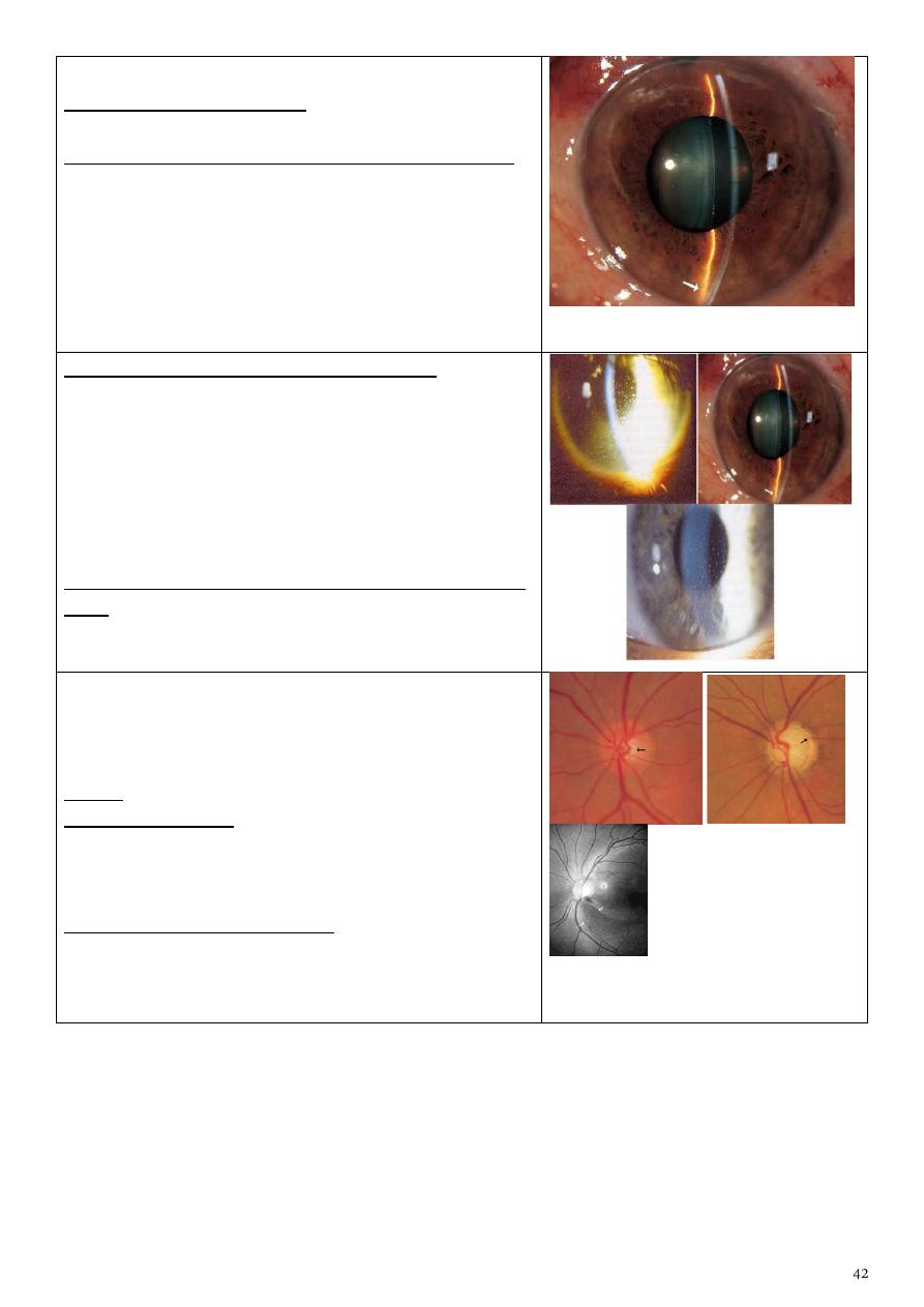
Buphthalmos
Surgical procedures:
1) Goniotomy.
2) Trabeculotomy.
Bitemporal hemianopia
Caused by: Optic chiasma lesions (nasal fibers
damage) e.g. Pituitary gland tumor.
Myopia
Correction is by: Concave lenses (-) & refractive eye
surgery.
Complications: Retinal detachment, macular hole,
open angle glaucoma.
Hypermetropia
Correction is by: Convex lenses (+) & refractive eye
surgery.
Significance: Angle closure glaucoma as a
complication.
Error of refraction: High myopia.
Complications:
1) Chorio-retinal degenerations.
2) retinal tears.
3) retinal detachment.
Symblepharon
Causes:
1) Post-trachomatous.
2) Post-operative (Pterygium excision).
3) Ocular cicatricial pemphigoid.
Corneal foreign body.
Treatment: Surgical removal.
F.B. in the inner surface of upper lid
Left partial ptosis
Causes:
1) Oculomotor nerve palsy.
2) Horner syndrome.
3) Birth trauma.
4) Congenital.
Factors affecting prognosis & treatment:
1) Is the pupil covered by the ptotic lid or not?
2) Are there significant refractive errors between the
eyes?
The significance is developing of amblyopia (causes):
1) Strabismus amblyopia due to squint.
2) Anisometropic amblyopia due to deferent
refractive errors.
3) Deprivational amblyopia.
Iridodialysis
A dark crescentic gap at the edge of the iris where
the tear has occurred.
Causes: Trauma, Tumor excision.
Patient complaint: Uniocular diplopia.
Treatment: Surgical reduction and suturing of the
iris.

Rubeosis iridis
(Growth of blood vessels onto the
iris)
& Peripheral iridectomy
Causes:
1) Diabetic retinopathy.
2) Central retinal vein occlusion (CRVO).
3) Long standing retinal detachment.
4) Ocular ischemic syndrome.
5) Hyoptonus eye.
6) Long standing uveitis.
7) Uveal melanoma, Retinoblastoma.
Complication: Rubeotic glaucoma & prognosis is very
bad.
Posterior Synechiae
Cause: Anterior uveitis (iritis).
Complications:
1) Band keratopathy.
2) Bulus keratopathy.
3) Cataract, Glaucoma.
Treatment:
1) Atropine & corticosteroid drops.
2) Surgical separation (synechialysis).
Mucopurulent conjunctivitis
Diffuse redness of the sclera with yellowish
mucopurulent discharge & eyelid edema.
Cause: Trachoma.
Complications:
1) Membrane formation.
2) Subsequent scarring of the punctum.
3) Corneal ulcer.
Treatment:
1) Eye lotions, Hot foments.
2) Antibiotics ointments e.g. tobramycin at night.
3) Antibiotic eye drops (tetracycline).
Phlyctenular conjunctivitis
Small pinkish-yellow nodule surrounded by a zone of
dilated blood vessels at corneoscleral junction.
Causes:
Hypersensitivity reaction (type 4) to endogenous
antigens e.g. bacterial antigens as T.B & Chlamydia.
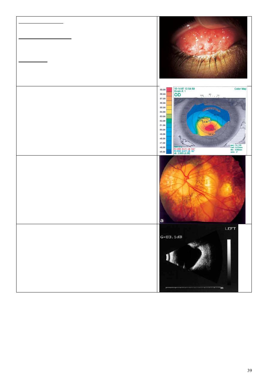
Ulcerative blepharitis
Erythema & crusting of the lashes and lid margins &
adherence of the lashes with each other by oily
debris.
Causes:
1) Herpes simplex & Varicella zoster dermatitis.
2) Allergic or contact dermatitis.
3) Bacterial infection (Staph.) is the usual pathogen.
4) Exposure to smoke, fumes & other irritants.
5) Sjogren syndrome may be present as blepharitis.
Complications:
Chronic conjunctivitis, Madarosis, trichiasis, Poliosis,
epiphora, Ectropion, corneal ulcer.
Treatment:
1) Eyelid margin hygiene (warming, washing &
application of antibiotic ointment to the lid margins).
2) Antibiotic – corticosteroid solutions to reduce
inflammation if the cause is bacterial.
3) Antivirals.
4) Avoiding exposure to irritant materials.
Bulbar spring catarrhal
Treatment:
Avoiding exposure to the causative allergen, topical
steroids, mast cell stabilizers, anti-histaminic dark
glasses, cold compresses.
Ophthalmia neonatorum
(neonatal conjunctivitis)
Causes:
1) Chemical (silver nitrate).
2) Bacterial (1st 3 days=Staph.aureus , after the 3rd
day= Strept.peumonia , after the 1st week=Neisseria
gonorrhea , after 12 days=Chlamydia).
3) after 15 days= H. influenza & herpes simplex
virus).
Treatment:
1) Gonococcal infection treated by cefotaxime for 7
days.
2) Chlamydial infection by systemic erythromycin.
3) Herpes by I.V acyclovir.
4) Topical antibiotic for staph.
Stye
Red swelling of the upper eyelid due to
inflammation of eyelash follicles.
Causes:
Bacterial infection staph. Aureus infection.
Treatment:
1) Local antibiotics & eye drops.
2) Bathing in warm water.
3) Removal of the eyelash involved.
Entropion with trichiasis & corneal ulceration
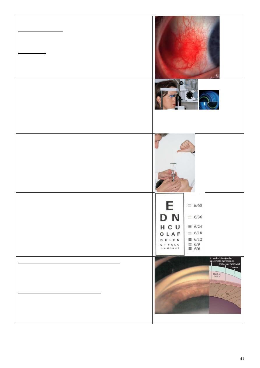
Retinopathy of prematurity
• in premature babies peripheral retina is not
vascularised at birth
• Under certain conditions instead of the
vascularisation proceeding normally, there is a
growth of new vessels and scarring
• 100% oxygen, crucial period 28-36weeks,
especially low birth weight
• vascularisation
• Laser peripheral retina
• Without laser,retinal detachment/blindness;
have to check (screen) all appropriate neonates
Cicatricial ectropion
Treatment: V to Y plasty or Z plasty
Phlycten
Causes:
Hyper sensitivity to an endogenous antigen e.g.
tuberculo-protein, Intestinal parasites,
staphylococcal blepharoconjunctivitis.
Acute congestive glaucoma
2 Medications for emergency: hyper-osmotic agent,
topical miotics, topical steroids
Treatment: essentially surgical recent….surgical
iridectomy late….their is PAS, an external fistulizing
operation.
Nerve& muscle affected: Right Abducent nerve -
right lateral rectus
Direction of gaze: To the right
Main complaint: Binocular Diplopia
Type of squint: exotropia
angle: 30
Confirmatory test: Cover test
After cataract
Treatment:
- No interference if vision is not affected
- If thick : surgical intervention
Senile ectropion
Possible complications:
1- xerosis
2- corneal ulcer
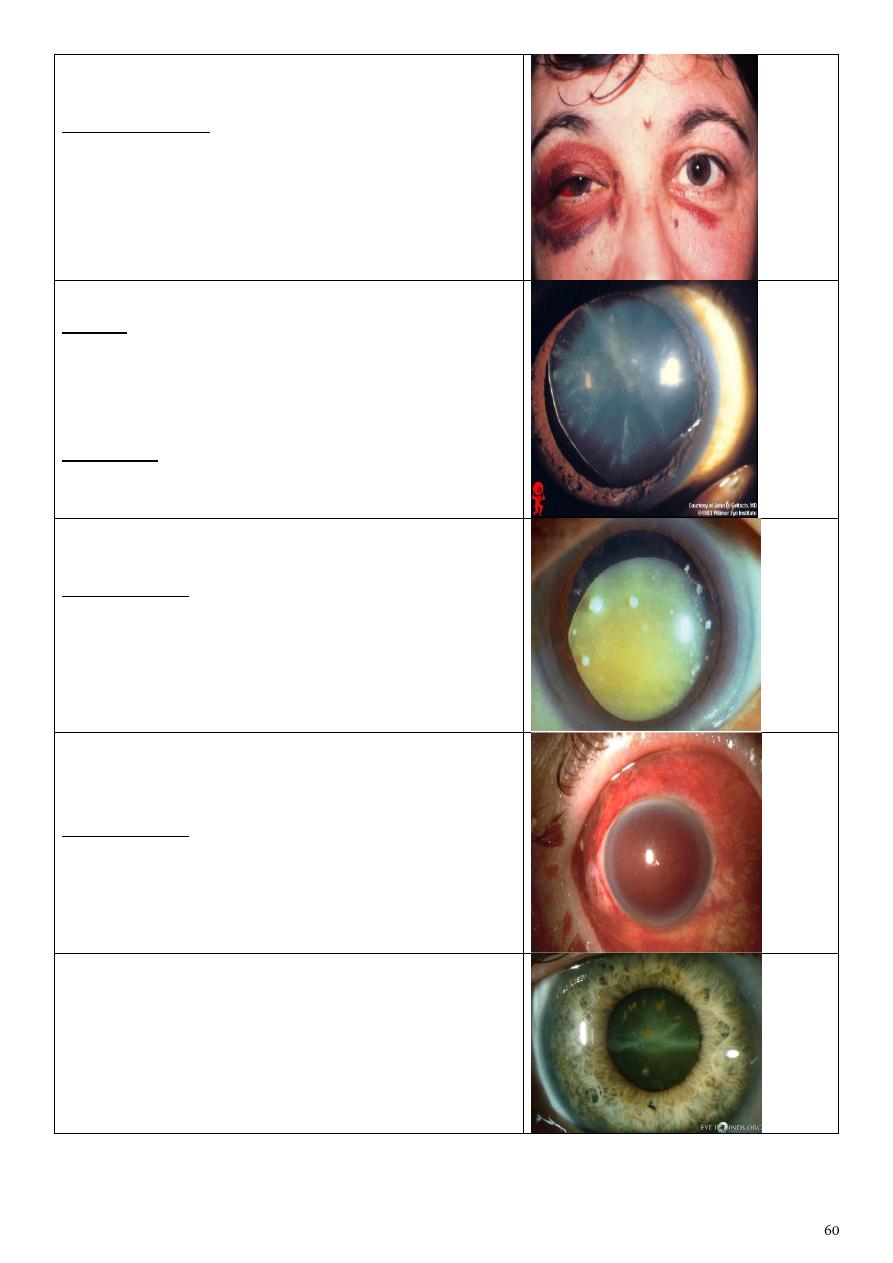
Ecchymosis & subconjunctival haemorrhage
Possible findings:
Concussion cataract " Rosette-shaped"
Lens subluxation or dislocation
Lens subluxation
Causes:
1- Marfan's syndrome
2- Homocystenuria
3- Trauma
4- High myopia
Symptoms:
Blurred vision, diplopia, increase IOP, pupillary block
(glaucoma), affected visual fields.
S
ublaxated & cataractous lens
Complications:
lens dislocation
2ry Glaucoma
Iridocyclitis
Blood staining of the cornea
“total hyphema or
8-ball hyphema”
Complications:
Elevation of IOP
Corneal staining
Sederosis bulbi
Patient with foreign body in his eye from one year
In picture you will see one eye normal &other
eye(black iris)

Scotoma
Area of blindness surrounded by area of vision due
to lesions in specific regions of retina.
Tunnel vision
Only see the central part.
For more photos https://goo.gl/YCZx7a