The Red Eye
Differential Diagnosis

Differential Diagnosis of “red eye”
Conjunctiva
Pupil Cornea
Anterior
chamber
IOP
Subconjunctival
Haemorrhage
Bright red
Normal Normal Normal
Normal
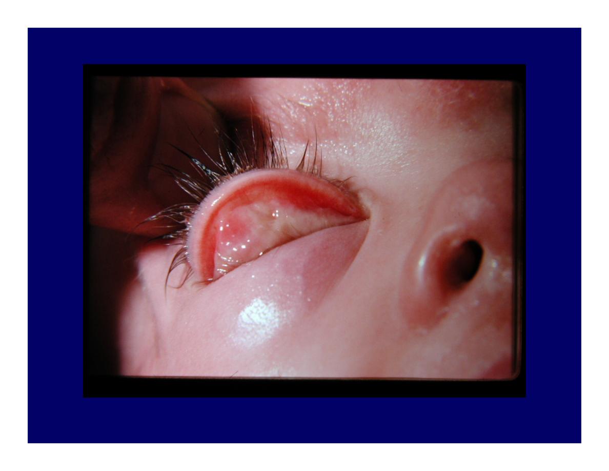
Conjunctivitis
Injected
vessels,
fornices.
Discharge
Normal Normal Normal
Normal
Iritis
Injected
around cornea
Small,
fixed,
irregular
Normal,
KPs
Turgid,
deep
Normal
Acute glaucoma
Entire eye red
Fixed,
dilated,
oval
Hazy
Shallow
High
Conjunctivitis
Follicles
Purulent discharge
Papillae
Chemosis
Redness
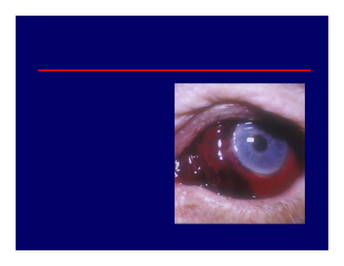
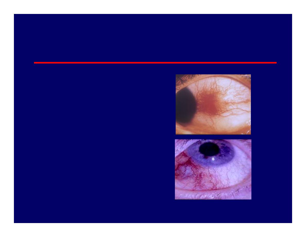
Subconjunctival Haemorrhage
• Diffuse or localised
area of blood under
conjunctiva.
Asymptomatic
• Idiopathic, trauma,
cough, sneezing,
aspirin, HT
• Resolves within 10-14
days
Dry Eye Syndrome
• Poor quality
– Meibomian gland disease,Acne rosacea
– Lid related
– Vitamin A deficiency
• Poor quantity
– KCS
• Sjogren Syndrome
• Rheumatoid Arthritis
– Lacrimal disease ie, Sarcoidosis
– Paralytic ie, VII CN palsy

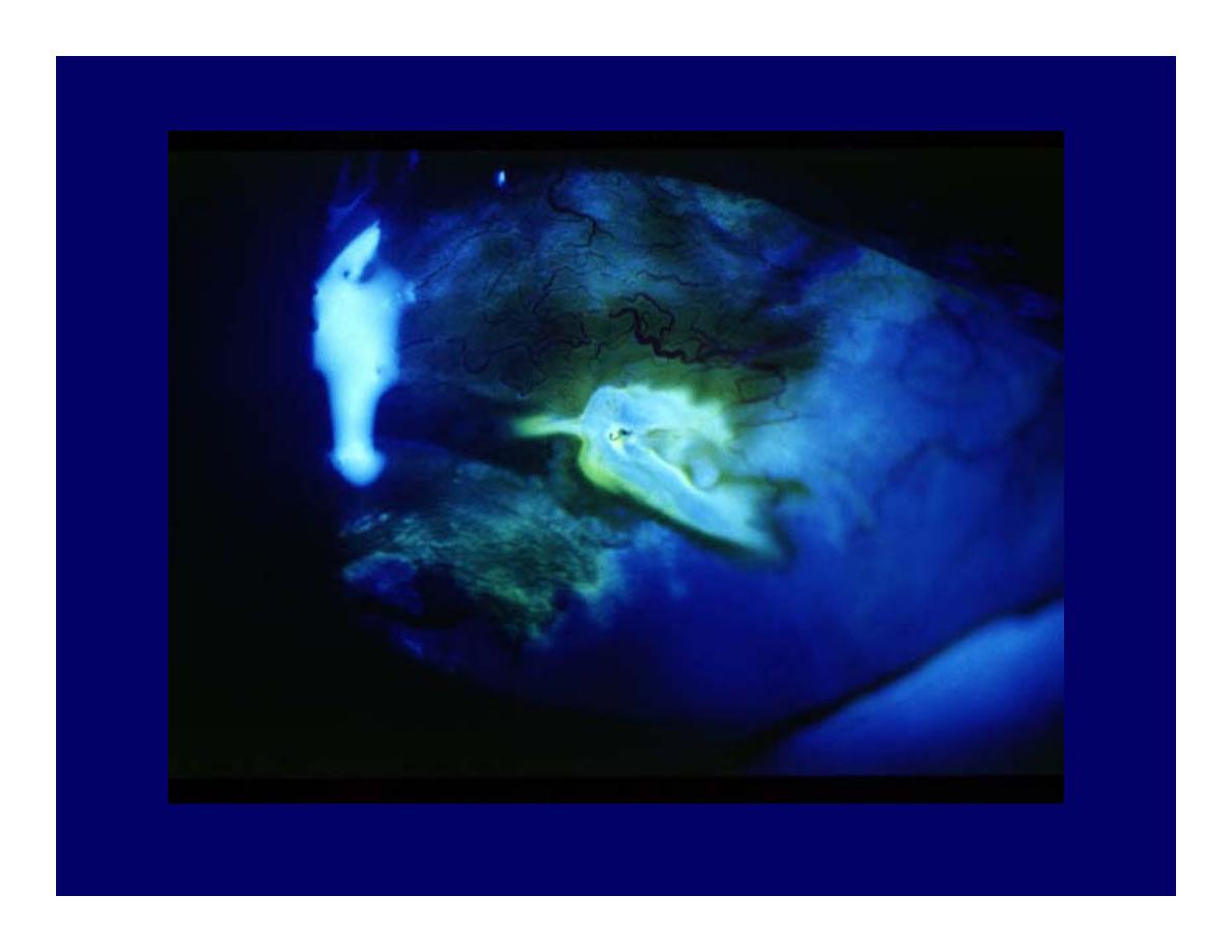
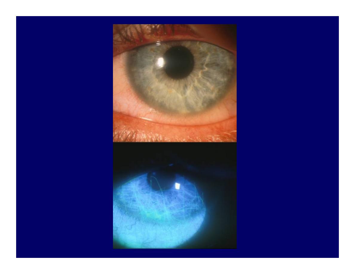
Corneal Abrasion
• Surface epithelium sloughed off.
• Stains with fluorescein
• Usually due to trauma
• Pain, FB sensation, tearing, red eye

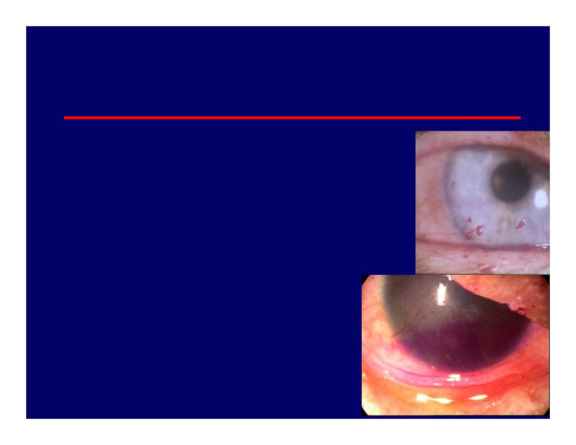
Corneal Ulcer
• Infection
– Bacterial: Adnexal infection, lid malposition,
dry eye, CL
– Viral: HSV, HZO
– Fungal:
– Protozoan: Acanthamoeba in CL wearer
• Mechanical or trauma
• Chemical: Alkali injuries are worse than acid
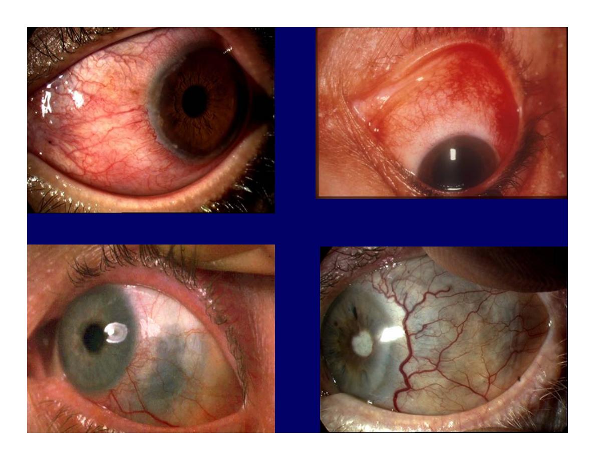
Episcleritis
• Superficial
• Idiopathic, collagen
vascular disorder (RA)
• Asymptomatic, mild
pain
• Self-limiting or topical
treatment

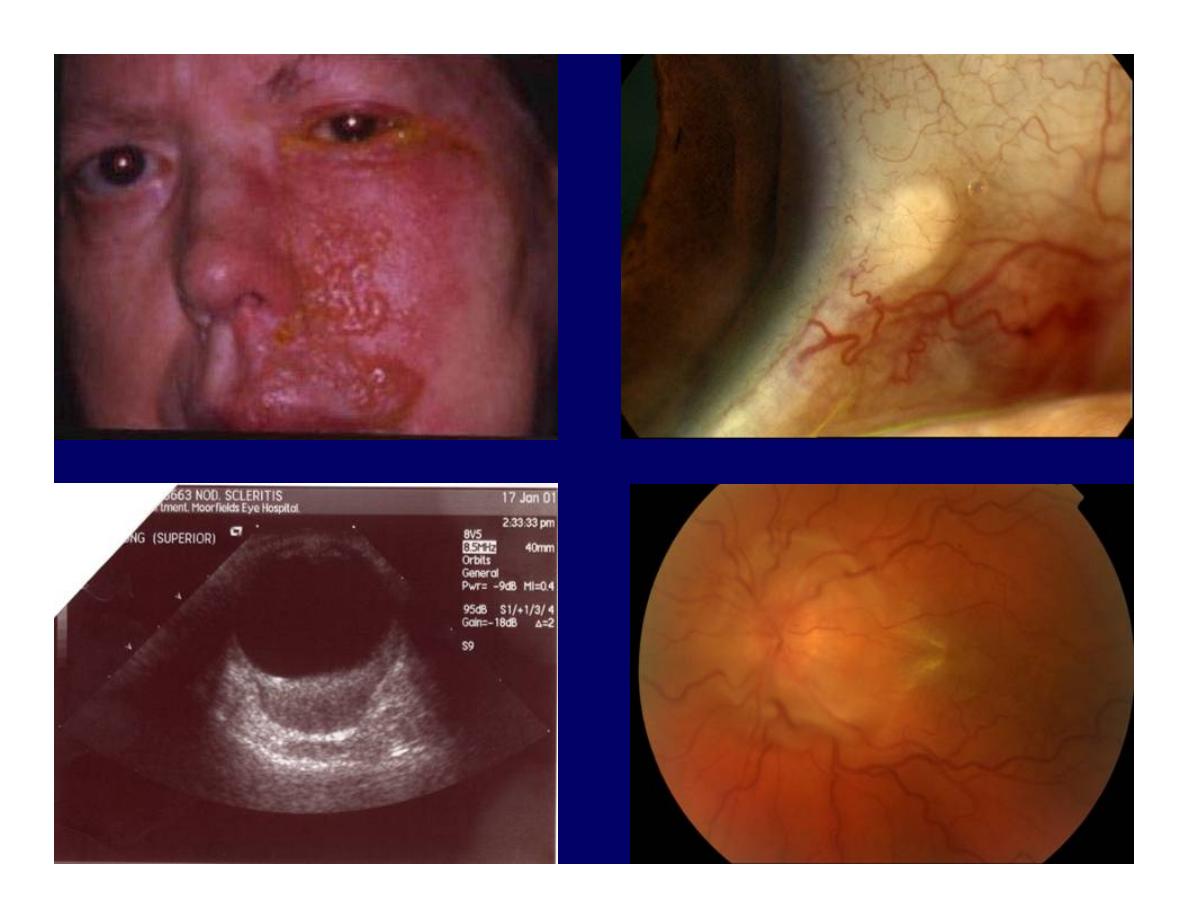
Scleritis
• Deep
• Idiopathic
• Collagen vascular disease (RA,AS, SLE,
Wegener, PAN)
• Zoster
• Sarcoidosis
• Dull, deep pain wakes patient at night
• Systemic treatment with NSAI or Prednisolone if
severe

Uveitis
Anterior: acute recurrent and chronic
Posterior: vitritis, retinal vasculitis, retinitis,
choroiditis
Panuveitis:anterior and posterior

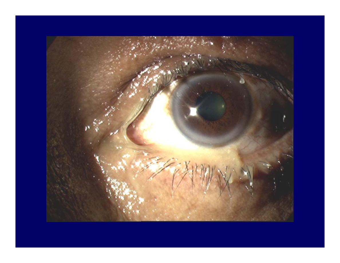
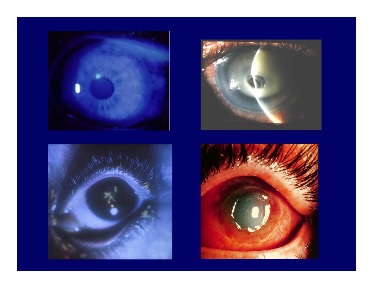
Anterior uveitis (iritis)
• Photophobia, red eye, decreased vision
• Idiopathic. Commonest
• Associated to systemic disease
– Seronegative arthropathies:AS, IBD, Psoriatic
arthritis, Reiter’s
– Autoimmune: Sarcoidosis, Behcets
– Infection: Shingles, Toxoplasmosis, TB,
Syphillis, HIV
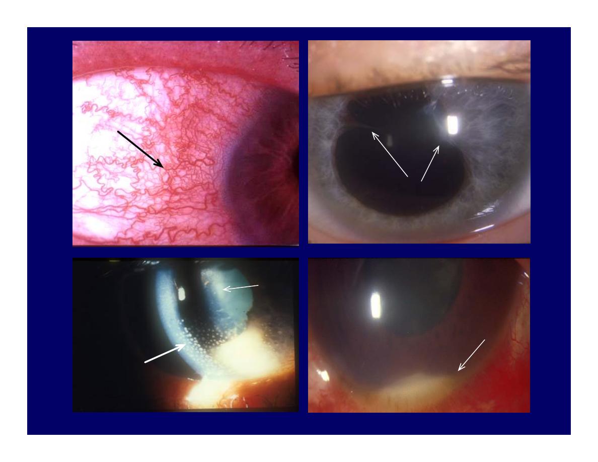
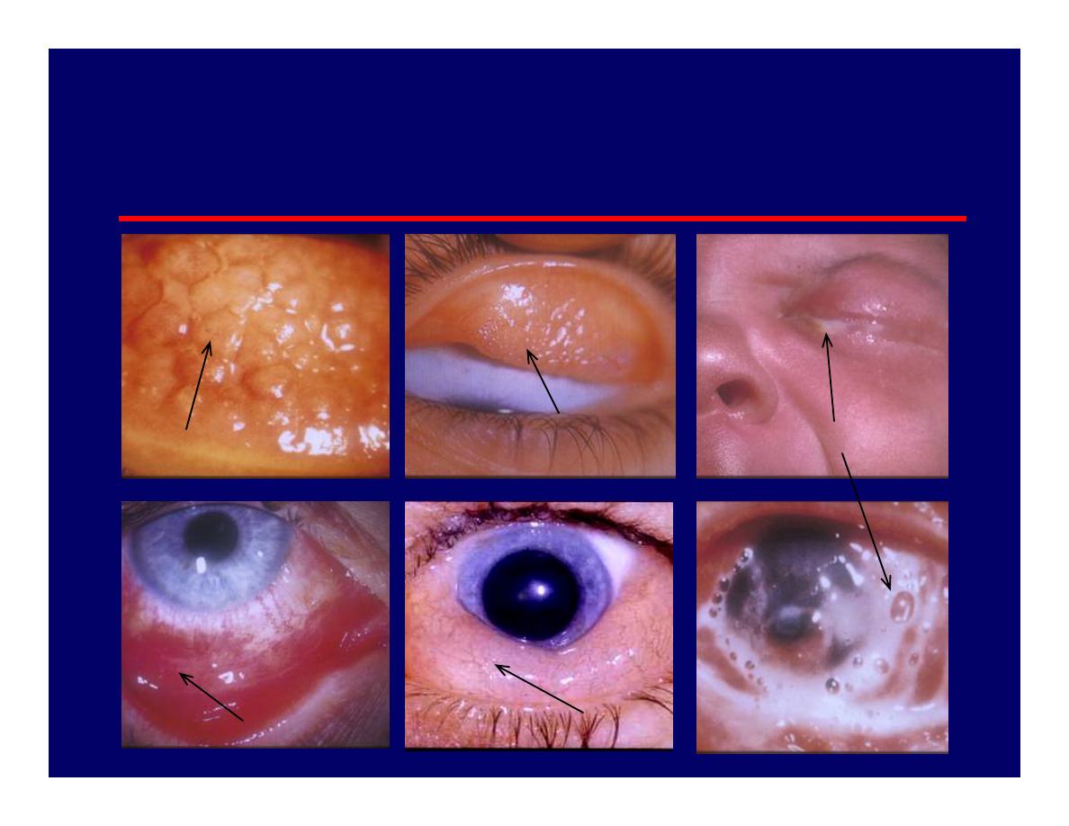
Ciliary flush
Posterior synechiae
KPs
Fibrin
Hypopyon
Flare
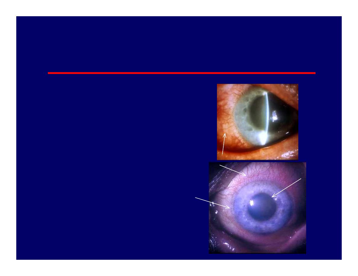
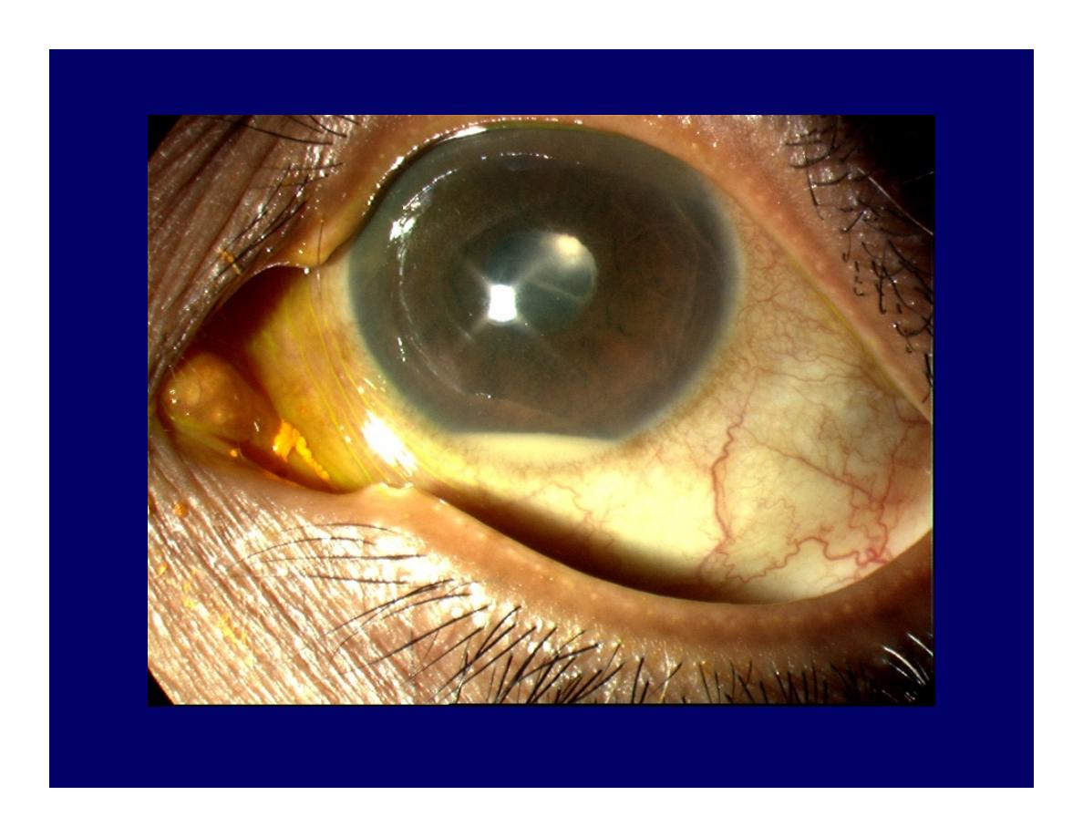
Acute Angle-closure Glaucoma
• Symptoms
– Pain, headache,
nausea-vomiting
– Redness, photophobia,
– Reduced vision
– Haloes around lights
Corneal oedema
Ciliary hyperaemia
Dilated pupil