The HandLab Session 10
Dr. Hayder Jalil Al-AssamMBChB (Iraq), MRes Anatomy (UK)
Email: dr_hayder_anatomy@yahoo.com
Palmar Aponeurosis
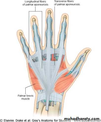
The Palmar Aponeurosis: is triangular thickening of the deep fascia that occupies the central area of the palm.
The medial and lateral borders of the palmar aponeurosis are continuous with the thinner deep fascia covering the hypothenar and thenar muscles.
The function of the palmar aponeurosis is to give firm attachment to the overlying skin and so improve the grip and to protect the underlying tendons.
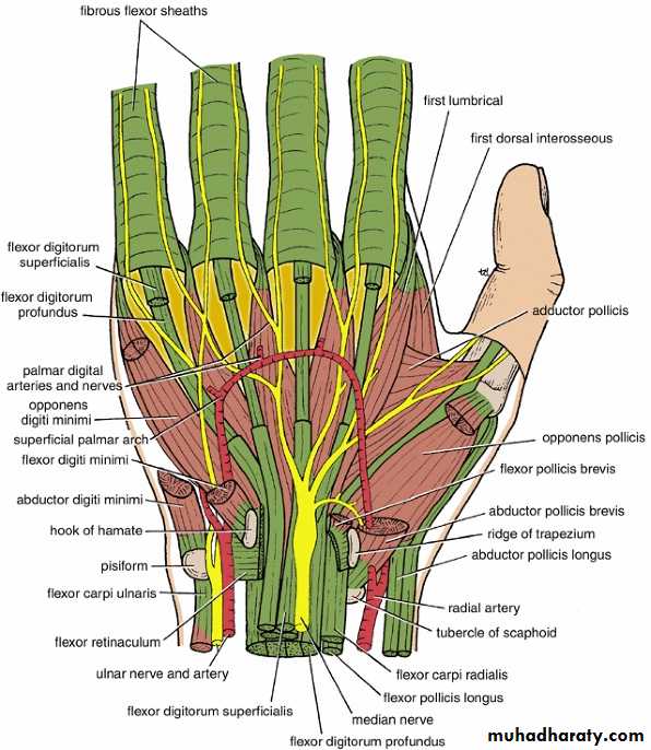
Synovial Flexor Sheaths
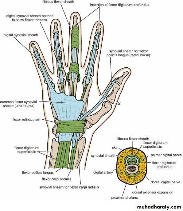
The tendons of the flexor digitorum superficialis and profundus muscles have a common synovial sheath.
The medial part of this common sheath extends distally without interruption on the tendons of the little finger.
The flexor pollicis longus tendon has its own synovial sheath that passes into the thumb.
The synovial sheath of the flexor pollicis longus communicates with the common synovial sheath at the level of the wrist in about 50% of subjects.
Fibrous Flexor Sheaths
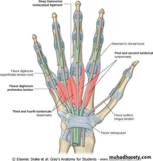
Each tendon of the flexor digitorum superficialis enters the fibrous flexor sheath & divides into two halves, which pass around the profundus tendon and meet on its deep or posterior surface.
With partial decussation of the fibers, the superficialis tendon divides into two further slips and attached to the borders of the middle phalanx.
Each tendon of the flexor digitorum profundus pass through the division of the superficialis tendon to be inserted into the base of the distal phalanx
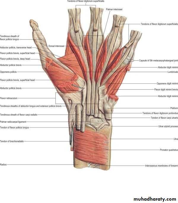
Small Muscles of the Hand
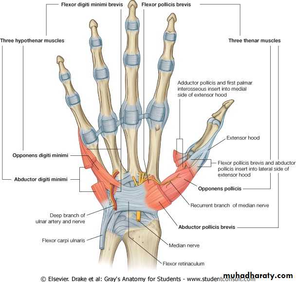
The small muscles of the hand include the four lumbrical muscles, the eight interossei muscles, the short muscles of the thumb, and the short muscles of the little finger.The short muscles of the thumb are the abductor pollicis brevis, the flexor pollicis brevis, the opponens pollicis, and the adductor pollicis. The first three of these muscles form the thenar eminence.
The short muscles of the little finger are the abductor digiti minimi, the flexor digiti minimi brevis, and the opponens digiti minimi, which together form the hypothenar eminence.
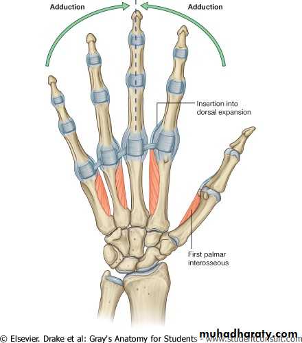
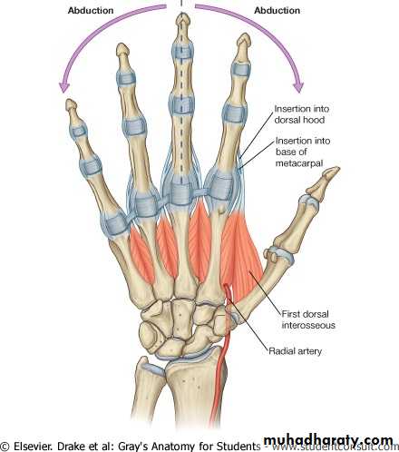
Dorsal & Palmar interossei (8)
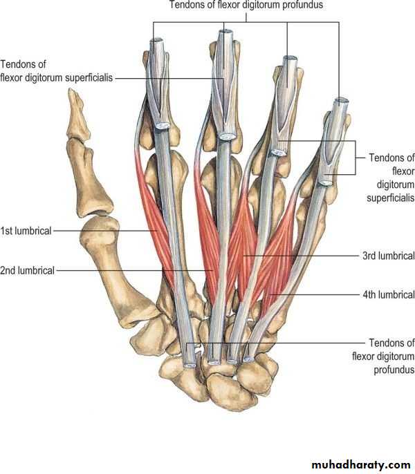
Lumbricals (4)
Short muscles of the thumb & little finger
Thumb muscles
• abductor pollicis brevis• The flexor pollicis brevis
• The opponens pollicis
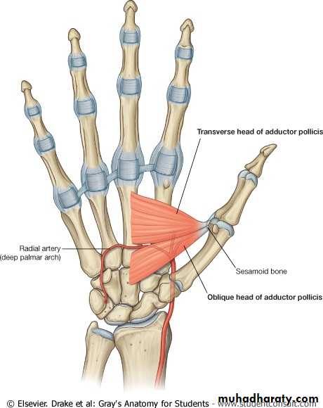
• The adductor pollicis
Little finger muscles
• The abductor digiti minimi
• The flexor digiti minimi brevis
• The opponens digiti minimi
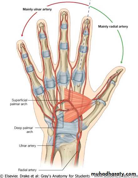
Arteries of the Palm
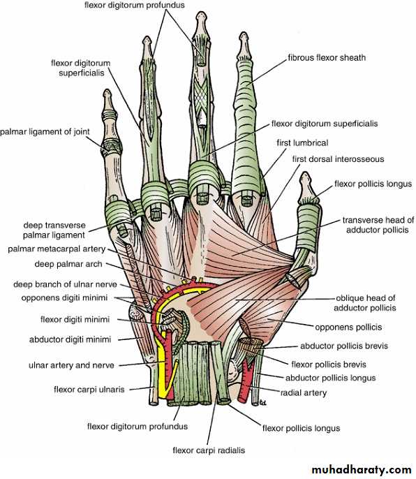
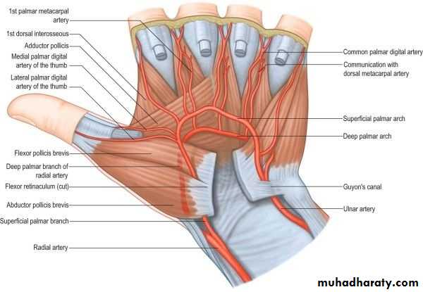
Ulnar Artery: enters the hand anterior to the flexor retinaculum on the lateral side of the ulnar nerve and the pisiform bone. The artery gives off a deep branch and then continues into the palm as the superficial palmar arch.
The deep branch of the ulnar artery arises in front of the flexor retinaculum, passes between the abductor digiti minimi and the flexor digiti minimi, and joins the radial artery to complete the deep palmar arch.
Radial Artery: leaves the dorsum of the hand by turning forward between the proximal ends of the first and second metacarpal bones and the two heads of the first dorsal interosseous muscle.
The deep palmar arch sends branches superiorly, which take part in the anastomosis around the wrist joint, and inferiorly, to join the digital branches of the superficial palmar arch.
Nerves of the Palm
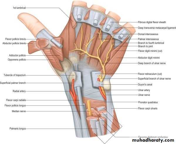
Median Nerve: The median nerve supplies the muscles of the thenar eminence (the abductor pollicis brevis, the flexor pollicis brevis, and the opponens pollicis) and the first 2 lumbrical muscles.
Ulnar Nerve: divides into a superficial and a deep terminal branch.
Superficial Branch of the Ulnar Nerve: The superficial branch of the ulnar nerve gives off the following branches: a muscular branch to the palmaris brevis and cutaneous branches to the palmar aspect of the medial side of the little finger and the adjacent sides of the little and ring fingers.
Deep Branch of the Ulnar Nerve: gives off muscular branches to the three muscles of the hypothenar eminence, namely, the abductor digiti minimi, the flexor digiti minimi, and the opponens digiti minimi. It supplies all the palmar and dorsal interossei, the third and fourth lumbrical muscles, and both heads of the adductor pollicis muscle.
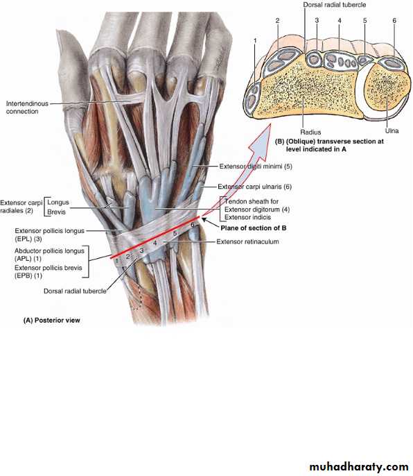
Dorsum of the hand
The four tendons of the extensor digitorum, tendon of the extensor indicis and the two tendons of the extensor digiti minimi.
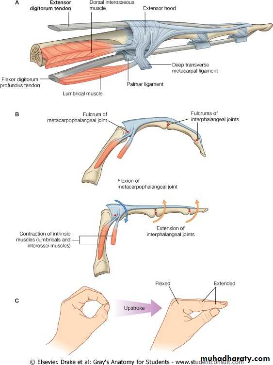
On the posterior surface of each finger, the extensor tendon joins the fascial expansion called the extensor expansion.
Dorsal Extensor Expansion
Near the proximal interphalangeal joint, the extensor expansion splits into three parts: a central part, which is inserted into the base of the middle phalanx, and two lateral parts, which converge to be inserted into the base of the distal phalanxThe dorsal extensor expansion receives the tendon of insertion of the corresponding interosseous muscle on each side and farther distally receives the tendon of the lumbrical muscle on the lateral side .