Embryology & Anatomy of Female Genital Tract
2016-2017EMBRYOLOGY
Development of the genital organsDevelopment of the uterus and Fallopian tubes
Development of the vagina
Development of the ovary
Development of external genitalia
ANATOMY:
EXTERNAL GENITALIA
Bartholin's glands
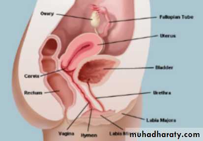
INTERNAL REPRODUCTIVE ORGANS
The vagina
Uterus
The cervix
The Fallopian tubes (oviducts)
The ovaries
The blood supply
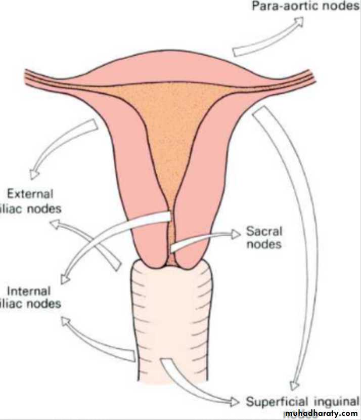
The lymphatic drainage
The nerve supply of pelvis
EMBRYOLOGY
• Development of the genital organs
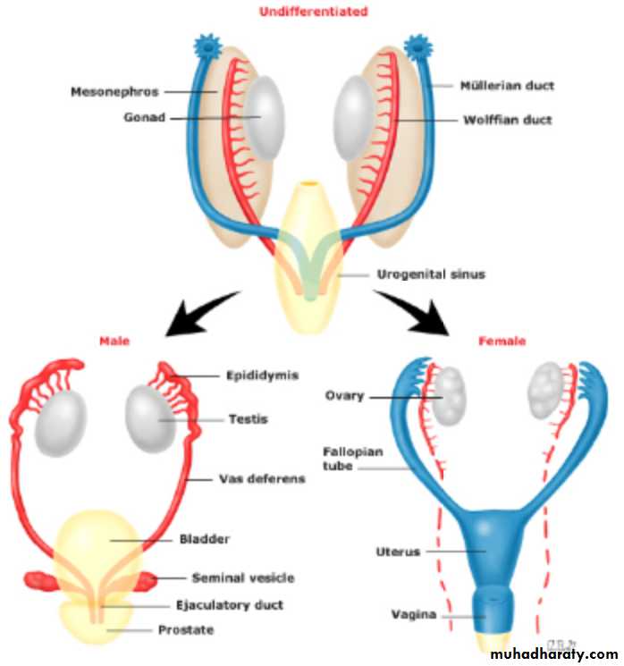
• Following fertilization, the normal embryo contains 23 sets of chromosomes, including 22 autosomes and one sex chromosomes from each parent.
46xy embryo will develop as a male
46xx embryo will develop as a female.
The presence or absence of the y chromosome which determines whether the undifferentiated gonad becomes a testis or an ovary.
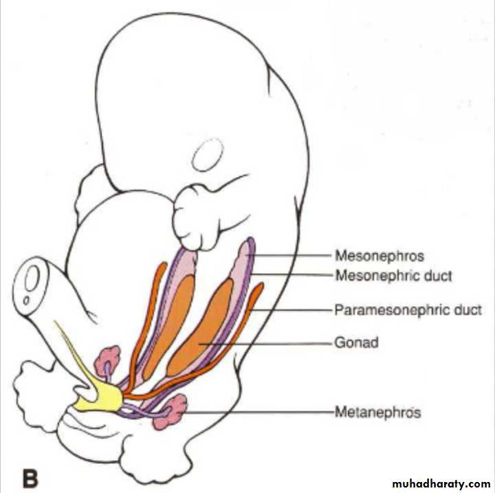
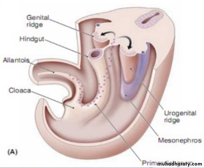
By the 5th week of embryonic life, both male and female embryos start to develop the following structures on either side of the midline.
1-genital ridge.
2-mesonephric (wolffian) duct.3-para meso-nephric (Mullerian)duct.
Development of the uterus and Fallopian tubes
The lower end of the Mullerian ducts come together in the mid line, fuse and develop into uterus and cervix.
The upper parts of both ducts form the Fallopian tube .The lower end of the fused Mullerian ducts beyond the uterin luman remains solid forms a cord.
Development of the vagina
The Mullerian ducts reach down to the urogenital sinus and at the meeting point, form the Mullerian tubercle which meet apair of sino-vaginal bulb.These sino-vaginal bulbs, proliferate and form a solid vaginal plate. By the 5th month, the vaginal plate is entirely canalized to form vagina.The luman of the vagina remains separated from the urogenital sinus by a thin tissue plate, the hymen.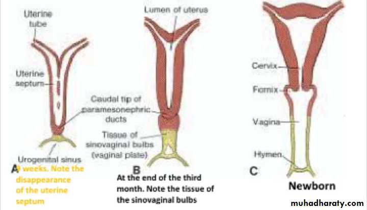
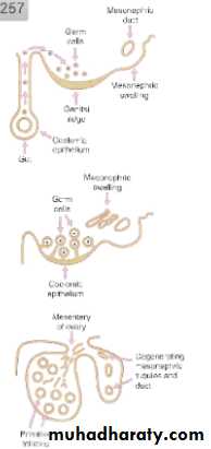
Development of the ovary
• If there is no y chromosome, the gonads, forms an ovary .The primitive gonad is first evident in embryos at 5 weeks. It forms as a bulb on the medial aspect of the meso-nephric ridge & is of triple origin.*Soelomic epithelium of the genital ridge
*The underlying mesmesoderm
*The primitive germ cells.
Approximately 7 million germ cells are present at 5 month, but at birth this has fallen to 2 million.
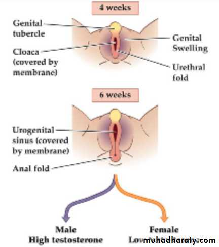
Development of external genitalia
The female development is a simple progression from these structures ;
*genital tubercle_______clitoris*genital folds_______labia minora
*genital swellings________labia majora.
ANATOMY
EXTERNAL GENITALIA(the vulva) include ;
1-The mons pubis
2- The labia majora3- The labia minora
4- The clitoris
5- The vestibule and the vestibular orifice
6- The greater vestibular glands
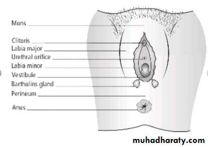
Bartholin's glands, each about the size of a small pea, lie at the base of each bulb and open via a 2 cm duct into the vestibule between the hymen and the labia minora. Some times the ducts of this gland obstruct leading to Bartholin cyst and if infection develop it may lead to Bartholin abscess.
INTERNAL REPRODUCTIVE ORGANS
1-The vagina2-The uterus
3-The fallopian tubes
4-The ovaries
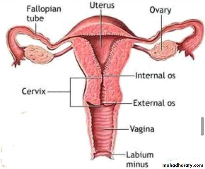
The vagina
* It is a fibromuscular canal lined with stratified squamous epithelium . It islonger in the posterior wall (9 cm) than anteriorly (7 cm). The vault of the vagina is divided into 4 fornices ; posterior, anterior and two lateral.
*The vaginal walls are rugose with transverse folds
*It has no glands.
*The epithelium is thick and rich in glycogen, the growth of Doderlein's bacillus a normal commensal of the vagina that breaks down the glycogen to form lactic acid, producing a PH of around 4.5
Age changes
#At birth, the vagina is under the influence of maternal oestrogens, so the epithelium is well developed. After a couple of weeks, the effects of oestrogen disappear and the PH rises to 7 and the epithelium atrophies.# At puberty, the reverse occurs.
# At the menopause,the vagina tends to shrink and the epithelium atrophies.
Uterus
It is a fibromuscular organ & in non-pregnant state is situated entierly within the pelvis.Pre-pregnancy; 7*5*3 cm; weight 70 g.
Full term; 30*25*20 cm; weight 1000 g.It consist of
The corpus (body)
The cervix
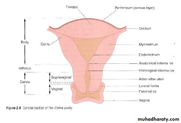
The corpus
It is the upper part of the uterus consist of :1-the fundus, the upermost part.
2-cornu, which is the site of insertion of the Fallopian tube.
3-the isthmus, when uterus tapers to a small central constricted area, ends by attaching to the cervix.
The constriction at the isthmus where the corpus joins the cervix is the anatomical internal os.
Uterus is in anteflexion and anteversion position.
In around 20% of women, this tilt is not forwards but backwards-retroversion and retroflexion.
The uterus consists of 3 layers
*peritoneum; the outer aerosol layer; laterally it spreads out to form the leaves of the broad ligament.*myometrium; the middle muscular layer;
*endometrium; the inner mucous layer; covered by a single layer of columnar epithelium . This epithelium is under go cyclical changes and lost due to effects of pregnancy & menstruation.
Uterus is supported by ligaments
1-transverse cervical( cardinal ligaments)2-round ligament
3-uterosacral ligaments.
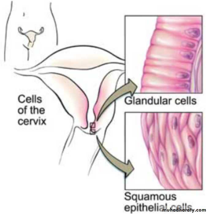
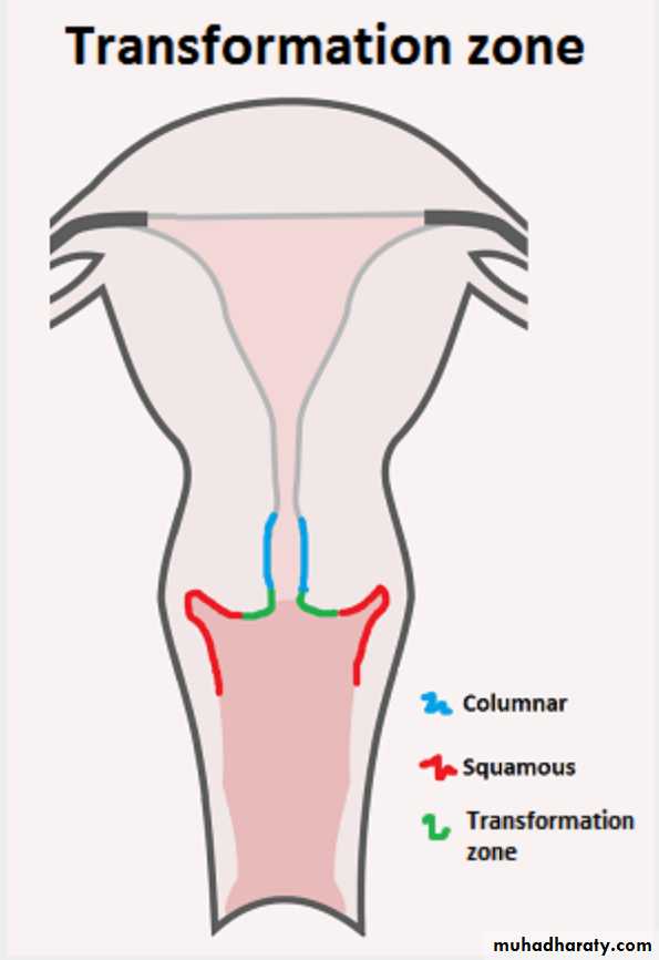
The cervix
Is a narrower than the body of the uterus approximately 2.5-3 cm in length It consist of two parts :* supra vaginal part: lined with columner ep.
*vaginal part :protruding into the vagina, lined with stratified sequamous ep.
The squamocolumner junction is also known as the transformation zone.
Age changes
* After birth, the disappearance of maternal oestrogens causes the uterus to decrease in length by around one third and cervix is then twice the length of the uterus.*At puberty, the corpus grows much faster and the size ratio reverses.
*After menopause, the uterus atrophied, the mucosa becomes very thin, the glands almost disappear & the wall becomes less muscular.
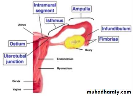
The Fallopian tubes (oviducts)
Each tubeis about 10 cm longThe tubes convey the ovum from the ovary towards the uterus & its the site of fertilization which provides oxygenation & nutrition for sperm, ovum & zygote.
It is distiguished in to 4 parts;
• 1-interstitial part
• 2-the isthmus• 3-ampulla
• 4-infundibulum
The ovaries
In the young adult, they are almond shaped, solid, a greyish pink and approximately 3 ×1.5 ×1cm,10g weights. It has a twin functions both steroid production and gametogenesis.The ovary is the only intra-abdominal structure not covered by peritonum.
Age changes
*In the child, they are small structures, 1.5 cm long. They have a smooth surface & at birth contain between1 and 2 million primordial follicles.
*At puberty, the ovaries increase in size as a result of proliferation of the stromal cells.
*After the menopause, no active follicles are present and the ovary becomes a small, shrunken with a wrinkled surface.
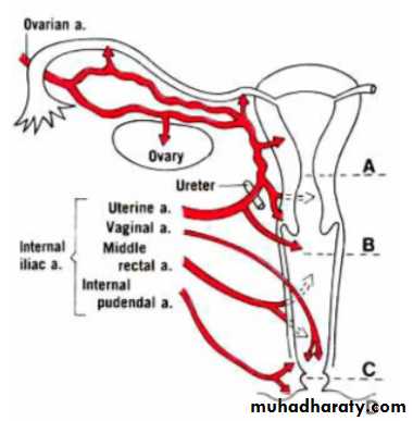
The blood supply
1-Ovarian A.2-Uterine A.
3-Vaginal A.
4-Internal pudendal A.
The lymphatic drainage
***Ovaries and tubes_____para aortic LN.***Uterus, cervix and upper⅔ of vagina____internal iliac, obturator and external iliac LN, common iliac and para aortic LN.
***Vulva, lower⅓ of vagina.___superficial inguinal and femoral LN
The nerve supply of pelvis
*Pudendal nerve (S2,3,4)Sensory fibres from the mons and labia also pass in the ilioinguinal and genitofemoral n. to the 1st lumber root.
*Posteriofemoral cutaneous n.
*Sympathetic n.fibers of the pre aortic plexus
*Para sympathetic fibers from S2, S3, S4