Fifth stage
PediatricLec-3
د.خليل
19/10/2016
The cardiovascular systemCyanotic Congenital Heart Disease
Lesions Associated with Decreased Pulmonary Blood Flow
Tetralogy of Fallot
Epidemiology and pathology
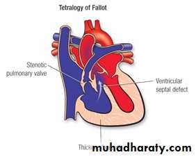
Tetralogy of Fallot is the most common cyanotic congenital heart defect, representing about 10% of all congenital heart defects .
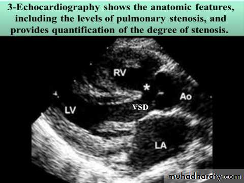
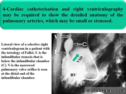
Anatomically, there are four structural defects: VSD, pulmonary stenosis, overriding aorta and right ventricular hypertrophy.
The VSD is large and the pulmonary stenosis is most commonly
subvalvularor (infundibular).
Clinical features
A- Symptoms
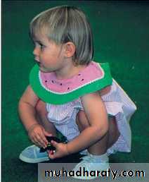
Most patients are symptomatic with cyanosis at birth or shortly thereafter. Dyspnea on exertion, squatting, or hypoxic spells develop later, even in mildly cyanotic infants .
Occasional infants with acyanotic TOF may be asymptomatic or may show signs of CHF from a large left-to-right ventricular shunt.
Immediately after birth, severe cyanosis is seen in patients with TOF and pulmonary atresia.
Treatment of spells

Placement of the infant on the abdomen in the knee-chest position while making certain that the infant's clothing is not constrictive. Premature attempts to obtain blood samples may cause further agitation and –ve effect.
O2 adminstration
S.c morphine not > 0.2mg/kg.
B-adrenergic blockers e.g propranolol (0.1-0.2) mg /kg i.v and slowly.
Physical examination
Growth and developmental delay in patients with severe untreated tetralogy of Fallot, particularly when oxygen saturation is chronically <70%. Puberty may also be delayed in patients who do not undergo Surgery.Varying degrees of cyanosis, tachypnea, and clubbing (in older infants and children) are present.
The pulse is usually normal, as is venous and arterial pressure.
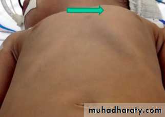
The left anterior hemithoraxmay bulge anteriorly because of RVH.
In about half the cases, a systolic thrill is felt along theleft sternal border .
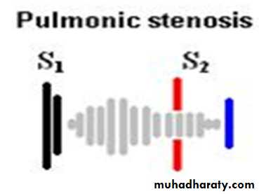
The murmur is usually loud, systolic, most intense at the left sternal border. It is due to PS and generally ejection in quality , but it may sound more holosystolic toward the lower sternal border.
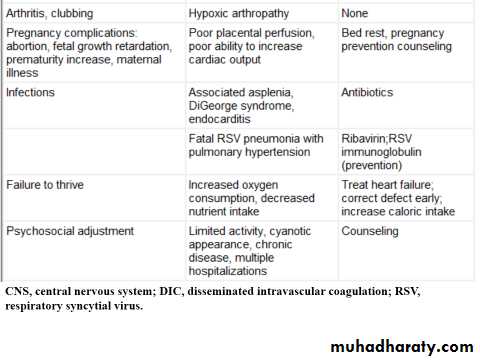
Natrural history and complications
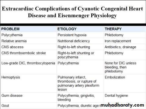
Worsening of cyanosis with time.Physicians need to watch for the development of relative iron-deficiency state (i.e., hypochromia).
Polycythemia develops secondary to cyanosis.
Growth retardation may be present if cyanosis is severe.
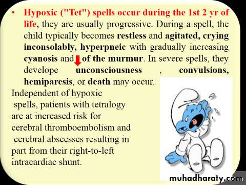
Hypoxic spells may develop in infants.
Brain abscess and cerebrovascular accident rarely occur
SBE is occasionally a complication.
8.Coagulopathy is a late complication of a long-standing cyanosis.
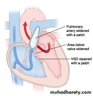
Management
Initial management is medical, with corrective surgery at around 6 months of age. It involves closing the VSD and relieving right ventricular outflow tract obstruction with an artificial patch, which sometimes extends across the pulmonary valve.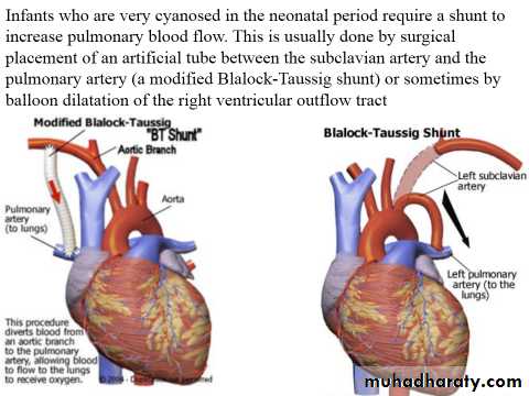
TRICUSPID ATRESIA
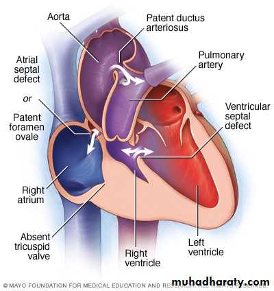
Etiology, Epidemiology and pathophysiology
Tricuspid atresia accounts for approximately 2% of all congenitalheart defects . The absence of the tricuspidvalve results in a hypoplastic right ventricle. All systemic venous return must cross the atrial septum Into the left atrium. A PDA or VSD is
necessary for pulmonary blood flow andsurvival.
Clinical Manifestations
Severe cyanosisIf a VSD is present, there may be a murmur. A diastolic murmur across the mitral valve may be audible.
*Frequently there is no significant murmur.
Diagnosis
ECG shows LVH and Lt axis deviation.
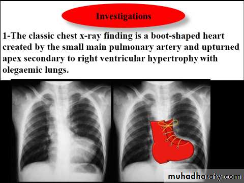
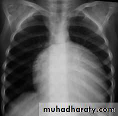
CXR shows cardiomegaly with decreased pulmonary blood flow
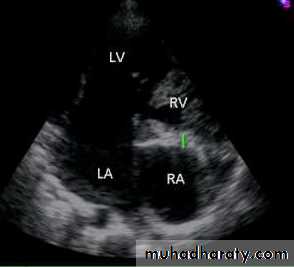
Echo is diagnostic and shows the abnormalities.
Treatment
If there is no VSD PGE1 is infused to maintain ductal patency and pulmonary flow.
Surgical correction involve (Blalock-Taussig procedure) then Fontan opertation.
Ebstein Anomaly
Ebstein anomaly consists of downward displacement of an abnormal tricuspid valve into the right ventricle.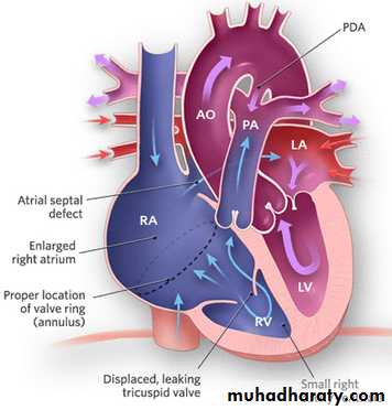
Pathophysiology
The right ventricle is divided into 2 parts by the abnormal tricuspid valve: the 1st, a thin-walled “atrialized” portion, is continuous with the cavity of the right atrium; the 2nd, often smaller portion consists of normal ventricular myocardium.
RV output is decreased due to a combination of the poorly
functioning small right ventricle and tricuspid valve regurgitation,
Clinical Manifestations
SymptomsSeverity is variable depending on the severity of pathology.
Mild cases present at teenage or adulthood with fatigue and
palpitation as a result of cardiac dysrhythmias.
There may be atrial right-to-left shunt through an ASD
causing cyanosis and polycythemia.
Physical signs
High JVP.
A holosystolic murmur caused by tricuspid regurgitation is
audible over most of the anterior left side of the chest.
*Neonates with severe form present with severe cyanosis, heart failure and pulmonary hypoplasia and may die.
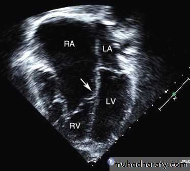
Diagnosis
ECG shows RBB, large tall p wave, prolonged PR interval and SVT due to Wolf-Parkinson white syndrome.
CXR shows cardiomegaly
Echo is diagnostic and shows the abnormalities.
Treatment
In non severe cases by surgical repair of TV
In severe cases by single ventricle repair ( Fontan opertation)
Lesions Associated with Increased Pulmonary Blood Flow
D-Transposition of the Great ArteriesEtiology and Epidemiology
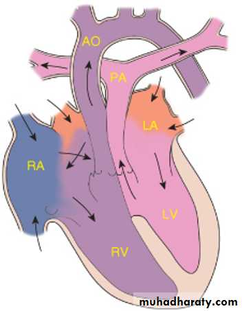
T.G.A represents about 5% of congenital heart defects, and it is the most common cyanotic lesion to present in the Newborn period . T.G.A is ventriculoarterial discordance secondary to abnormalities of septation of the truncus arteriosus, the aorta arises from the right ventricle, anterior and to the right of the pulmonary artery, which arises from the left
ventricle. This transposition results in desaturated blood returning to the right heart and being pumped back out to the body, while well-oxygenated blood returning from the lungs enters the left heart and is pumped back to the lungs. Without mixing of the two circulations, death occurs quickly. Mixing can occur at the atrial (patent foramen ovale/ASD), ventricular (VSD), or great vessel (PDA) level
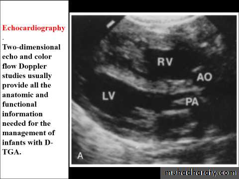
CLINICAL MANIFESTATIONS
History of cyanosis from birth is always present.
Symptoms of congestive heart failure (CHF) with dyspnea and feeding difficulties may develop during the newborn period
On examination:-
Moderate to severe cyanosis and tachypnea.
No VSD no murmur, VSD holosystolic murmur, and there may be murmur of RVOT or LVOT obstruction.
There may be features of heart failure.
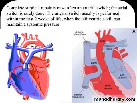
Treatment
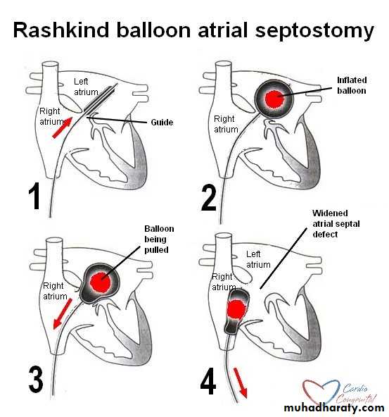
Initial medical management includes prostaglandin E1 to maintain ductal patency.
If the infant remains significantly hypoxic on prostaglandintherapy, a balloon atrial septostomy is performed to improve mixing between the two circulations.

HYPERLINK "https://www.muhadharaty.com/lecture/13484" https://www.muhadharaty.com/lecture/13484