Lab3:
Why we need to stain bacteria?Bacteria are transparent and colorless , so they would be invisible to naked eye if observed under a microscope
thus
bacteria should be stained with certain dyes in order to visualize bacterial cell or their internal structures using the light microscope.
Dye (stain):
colored organic compound in the form of salt, composed of positive and negative ion, one of these ions is responsible for colour called chromogen.Types of Dyes:
Basic dyes
Acidic dyes
Basic dyes:
In which chromogen is the positive ion (cation).Basic dye has the form: dye+ Cl-
examples include crystal violet, methylene blue and safranin.
Acidic dyes:
In which chromogen is the negative ion (anion).Acidic dye has the form: Na+dye-
Examples include nigrosin and Indian ink
Simple Staining:
Principle
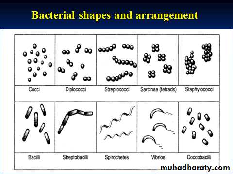
The simple stain can be used as a quick and easy way to determine cell shape, size and arrangements of bacteria. True to its name, the simple stain is a very simple staining procedure involving single solution of stain. Any basic dye such as methylene blue, safranin, or crystal violet can be used to color the bacterial cells.
These stains will readily give up a hydroxide ion or accept a hydrogen ion, which leaves the stain positively charged. Since the surface of most bacterial cells and cytoplasm is negatively charged, these positively charged stains adhere readily to the cell surface. After staining, bacterial cell morphology (shape and arrangements) can be appreciated.
Simple Staining Procedure
Preparation of a smear and heat fixingUsing a sterilized inoculating loop, transfer loopful of liquid suspension containing bacteria to a slide or transfer an isolated colony from a culture plate to a slide with a water drop.
Disperse the bacteria on the loop in the drop of water on the slide and spread the drop over an area the size of a dime. It should be a thin, even smear.
Allow the smear to dry completely.
Heat-fix the smear carefully by passing the underside of the slide through the burner flame two or three times. This kills the bacteria, attaches the cells to the slide , enhances the stain uptake, minimize any changes in bacteria cells and keep the shape of bacteria after fixation . Do not overheat the slide as it will distort the bacterial cells.
Staining
Cover the smear with methylene blue and allow the dye to remain in the smear between one minute to 2 minutes .Using distilled water in wash bottle, to gently wash off the excess methylene blue from the slide by directing a gentle stream of water over the surface of the slide.
Wipe the back of the slide and blot the stained surface with bibulous paper or with a paper towel.
Place the stained smear on the microscope stage smear side up and focus the smear using the 10X objective.
Choose an area of the smear in which the cells are well spread in a monolayer. Center the area to be studied, apply immersion oil directly to the smear, and focus the smear under oil with the 100X objective.