1
Fifth stage
Pediatric
Lec-4
د. خليل
23/11/2016
Acquired Heart Diseases
Acute rheumatic fever; cardiomyopathies; cardiovascular infections, including myocarditis
and infective endocarditis; and non congenital valvular heart disease are the most common
acquired heart diseases among the pediatric age group patients.
Rheumatic Fever
Definition
Rheumatic fever is a delayed autoimmune reaction in genetically predisposed individuals
to group A, β-hemolytic, streptococcal pharyngitis. It is a self-limited disease that may
involve joints, skin, brain, serous surfaces, and the heart Despite the dramatic nature of
the acute episode, ARF leaves no lasting damage to the brain, joints or skin.
Epidemiology
The incidence of acute rheumatic fever is 3 to 61 per 100,000 school children.
ARF is predominantly a disease of children aged 5-14 years and generally does not
affect children less than 3 years old or adults.
Acute rheumatic fever is most common during winter and spring, a seasonal variation
similar to that of streptococcal pharyngitis.
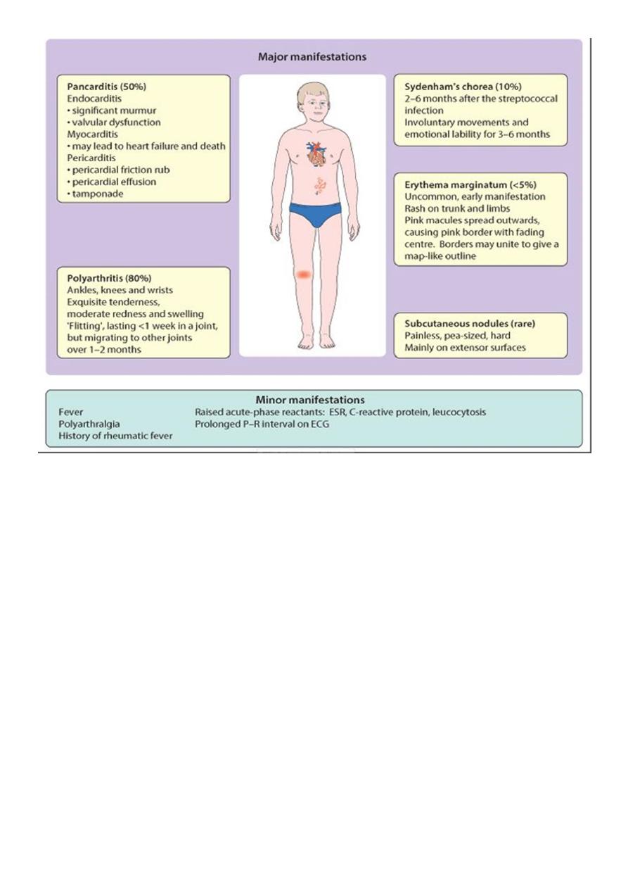
Clinical manifestation
• Acute rheumatic fever is diagnosed using the revised Jones criteria, which consist of
clinical and laboratory findings.
• The presence of either two major criteria or one major and two minor criteria, along
with evidence of an antecedent streptococcal infection, confirm a diagnosis of acute
rheumatic fever.
• The infection often precedes the presentation of rheumatic fever by 2 to 6 weeks.

2
Supporting evidence of a preceding streptococcal infection
1. Elevated or rising antistreptolysin-O or other streptococcal antibody, or
2. Rapid antigen test for group A streptococci, or
3. A positive throat culture, or
4. Recent scarlet fever.
Laboratory Findings
1. High ESR.
2. Anemia, leucocytosis .
3. Elevated C-reactive protein.
4. ASO titre >200 Todd units
5. Anti-DNAse B test .
6. Throat culture-GABH streptococci.

3
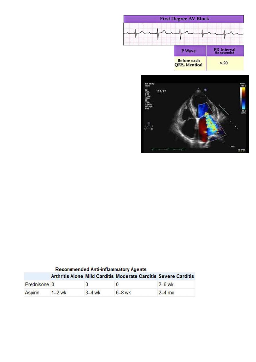
7. ECG-
prolonged PR interval,
2nd or 3rd degree blocks ,
ST depression or T inversion.
8.
2 D Echo cardiography –
valve edema ,mitral regurgitation, LA & LV
dilatation ,pericardial effusion ,decreased
contractility.
Management
1. Benzathine penicillin G, 0.6 to 1.2 million units intramuscularly, is given to eradicate
streptococci. This serves as the first dose of penicillin prophylaxis as well .
In patients allergic to penicillin, erythromycin, 40 mg/kg per day in two to four doses for
10 days, may be substituted for penicillin.
2. Bed rest of varying duration is recommended. The duration depends on the type and
severity of the manifestations and may range from a week (for isolated arthritis) to
several weeks for severe carditis. Bed rest is followed by a period of indoor ambulation
of varying duration before the child is allowed to return to school.
3. Therapy with anti-inflammatory agents should be started as soon as acute rheumatic
fever has been diagnosed as follows.
Prednisone, 2 mg/kg/24 hours in 4 divided doses.
Aspirin, 100 mg/kg/d in4-6 divided doses.

4
4. Treatment of CHF includes the following:
a. Complete bed rest with O2.
b. Prednisone for severe carditis of recent onset
c. Digoxin, used with caution, beginning with half the usual recommended dose,
because certain patients with rheumatic carditis are supersensitive to digitalis.
d. Furosemide,1 mg/kg every 6 to 12 hour.
5. Management of Sydenham's chorea:
a. Reduce physical and emotional stress.
b. For severe cases, any of the following drugs may be used: phenobarbital , haloperidol
,valproic acid, chlorpromazine (Thorazine), diazepam (Valium), or steroids.
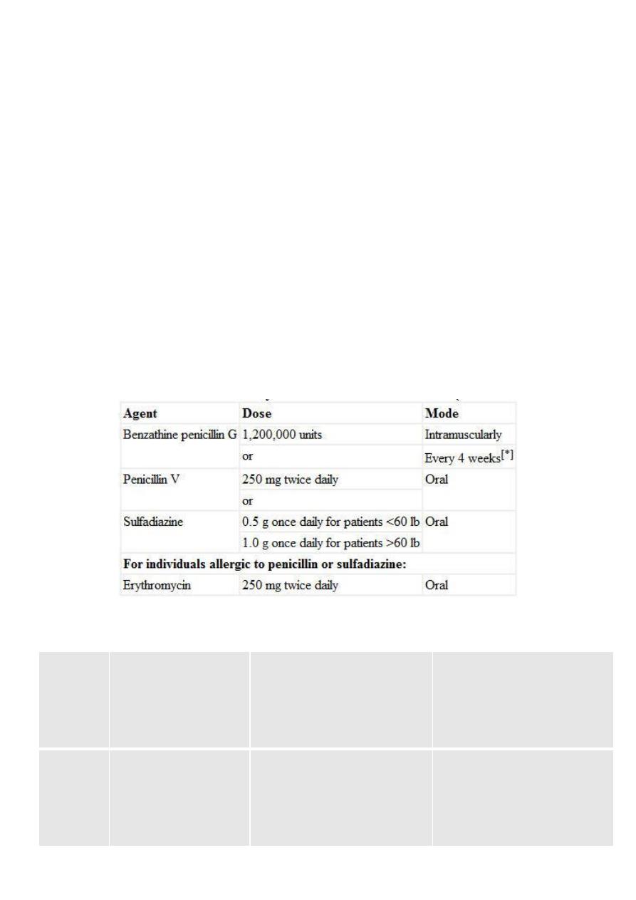
Prevention (prophylaxis)
A patient who has had acute rheumatic fever is susceptible to recurrent rheumatic fever for
the rest of his life.
Recommended Duration of Prophylaxis for Rheumatic Fever
Rheumatic fever with
carditis and residual heart
disease (persistent valvular
disease)
Rheumatic fever with
carditis but without
residual heart disease (no
valvular disease)
Rheumatic fever
without carditis
Category
At least 10 yr since last
episode and at least until
age 40 yr; sometimes
lifelong prophylaxis
At least for 10 yr or well
into adulthood, whichever
is longer
At least for 5 yr or
until age 21 yr,
whichever is longer
Duration

5
Cardiomyopathies
Primary myocardial disease, or cardiomyopathy, is a disease of the heart muscle itself, not
associated with congenital, valvular, or coronary heart disease or systemic disorders.
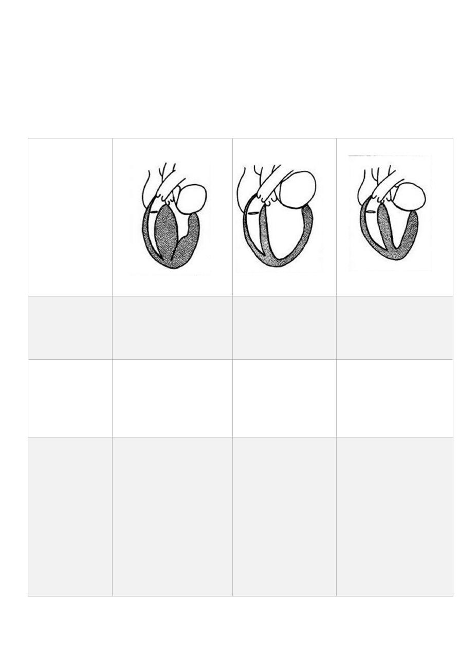
Cardiomyopathy has been classified into three types based on anatomic and functional
features: hypertrophic, dilated (or congestive), and restrictive
Features of different types
Restrictive
Dilated
Hypertrophic
Feature
Myocardial fibrosis,
hypertrophy, or
infiltration (amyloid,
hemochromatosis)
Pluricausal (e.g.,
toxic, metabolic,
infectious, alcohol,
doxorubicin)
Inherited (AD in about
50%) Sporadic (new
mutation ±)
Etiology
Diastolic dysfunction
(with normal systolic
function) (abnormally
stiff LV with impaired
ventricular filling)
Systolic contractile
dysfunction (↓
cardiac output, ↓
stroke volume, ↑
LVEDP)
Diastolic dysfunction
(rigid ventricular walls
impede ventricular
filling)
Hemodynamic
dysfunction
Exercise
intolerance,
weakness and dyspnea,
or chest pain.
signs and symptoms
of inadequate cardiac
output and CHF.
Infants, frequently
present with signs of
CHF .
Older children may be
asymptomatic, with
sudden death as the
initial presentation.
Dyspnea, fatigue, chest
pain, syncope or near-
syncope, and
palpitations may be
present.
Symptoms

6
Restrictive
Dilated
Hypertrophic
Feature
Jugular venous
distention, gallop
rhythm, and a systolic
murmur of AV valve
regurgitation may be
present.
Features of CHF. Rales
may be audible on
pulmonary
auscultation. Heart
sounds may be muffled,
and S3 is often present.
Concurrent infectious
illness may result in
circulatory collapse and
shock in children with
dilated
cardiomyopathies
A murmur is heard in
more than 50% of
children.
On examination
Biatrial enlargement
Biventricular dilatation
and ↓ EF%
Thickened LV (and
occasionally RV) wall
Echocardiography
Atrial hypertrophy. It
may show atrial
fibrillation and
paroxysms of
supraventricular
tachycardia.
Sinus tachycardia, LVH,
and ST-T changes are
the most common
findings.
(LVH), ST-T changes,
and abnormally deep
Q waves (owing to
septal hypertrophy)
Electrocardiography
cardiomegaly,
pulmonary venous
congestion, and
occasional pleural
effusion.
Generalized
cardiomegaly is usually
present, with or
without signs of
pulmonary venous
hypertension or
pulmonary edema.
Mild left ventricular
enlargement with a
globular-shaped
heart.
Chest x ray
-Diuretics
-Anticoagulants (±)
-Corticosteroids (±)
-Permanent
pacemaker for
advanced heart block
(±)
-Cardiac
transplantation (±)
-Vasodilator therapy
-Digitalis plus diuretics
-β-Adrenoceptor
blockers (±)
Antiarrhythmics (±)
Cardiac transplantation
(±)
-β-Adrenoreceptor
blockers
-Calcium antagonists
-(Digitalis/catechols
,Diuretics and
nitrates are
contraindicated).
Treatment
