
1
Fifth stage
radiology
Lec-3
د.هديل
12/12/2016
Joint Diseases
ARTHRITIS
TYPES OF ARTHRITIS
There are three types of arthritis(which often can be distinguished radiologically):
1 -
Degenerative joint disease
Osteophytes ,
Subchondral sclerosis
Uneven loss of articular space
2 -
Inflammatory arthritis
Unrnarginated erosions
Periarticular osteoporosis is common
Soft tissue swelling
Uniform loss of articular space
3 -
Metabolic arthritis
Lumpy bumpy soft tissue swelling
Marginated bony erosions with
overhanging edges
Degenerative Arthritis
Degenerative joint disease (DJD) = osteoarthritis (OA).
80% of population > 50 years have radiological evidence of OA.
types:
--Primary OA
*No underlying local etiological factors
*Abnormally high mechanical forces on normal joint
*Age related
2
--Secondary OA
Underlying etiological factors :
*trauma, inflammatory arthritis, hemochromatosis, acromegaly, congenital hipdysplasia,
osteonecrosis, loose bodies
*Normal forces on abnormal joint
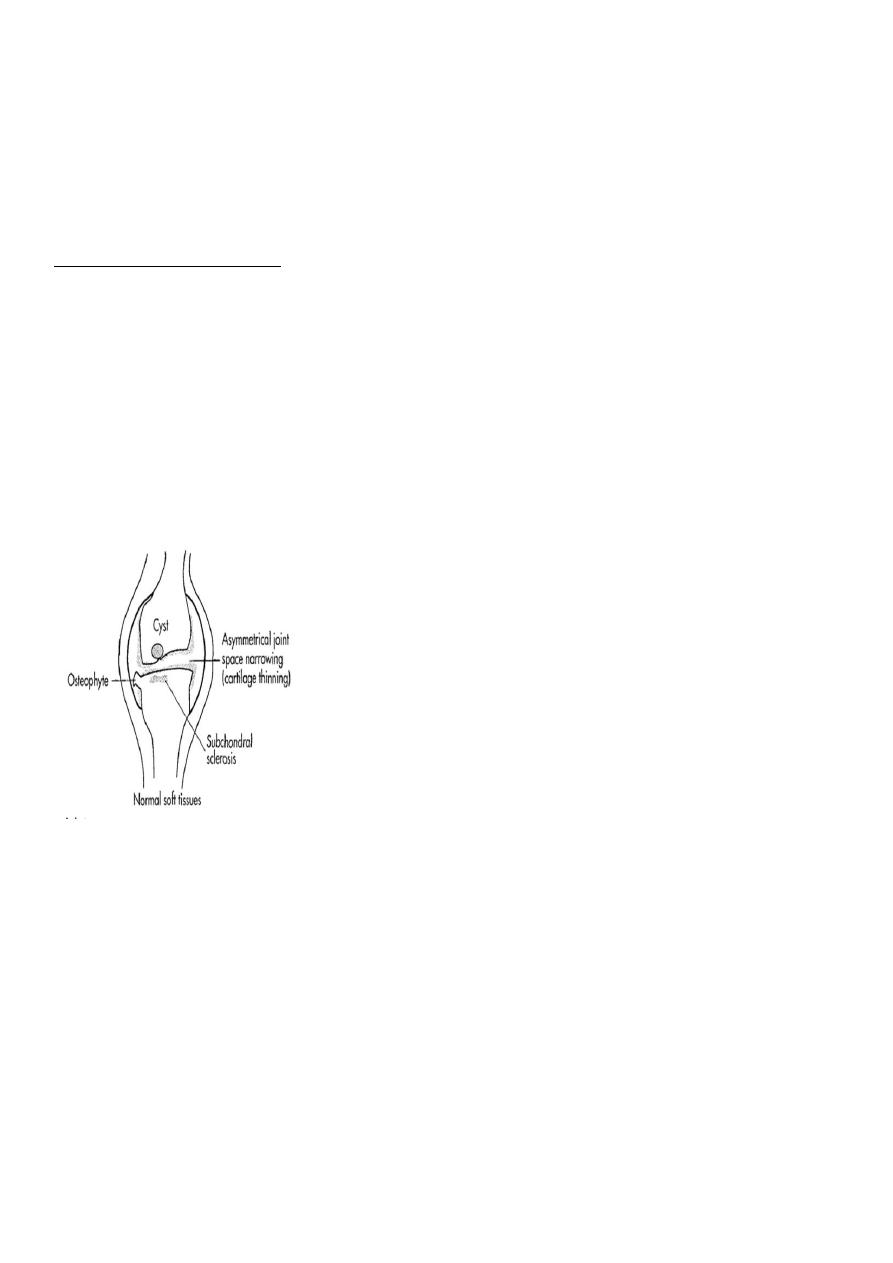
Radiographic features:
Five hallmarks:
*Narrowing of joint space, usually asymmetrical
*Subchondral sclerosis
*Subchondral cysts (true cysts or pseudocysts)
*Osteophytes
* Lack of osteoporosis
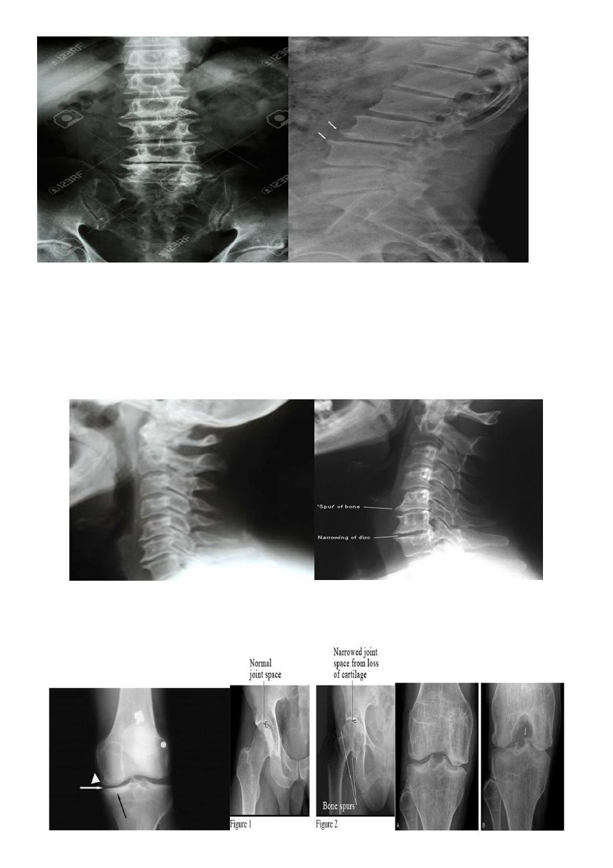
In the Spine :
*Lower cervical and low lumbar spine are most comonly affected.
*Osteophytes may encroach on neural foramina (best seen on oblique views).
*Vacuum phenomenon: gas (N2),is pathognomonic of the degenerative process.
* OA of the spine occurs in the apophyseal joints .
* Degenerative spondylolisthesis (pseudospondylolithesis)
3
Lumbar spondylosis. There is distal narrowing and a vacuum
phenomenon is present in the degenerative discs. Marginal osteophytes are
present. Inferiorly the facet joints show features of degeneration and, with the increase in
lordosis, the spinous processes are in contact
Cervical spondylosis

4
Inflammatory arthritis
There are three types of inflammatory arthritis
1 -
Autoimmune arthritis
RA
Scleroderma
Systemic lupus erythematosus (SLE)
Dermatomyositis
2 -
Seronegative spondylarthropathies
Ankylosing spondylitis
Reiter's syndrome
Psoriasis
Enteropathic arthropathies
3-
Erosive OA
RHEUMATOID ARTHRITIS
Female:male = 3:l
Radiographic features:
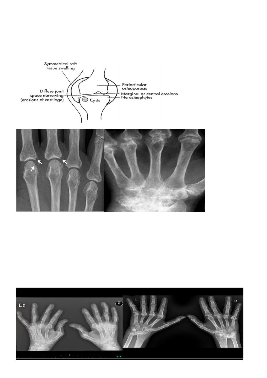
Early changes
*Peri articular soft tissue swelling (edema, synovial congestion)
*Peri articular osteoporosis in symmetrical distribution (hallmark)
*Preferred sites of early involvement
Hands: 2nd and 3rd MCP joint
Feet: 4th and 5th MTP joint
Late changes
* Erosions (pannus formation, granulation tissue) first attack joint portions in which
protective cartilage is absent (i.e., capsular insertion site).
*Erosions of the ulnar styloid and triquetrum are characteristic.
5
*Subchondral cysts formation results from synovial fluid, which is pressed into bone
marrow through destroyed cartilage.
Subluxations , Carpal instability and ulnar deviation
.
* Fibrous ankylosis is a late finding.
(A) The initial radiograph shows a hint of early trabecular loss around the proximal
interphalangeal joint of a finger with preservation of the joint space and early marginal
cortical loss at the
base of the middle phalanx .
(B) The subsequent radiograph shows established erosive change in the area of ill-defined
demineralisation in association with joint space narrowing .
6
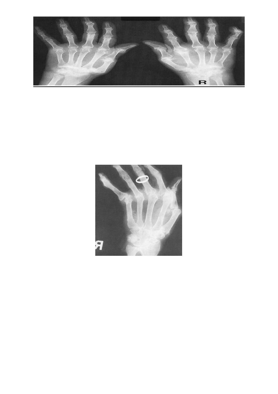
Rheumatoid arthritis. Bilateral changes are fairly symmetrical. Soft-tissue swelling is
demonstrated, especially over the ulnar styloids. Erosions are demonstrated at the carpus,
distal radius and ulna, with joint space narrowing and collapse of bone.
Metacarpophalangeal erosions are also seen associated with joint space narrowing. There is
a swan-neck deformity of the right fifth distal interphalangeal joint
Gross rheumatoid arthritis at the carpus with ulnar deviation,
subluxation and joint narrowing at the metacarpophalangeal joints.
Boutonniere deformities are present at the index and little fingers.
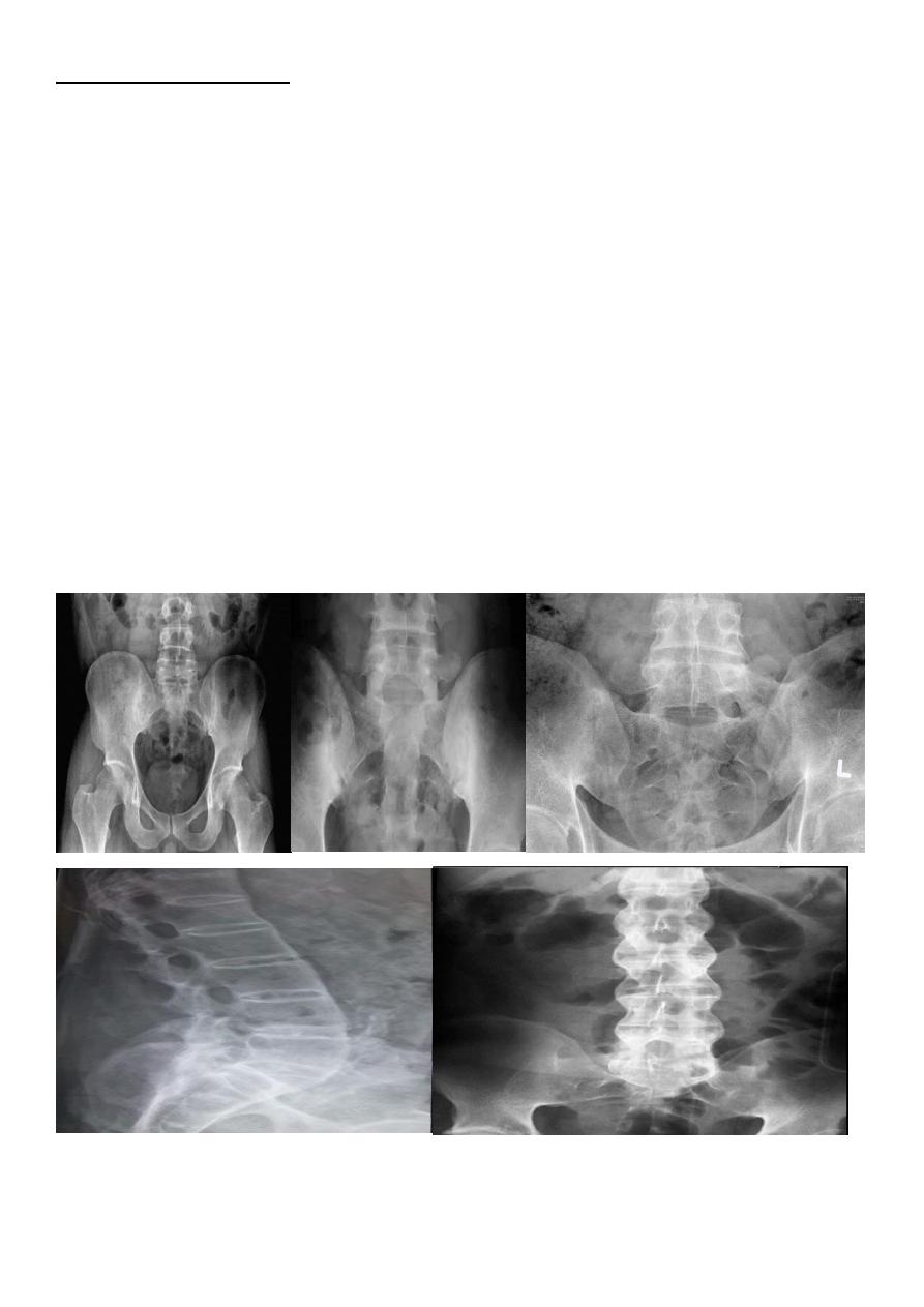
ANKYLOSING SPONDYLITIS (AS)
Seronegative spondyloarthropathy of the axial skeleton and proximal large joints.
Clinical: males >> females. HLA-B27 in 95%. Insiduous onset of back pain and stiffness.
Onset: 20 years.
7
Radiographic features:
*SI joint is the initial site of involvement:
bilateral, symmetrical
Erosions: early ,Sclerosis: intermediate , ankylosis: late
*Contiguous thoracolumbar involvement
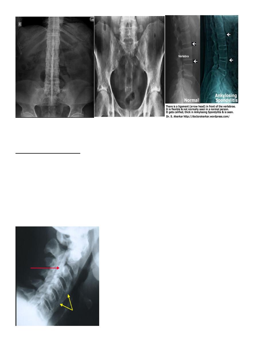
Vertebral body "squaring": early osteitis
*Syndesmophytes
*Bamboo spine: late fusion and Bamboo spine
ligamentous ossification
*ankylosed spine (fracture)
*Enthesopathy is common(("whiskering of tuberosities
)
* Arthritis of proximal joints (hip > shoulder) in 50% ,erosions and osteophytes
8
DIFFUSE IDIOPATHIC SKELETAL HYPEROSTOSIS (DISH)
Radiographic features:
*Flowing osteophytes of at least four contiguous vertebral bodies
*Preserved disk height
*No sacroiliitis or facet ankylosis
*Calcification of ligaments and tendons
*Associated with hypertrophic DJD
9
EROSIVE OSTEOARTHRITIS
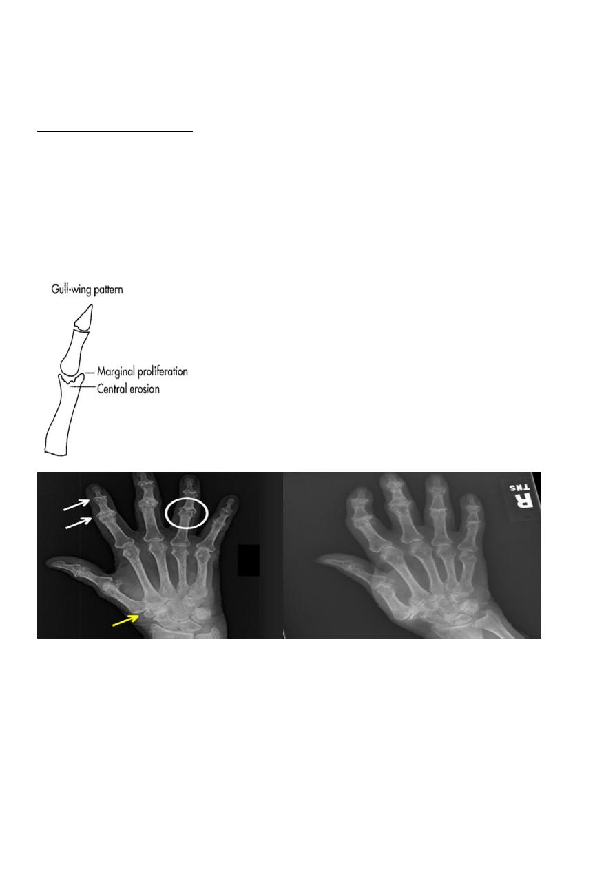
OA with superimposed inflammatory, erosive changes. Characteristically affects middle-
aged women.
Radiographic features:
*Erosive and productive changes of DIP and PIP
*Gull-wing pattern: secondary to central erosions and Marginal proliferation osteophytes .
Typical involvement of first CMC may help distinguish erosive OA from rheumatoid arthritis
(RA), psoriatic arthritis, and adult Still's disease.
* Interphalangeal fusion may occur.
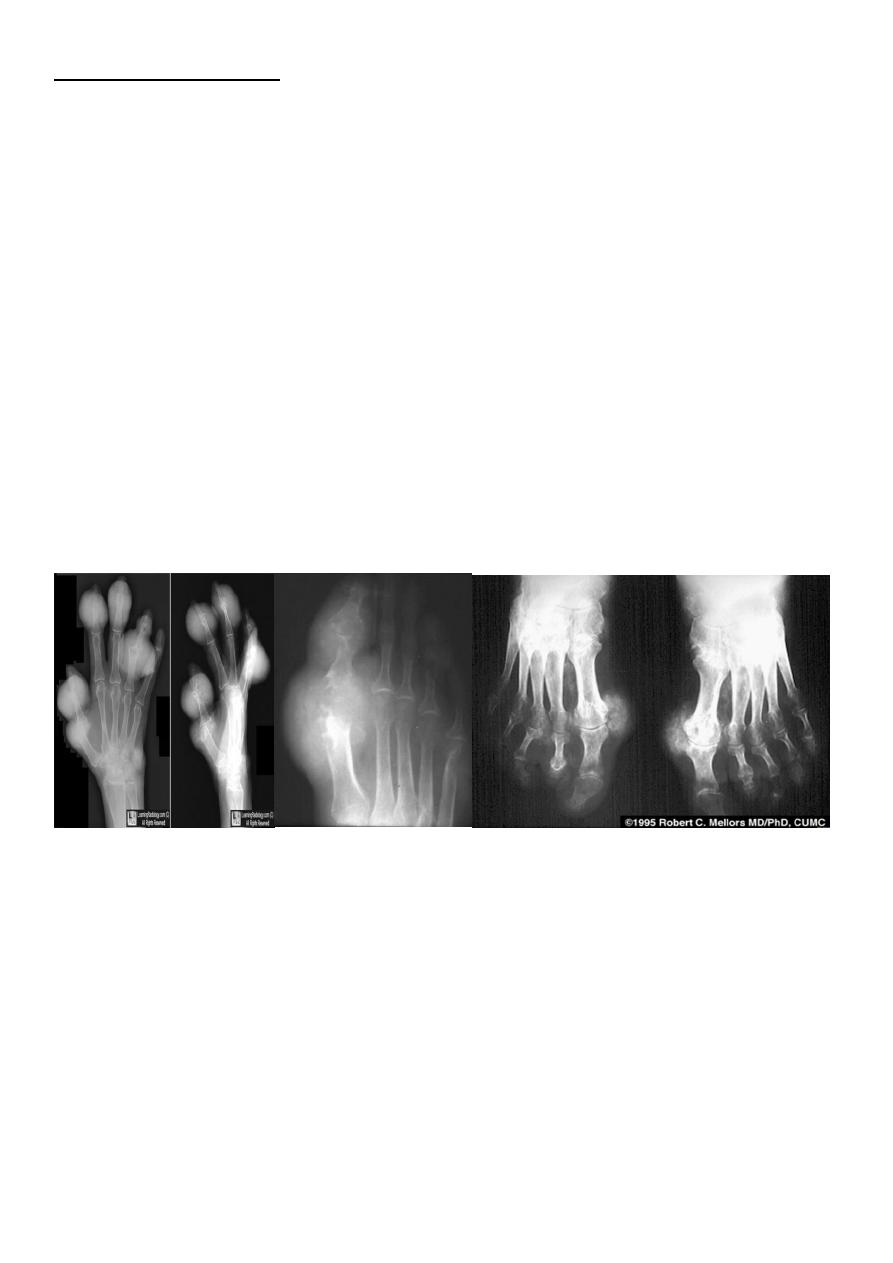
GOUT
Heterogeneous group of entities characterized by recurrent attacks of arthritis secondary to
deposition of sodium urate crystals in and around joints.
*
99
%
of patients are male
*Causes due to either Uric acid overproduction, 10% or underexcretion, 90%.
19
Radiographic features:
Lower extremity > upper extremity; small joints > large joints
*First MTP is most common site
*Marginal, peri articular erosions: overhanging edge
*Erosions may have sclerotic borders
*Joint space is preserved
*Soft tissue and bursa deposition
Tophi: juxtaarticular, helix of ear
Bursitis: olecranon, prepatellar
*Erosions and tophi only seen in longstanding disease
*Tophi calcification, 50%
*Chondrocalcinosis
Infectious arthritis (septic arthritis)
Infectious arthritis usually results from hematogenous spread to synovium and subsequent
spread into the joint .
Direct spread of osteomyelitis into the joint is much less common .
The diagnosis is made by joint aspiration.
Organism , Staphylococcus aureus (most common) ,B-Streptococcus in infants , Salmonella
is seen in sickle cell patients ; however, the most common infection in sickle cell patients is
Staphylococcus.
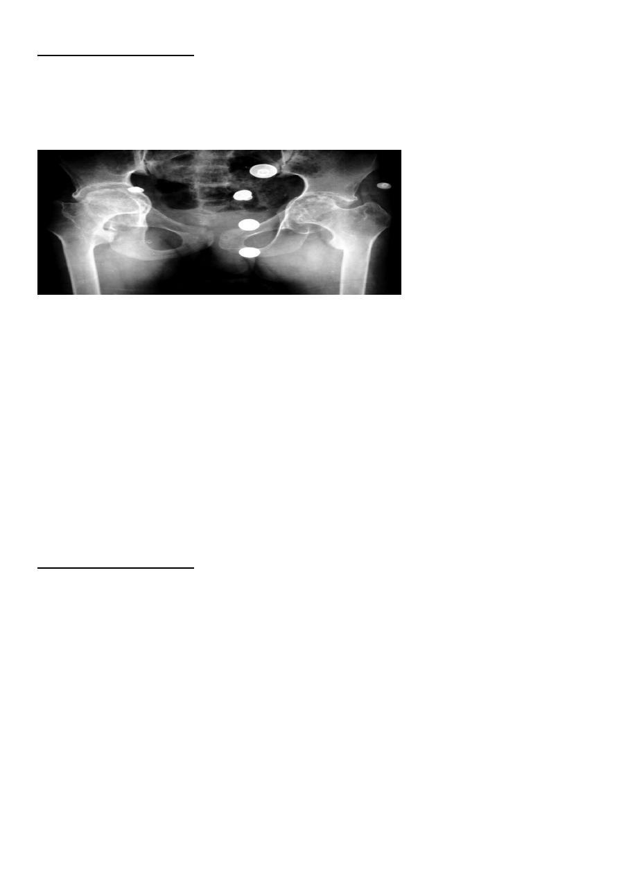
11
Radiographic features:
*Joint effusion
*Juxtaarticular osteoporosis
* Destruction of subchondral bone on both sides of the joint
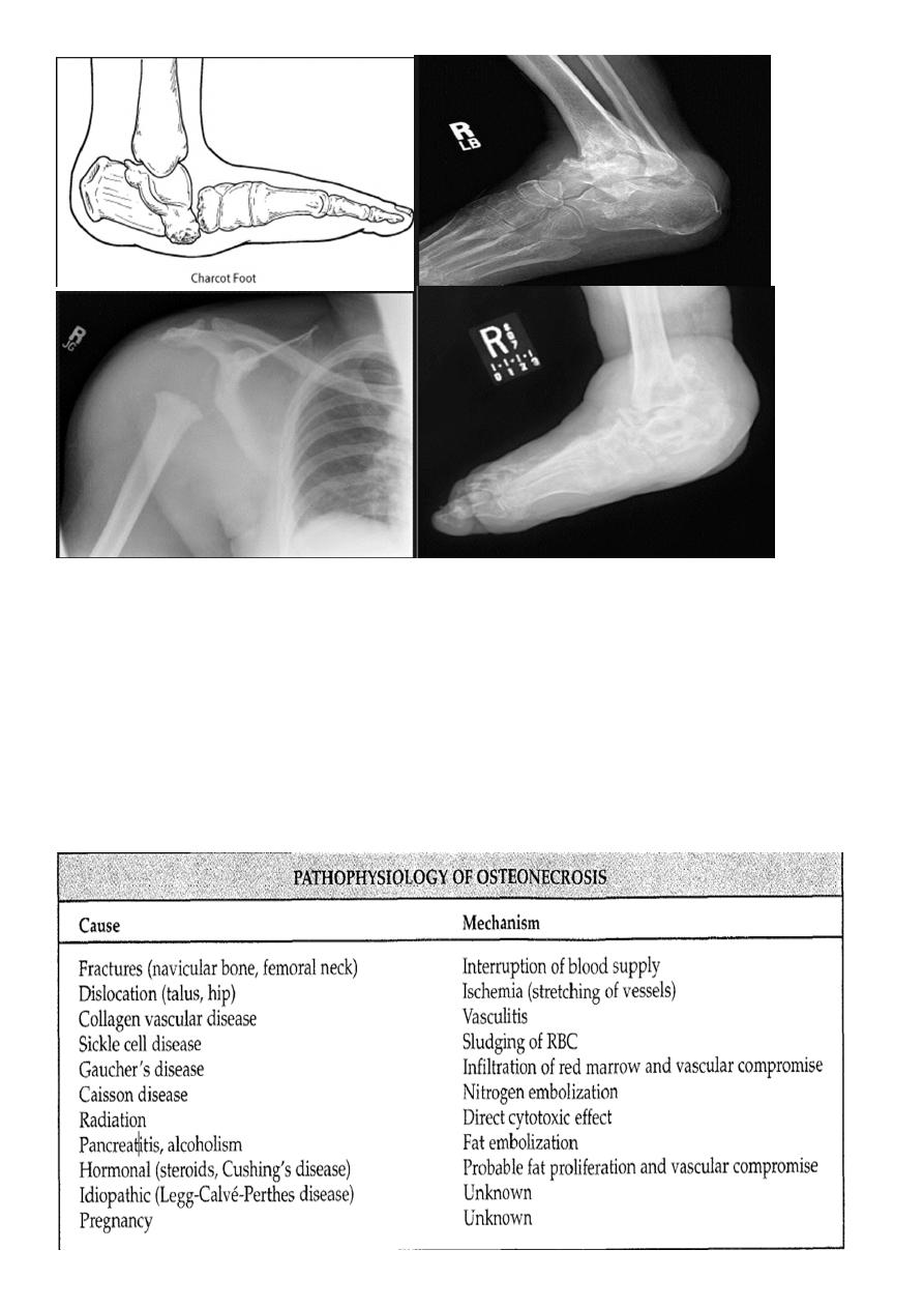
NEUROPATHIC ARTHRITIS (CHARCOT'S JOINT)
Primary loss of sensation in a joint leads to arthropathy. Distribution helps determine
etiology.
Causes
Diabetes neuropathy: usually foot
Tertiary syphilis : usually knee
Syringomyelia: usually shoulder
Radiographic features:
Common to all types
* Joint instability: subluxation or dislocation
* Prominent joint effusion
---
Hypertrophic type, 20%
Marked fragmentation of articular bone
Much reactive bone
---
Atrophic type, 40°/0
Bone resorption of articular portion
--- Combined type, 40%
12
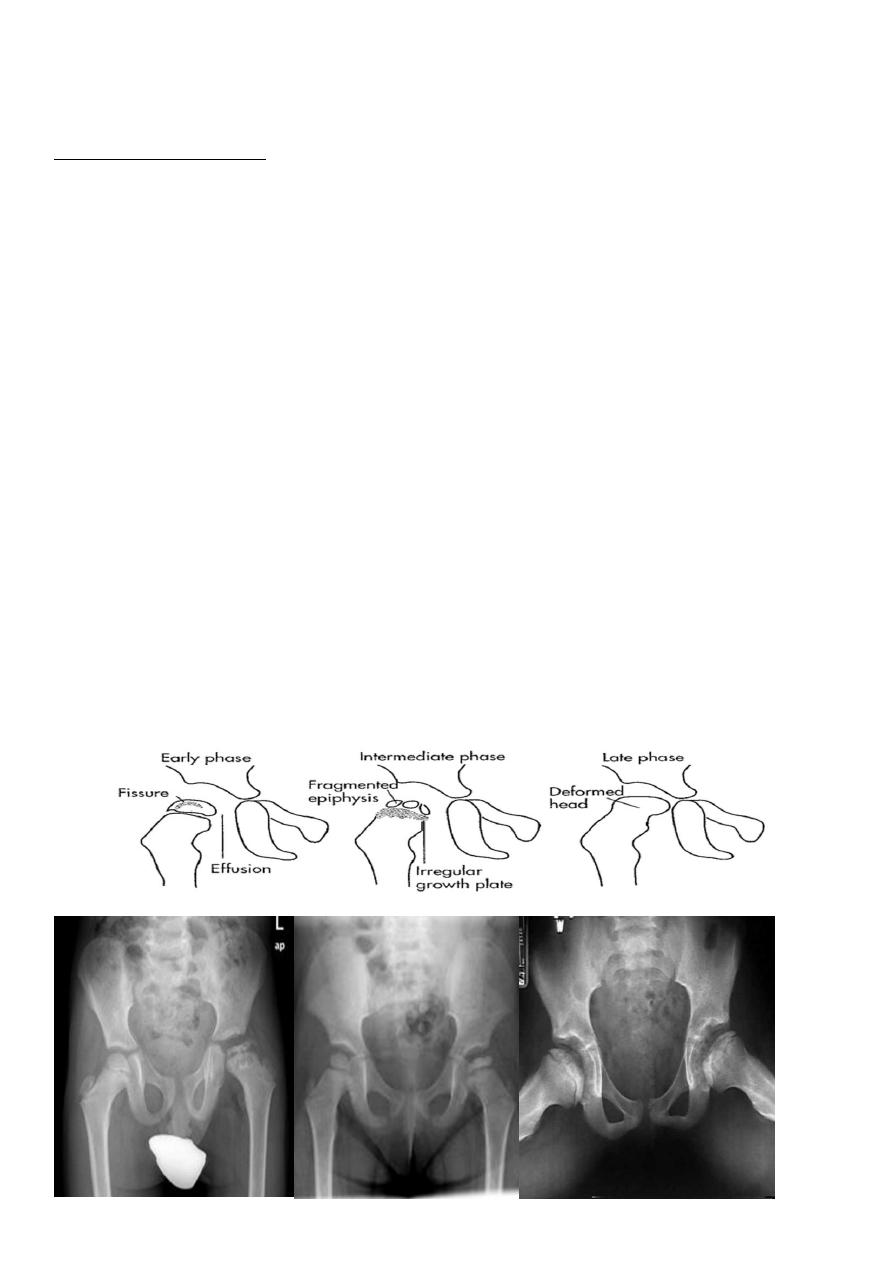
OSTEONECROSIS
Osteonecrosis (avascular necrosis, ischemic necrosis, aseptic necrosis) may be caused by
two mechanisms:
*Interruption of arterial supply
* Intra/extraosseous venous insufficiency
13
The pathophysiology of all osteonecrosis is the same :
Ischemia > revascularization >repair > deformity> osteoarthrosis
Radiological findings:
Findings lag several months behind time of injury .
These findings include areas of radiolucency, fissuring , fragmentation , bone collapse and
condensation , end with dense and flat bone with loss of bone contour and secondary
osteoarthritis
MRI
Most sensitive imaging modality: 95%-100% sensitivity
Osteochodrosis :
These osteonecroses are usually idiopathic in origin. The following are the most common
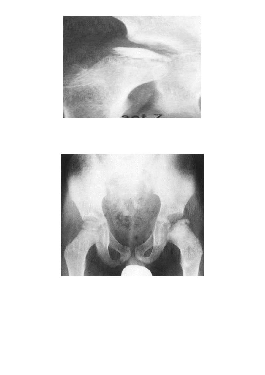
Legg-Calve-Perthes disease : osteochodrosis of the femur head
Usually affects 5-10 years , started as hip pain , if not treated it will ends with mushroom
deformity due to neglected and untreated perthis seen later on and is liable for early OA
chnges
.
14
Perthes' disease. A series of radiographs showing the stages of healing. (A) The initial
radiograph shows a flattened, sclerotic femoral head
The left femoral neck is broadened, the metaphysis sclerotic
with focal areas of lucency, the growth plate irregular and the femoral head
flattened and sclerotic. It is uncovered laterally. The joint space appears
widened
15
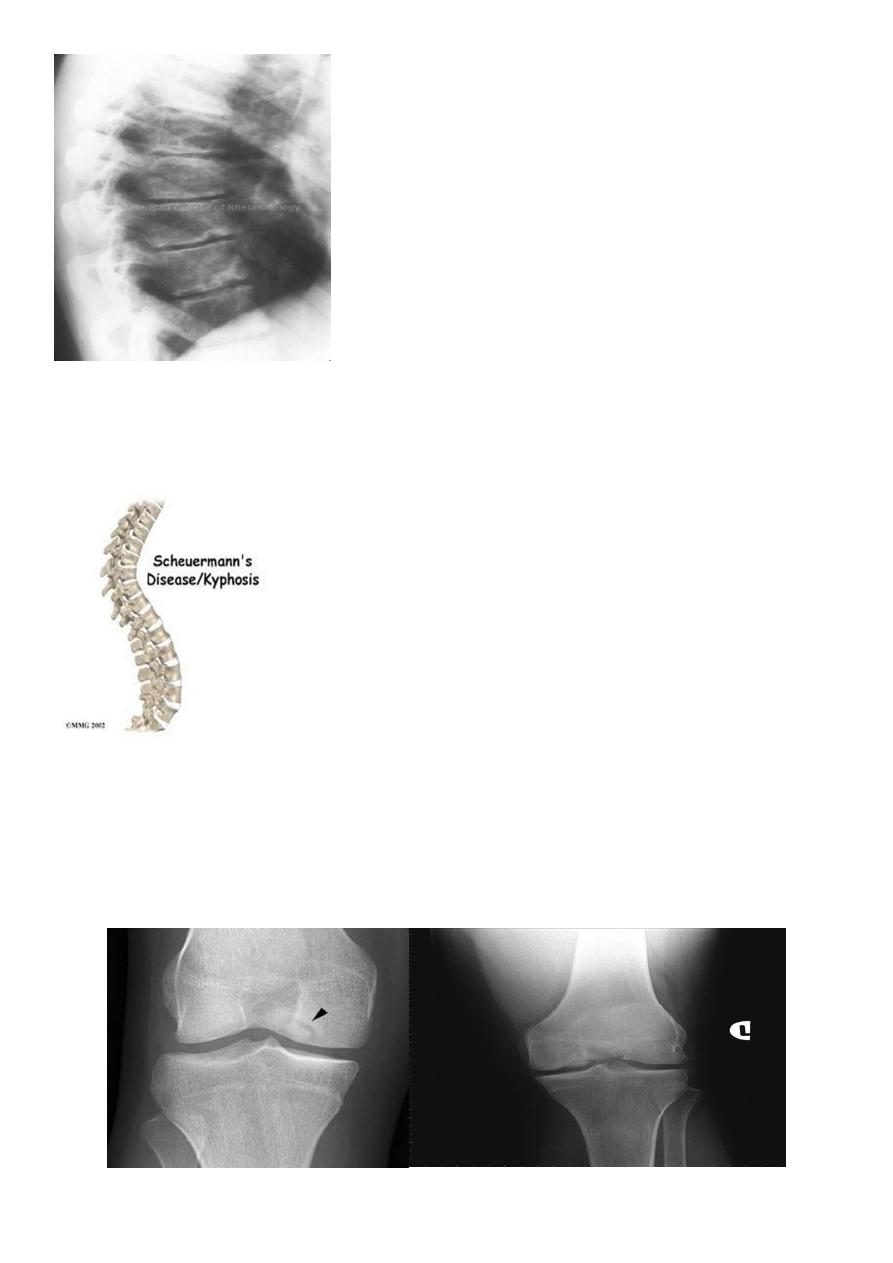
Scheuermann's disease : (adolecent Kyphosis).
osteochodrosis of the vertebral end plates
.
Usually affects 8- 10 years , characterized by erosion of anterior superior and inferior
vertebral margin resulting in decrease in the height of anterior part of the vertebra
(vertebral wedging ) ending with kyphosis.
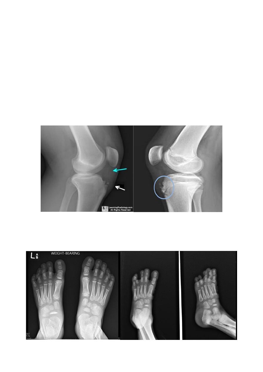
Osgood-Schlatter : 12-16 y,osteochodrosis of the tibial tuberocle.
Blount's disease: tibial epiphysis
Kohler's : 4-8 y ,osteochodrosis of the Navicular bone
.
Kienbock's: adults , osteochodrosis of the lunate bone .
Osgood-Schlatter disease.
Fragmentation of the tibial tuberosity
16
Osteochondritis of
lumbar vertebral bodies (advanced case).
Osteochondritis dissecans
affects the large articular surface , commomnly medial femural condylee ,talus and trochlea
.
Ends by separation of the affected part in to the joint space resulting in intra-articular loose
body .
Osteochondritis dissecans of the medial femoral condyle Osteochondritis dissecans of the medial part of the articular surface

17
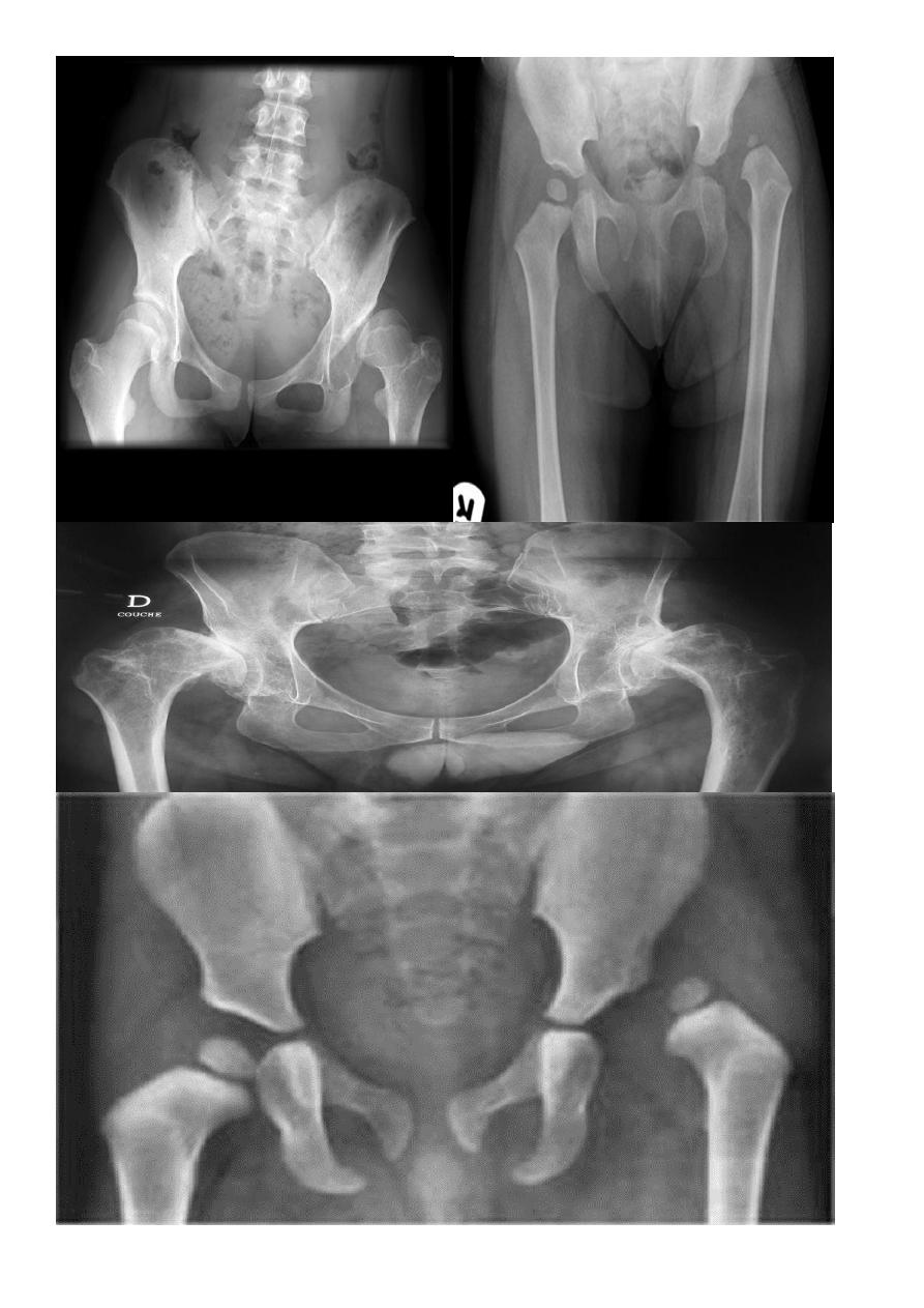
DEVELOPMENTAL DYSFLASIA OF HIP (CONGENITAL DISLOCATION OF THE HIP)
An abnormally lax joint capsule allows the femoral head to fall out of the acetabulum,
leading to deformation.
Predisposing factors for the development of CDH are:
*Abnormal ligamentous laxity (effect of estrogen; fema1e:male = 6:l)
*Acetabular dysplasia
.
CDH occurs most commonly (70%) in the left hip. Bilateral involvement is seen in 5%.
Radiographic features:
US (commonly used today) at 1-3 months
* Normal femoral head is covered at least 50% by acetabulum , In CDH < 50% of femoral
head is covered by acetabulum .
Plain film
At 3-6 months
:
By doing special veiw (Von Rosen veiw )by abduction of the thigh 45 degree and internal
rotation
.
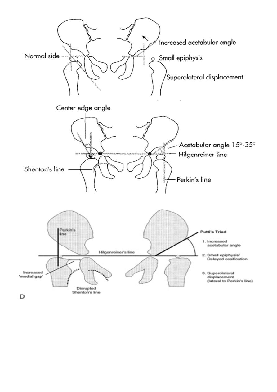
In DDH the lines that drown through the femura will meet in higher level than the normally
should at lumbosacral joint
.
6 months and later
*AP veiw ( femural epiphysis are visualized ) :
*Superolateral displacement of proximal femur (disturbed shenton’s line )
*Increase in acetabular angle
*Small capital femoral epiphysis
Femoral head is located lateral to Perkin's line
*Other features that are sometimes present
Abnormal sclerosis of the acetabulum
Shallow acetabulum
Formation of a false acetabulurn
18
19
