1
Fifth stage
Surgery
Lec-1
د
.
مثنى الحموشي
13/12/2016
Management of compound fractures
Patients with open fractures may have multiple injuries; a rapid general assessment is the
first step and any life threatening conditions are addressed.
The open fracture may draw attention away from other more important conditions and it is
essential that the step-by-step approach in advanced trauma life support not to be
forgotten.
When the fracture is ready to be dealt with, the wound is first carefully inspected;
1-arterial bleeding should be ligated
2- any gross contamination is removed .
3-the wound is photographed with aPolaroid or digital camera to record the injury
4-area then covered with a saline-soaked dressing to prevent desiccation. This is left
undisturbed until the patient is in the operating theatre .
5-The patient is given antibiotics, usually co-amoxiclav or cefuroxime, but clindamycin if
the patient is allergic to penicillin.
6- Tetanus prophylaxis is administered: toxoid for those previously immunized, human
antiserum if not.
7- The limb is then splinted until surgery is undertaken.
The limb circulation and distal neurological status will need checking repeatedly,
particularly after any fracture reduction maneuvers. Compartment syndrome is not
prevented by there being an open fracture
CLASSIFYING THE INJURY
Treatment is determined by
1- the type of fracture,
2- the nature of the soft-tissue injury (including the woundsize) and
3- the degree of contamination.
Gustilo’s classification of open fractures is widely used
Type 1 – The wound is usually a small, clean puncture through which a bone spike has
protruded. There is little soft-tissue damage with no crushing and the fracture is not
comminuted (i.e. a low-energy fracture).
Type II – The wound is more than 1 cm long, butthere is no skin flap. There is no much soft-
tissue damage and no more than moderate crushing or comminution of the fracture (also a
low- to moderate-energy fracture).
Type III – There is a large laceration, extensive damage to skin and underlying soft tissue
and, in the most severe examples, vascular compromise. The injury is caused by high energy
transfer to the bone and soft tissues. Contamination can be significant.

2
There are three grades of severity. In type III A the fractured bone can be adequately
covered by soft tissue despite the laceration. In type III B there is extensive periosteal
stripping and fracture cover is not possible without use of local or distant flaps. The fracture
is classified as type III C if there is an arterial injury that needs to be repaired, regardless of
the amount of other soft-tissue damage. The incidence of wound infection correlates
directly with the extent of soft-tissue damage, rising from less than 2 per cent in type I to
more than 10 percent in type III fractures.
PRINCIPLES OF TREATMENT
All open fractures, no matter how trivial they may seem, must be assumed to be
contaminated; it is important to try to prevent them from becoming infected. The four
essentials are:
• Antibiotic prophylaxis.
• Urgent wound and fracture debridement.
• Stabilization of the fracture.
• Early definitive wound cover.
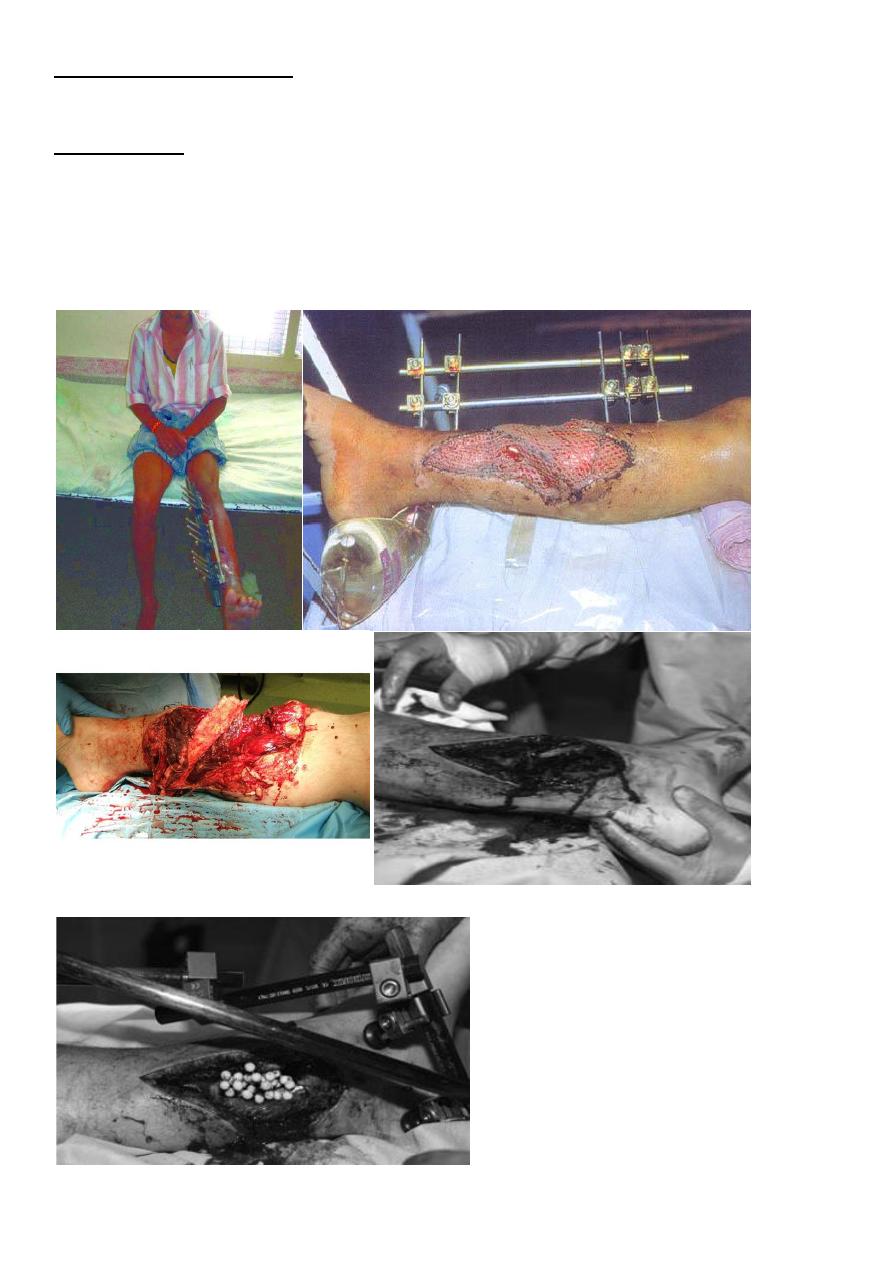
Debridement
The operation aims to render the wound free of foreign material and of dead tissue, leaving
a clean surgical field and tissues with a good blood supply throughout. Under general
anesthesia the patient’s clothing is removed, while an assistant maintains traction on the
injured limb and holds it still. The dressing previously applied to the wound is replaced by a
sterile pad and the surrounding skin is cleaned. The pad is then taken off and the wound is
irrigated thoroughly with copious amounts of physiological saline. The wound is covered
again and the patient’s limb then prepped and draped for surgery. It is advisable not to use
tourniquet in this condition unless if there is sever bleeding or arterial injury to deal with .
Wound excision The wound margins are excised, but only enough to leave healthy skin
edges.
Removal of devitalized tissue: Devitalized tissue provides a nutrient medium for bacteria.
Dead muscle can be recognized by a- its purplish colour, b-its mushy consistency, c-its
failure to contract when stimulated and d-its failure to bleed when cut. All doubtfully
viable tissue, whether soft or bony, should be removed. The fracture ends can be nibbled
away until seen to bleed
Wound cleansing : All foreign material and tissue debris is removed by excision or through
a wash with copious quantities of saline. A common mistake is to inject syringefuls of fluid
through a small aperture – this only serves to push contaminants further in; 6–12 L of saline
may be needed to irrigate and clean an open fracture of a long bone. Adding antibiotics or
antiseptics to the solution has no added benefit .
Nerves and tendons : as a general rule it is best to leave cut nerves and tendons alone at
the time of the wound excision,to be sutured by delay primary suture ; though if the wound
is absolutely clean and no dissection is required – and provided the necessary expertise is
available – they can be Sutured at the time of wound excision .

3
Stabilization of the fracture :
Stabilizing the fracture is important in reducing the likelihood of infection and assisting
recovery of the soft tissues. The stabilization of the fracture is usually by external fixation
Wound closure :
A small, uncontaminated wound in a Grade I or II fracture may (after debridement) be
sutured, provided this can be done without tension. In the more severe grades of injury,
immediate fracture stabilization and wound cover using split-skin grafts, local or distant
flaps is ideal, provided both orthopaedic and plastic surgeons are satisfied that a clean,
viable wound has been achieved after debridement.

4
Aftercare :
In the ward, the limb is elevated and its circulation carefully watched. Antibiotic cover is
continued but only for a maximum of 72 hours in the more severe grades of injury
GUNSHOT INJURIES
With high-velocity missiles (bullets, usually from rifles, travelling at speeds above 600 m/s)
there is
marked cavitation and tissue destruction over a wide area. The splintering of bone resulting
from the transfer of large quantities of energy creates secondary missiles,
causing greater damage.
With low-velocity missiles (bullets from civilian hand-guns travelling at speeds of 300–600
m/s) cavitation is much less, and with smaller weapons tissue damage may be virtually
confined to the bullet track. However, with all gunshot injuries debris is sucked into the
wound, which is therefore contaminated from the outset.
:
Emergency treatment
As always, the arrest of bleeding and general resuscitation
take priority. The wounds should each be covered with a sterile dressing and the area
examined for artery or nerve damage. Antibiotics should be given.
:
Definitive treatment
Traditionally, all missile injuries were treated as severe open injuries, by exploration of the
missile track and formal debridement. However, it has been shown that low-velocity
wounds with relatively clean entry and exit wounds can be treated as Gustilo type I injuries,
by superficial debridement, splintage of the limb and antibiotic cover; the fracture is then
treated as for similar open fractures. If the injury is to soft tissues only with minimal bone
splinters, the wound may be safely treated without surgery but with local wound
care and antibiotics. High-velocity injuries demand thorough cleansing of the wound and
debridement, with excision of deep damaged tissues and, if necessary, splitting of fascial
compartments to prevent ischaemia; the fracture is stabilized and the wound is treated as
for a Gustilo type III fracture. If there are comminuted fractures, these are best managed by
external fixation.
The method of wound closure will depend on the state of tissues after several days; in
some cases delayed primary suture is possible but, as with other open
injuries, close collaboration between plastic and orthopaedic surgeons is needed .
Close-range shotgun injuries, although the missiles may be technically low velocity, are
treated as highvelocity wounds because the mass of shot transfers large quantities of
energy to the tissues.
