
1
Surgery
(Orthopedics)
Fifth stage Lec-1
د.يقضان
3/1/2016
BONE TUMOURS
In general bone tumours can be classified into:
A- primary .
B- secondary .
Primary bone tumours classification
:
Most classification of bone tum.are based on the recognition of the dominant tissue in the
various lesion ; e.g. bone forming tum. Or cartilage forming tum. Etc.
Cell type
benign
malignant
Bone
osteoid osteoma
osteosarcoma
Cartilage
chondroma
chondrosarcoma
Fibrous t.
Fibroma
fibrosarcoma
b. Marrow
eosinophilic granuloma
Ewing sarcoma, myeloma
Vascular
haemangioma
angiosarcoma
Uncertain
giant cell tumor
malignant giant cell tumor
Tumor-like lesions: bone cysts , fibrous dysplasia , brown tumor
Surgical staging
Sarcoma are divided into :
Stage 1 :all low grade sarcoma .
Stage 2 :all high grade sarcoma .
Stage 3 : all metastatic tumors .
Enneking”s classification :
Type A : all the intracompartmental tumors .
Type B : all the extracompartmental tumors .
2
Radiological investigation
A- plain x- ray.
1- site of the lesion .
2- size of the lesion .
3- shape of the lesion .
4- density of the lesion .
5- homogenous or non-homogenous lesion .
6- boundary of the tumor and the transitional zone .
7- presence or absence of periosteal reaction .
Signs of aggressive lesions in x-ray
1- rapidly enlarged tumor .
2- wide transitional zone .
3- destruction of the cortex .
4- periosteal reaction .
B- bone scan e.g Tc99.
C- C.T scan.
D- MRI
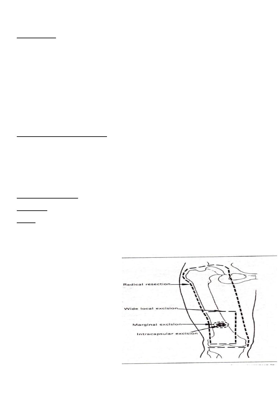
Principles of treatment
1- Benign asymptomatic lesion need
no treatment ; if the diagnosis is not
100% sure then biopsy is indicated .
2- Benign symptomatic :painful or
continue grow after arresting of
growth : it require biopsy and
marginal excision .
3- suspected malignant: it need
detailed investigation for knowing the
type and the staging to decide the
way of treatment.
3
Benign bone tumor
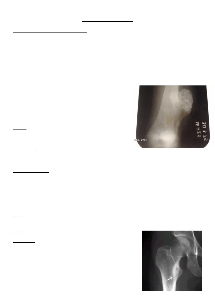
Osteochondroma(bone exostosis)
This is one of the commonest benign bone tumor ; it is developmental tumor start as small
over growth of cartilage at the edge of the physeal plate , and grow to boney protuberance
covered by cap of cartilage.
The commonest sites are the fast growing ends of the long bones , and it continue to grow
until the bone stop growing at the end of the growth period .
Any further increase in size after this period is mean malignant changes .
The patient is usually teenager or young adult
presented with a lump near the end of the long bones ,
some time there is pain .
Multiple lesions can be present as part of heritable
disorder (multiple exostosis) called diaphyseal acalasis.
X-ray : boney projection from the metaphysis of the
long bone, large lesion may have cauliflower – like
lesion .
Treatment : excision .
Osteoid osteoma
Small tumor of bone cause symptoms out of all proportion to its size .
Patient usually under 30 years old , male more than female , it can affect all bones except
the skull and mainly the femur and tibia .
The patient complain from persistent pain which typically revealed by salicylate .
X-ray :plain x-ray show small radiolucent area (nidus) in the diaphysis surrounded by area of
sclerosis and cortical thickening .
Tc99 : show localized increased activity .
Treatment : by complete excision of the nidus
4
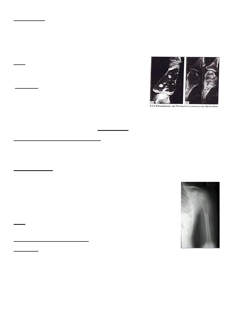
enchondroma
It is benign tumor of the cartilage , it seen in young people and it affect the tubular bone of
the hand and feet (inside the bone) .
The tumor is usually asymptomatic and discovered incidentally by x-ray or afterpathological
fracture .
X-ray show well defined centrally located , radiolucent area
and there is flecks of calcification within the lucent area (this
is a path gnomonic feature of the tumor) .
Treatment : removal of the tumor by curettage and bone
graft .
*The tumor has a possibility of 2% to change to malignant
Bone cysts
We have two main types of bone cysts :
1- simple bone cyst (solitary bone cyst) .
2- aneurysmal bone cyst .
Simple bone cyst
It is also called solitary or unicameral bone cyst .it appear during the
childhood , typically in the metaphysis of the long bones and mostly in the
metaphysis of the humerous or femur .
It is regarded as tumor – like lesion and it may heal spontaneously .
The condition discovered incidentally or after pathological fracture .
X-ray : show well defined homogenous radiolucent area in the metaphysis
,centrally located , the cortex is thin .
By aspiration by wide bore needle , it contain straw - colour fluid (yellow ) .
Treatment:
Small lesion can be treated conservatively By continuous follow up and advice the patient
not to exposed to trauma to avoid path. Fracture .
If no progression of healing occur or the cyst tend to be larger , we can do aspiration of the
fluid and injection of methylprednisolone 80-160mg inside the cyst this may induce healing.
If no improvement then surgical curettage and bone graft . This lesion is carry considerable
risk of recurrence .