Lecture 2 dental implant
Dr. Bara Sultan MinwahB.D.S, M.Sc, F.I.B.M.S
Axes of the lecture include:
1-Types of dental implant.
2- types of dental prosthesis on dental implant.
3-preoperative evaluation and preparation of a patient for dental implant.
• Mucosal Insert
• Endodontic Implant (Stabilizer)• Sub-periosteal implant
• Endosteal or Endosseous implant
• Plate-form implant
• Root-form implant
• Transosseous implant
Titanium Mucosal Insert

2. Endodontic Implant (Stabilizer)
Endodontic implants are similar to prosthodontic implants in many respects. However, they serve another purpose—the stabilization and preservation of remaining natural teeth, not the replacement of lost teeth.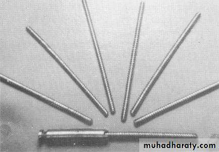
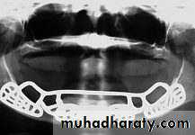
3- Subperiosteal Implants
• With this type of implant, a metal frame is placed under the periosteum but on top of the bone.Procedure Process of a Subperiosteal Implant
First surgeryThe alveolar ridge is exposed and impressions are taken.
The tissue is repositioned over the ridge and sutured back into place.
The impression is sent to the laboratory, where a metal frame with posts is fabricated.
(Cont’d)
Procedure Process of a Subperiosteal Implant
(Second surgery
The alveolar ridge is surgically exposed.
The metal frame is placed over the ridge.
With the frame in place, the tissues are repositioned and sutured into place.
Subperiosteal implant full-arch denture prosthesis.

Sub-periosteal implant
Subperiosteal Implants were already introduced in the 1940s. Of all currently used devices, it is the type of implant that has had the longest period of clinical application. These implants are not anchored inside the bone, such as Endosseous Implants, but are instead shaped to ride on the residual bony ridge of either the upper or lower jaw. They are usually not considered to be osseointegrated implants.Subperiosteal Implants have been used in completely edentulous as well as partially edentulous upper and lower jaws. However, the best results have been achieved in treatment of the edentulous lower jaw.
Indications:
Usually a severely resorbed, completely edentulous, lower jaw bone which does not offer enough bone height to accommodate root form Implants as anchoring devices.

4-Transosteal Implants
These implants are primarily used in patients with severely resorbed ridges. The implant is inserted through the inferior border of the mandible and into the edentulous area.Copyright © 2009, 2006 by Saunders, an imprint of Elsevier Inc. All rights reserved.
• These implants are not in use today , because they necessitate an extraoral surgical approach to their placement, which again translates into general anesthesia, hospitalization and higher cost.
In any case, these implants are used in mandibles only and are secured at the lower border of the chin via bone plates. These were originally designed to have a secure implant system, even for very resorbed lower jaws.
A typical Transosseous Implant. The plate on the bottom is firmly pressed against the bottom part of the chin bone, whereas the long screw posts go through the chin bone, all the way to the top of the jaw ridge inside the mouth. The two attachments that will eventually protrude through the gums can be used to attach an overdenture-type prosthesis.

The plate
long screw postsThe two attachments
5- Endosteal root-form implantsSince the introduction of the Osseointegration concept and the Titanium Screw by Dr. Branemark, these implants have become the most popular implants in the world today.
Termed “fixture”

• 1952 -- Osseointegration- founded by Professor Brånemark
• He discovered that bone can integrate with titanium components and the term derived from the Latin word:
• os - bone
• integrate - make whole.

Classification of dental implant prostheses
The four groups of implants prostheses are :1- over denture .
2- Single tooth replacement.
3- fixed prosthesis for the partially edentulous arch.
4- fixed prosthesis for the completely edentulous arch.
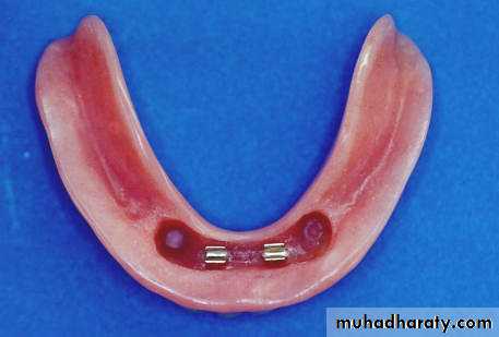
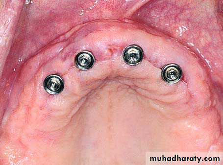
Over denture
Two types of over denture(ovd): 1- implant retained ovd and 2- implant retained and supported ovd.Implant retained ovd are held in place by implants only but their support comes from the soft tissues and underlying bone. They use individual attachments on the implant.
The implant retained and supported over denture are held in place by implants and get some or all of their support from the implants. this ovd uses a connecting bar with attachments for ovd retention.
Dr,salah hegazy


Dr,salah hegazy

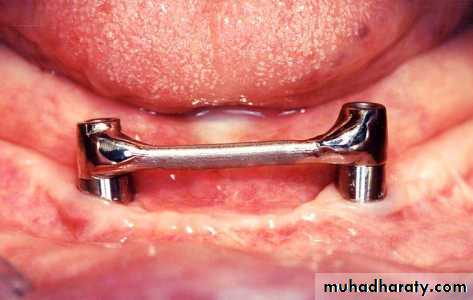

Two-Implant Bar Overdenture
Clinical photograph showing abutments immediatelyafter removal of sutures
Implants joined by over denture bar over whichprosthetic restoration is to be placed

Clips in denture which snap onto bar for retentionand support of denture

Clinical photograph showing prosthetic restorationadapted to implants

Single tooth prosthesis
The single tooth prosthesis is used to replace missing single tooth. Prosthesis support and retention come from the implant. Retention may be achieved through screws and cement.
Fixed prosthesis for the partially edentulous arch
The fixed -fixed bridge for the free end or bounded saddle area is a porcelain to metal restoration .it gains both its support and retention completely from the implants .
the retention may be in the form of screw retention or cement.
Fixed prosthesis for the completely edentulous arch
It can be made in a style similar to conventional crown and bridge .It gains it support and retention from the implants.
Retention may be gained through the screws or cement.

cooperation of the patient.
the patient must be informed about :1- the time necessary to complete the functional and aesthetic rehabilitation.
2- the importance of maintaining good oral hygiene.
3- the possibility, during the post treatment period, of discomfort or pain caused by edema, haematoma, bleeding.
Not to be ignored is also the patient psychological aspect and his/her expectations.
Pre operative PATIENT EVALUATION
PRE – OPERATION EVALUATIONS
the general health state of the patient .
The clinical examination of the oral cavity must be made so as to obtain information regarding
1- the conditions of the dental elements, their number and position and the presence of previous restoring interventions and their state of maintenance.
2- the state of the soft tissues must be evaluated, with particular attention for what concerns the quantity and quality of the present adherent mucosa
3- the degree of the oral hygiene.
4- to perform an accurate periodontal investigation .
5- the analysis of the bone base and that of the alveolar margins to verify their adequacy for the implant treatment.
6 - evaluate the intercortical distance of the bone (bone thickness)
7- determine the interocclusal distance both in the mouth and with study models mounted on an articulator.
Classification
DensityLocation
D1
Dense cortical bone more than 70%
Anterior lower jaw
D2
The compact bone 50-75 %
Ant./post. lower jaw
ant. upper jaw
D3
The dense cortical bone 25-50%
Ant./post. upper jaw
post lower jaw
D4
Spongeous bone, large structure. The dense bone less than 25%
Posterior upper jaw
Bone quality (according to MISCH)
• Misch classification
PRE – SURGICAL PLANNING Radiographic examinations
The measurement of the bone vertical height must be performed through a radiographic examination. This will supply also indispensable information about the anatomy of the patient, facilitating the selection of the implant to be used and its correct positioning, taking into consideration the variation in anatomic structures.
Panoramic radiography
It allows to have a view of the dental arches and bone structures, maxillary sinus and nasal cavity, of the existing conservative and prosthetic restorations.Moreover, it allows to evaluate the general conditions of the periodontium and to put in evidence eventual pathologies or anatomical anomalies.
When evaluating a panoramic radiography, it is necessary to consider its distortion factor.
1:1.25 magnification ratio.
Radiographic stent

Intraoral radiography
Periapical radiograph gives more fine details about the bone quantity of a determined site, previously individualized by a panoramic radiography and a therapeutic orientation. It enables also to collect information concerning eventual bone rarefaction and to localize the course of nerves, nasal cavity, maxillary sinuses or the root anatomy of the adjacent teeth.Prosthetic stent
Surgical stent
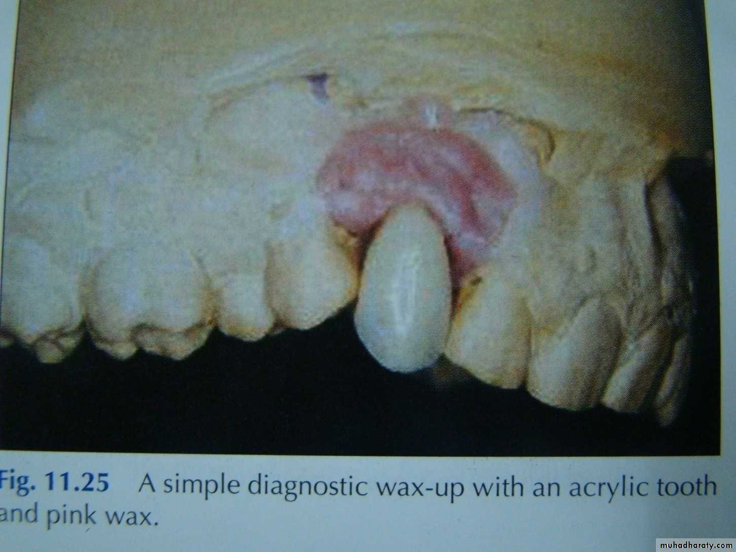
Once the position of the implants is determined by clinical, radio graphical , and diagnostic cast examinations, a surgical stent is then fabricated.The surgical stent should seat on the occlusal surfaces of the remaining dentition, and the ridge should be clearly visible through the occlusal surfaces where the wax-up has been removed.
Surgical stent
Holes are then drilled through the temporary stent on the cingulum of the anterior teeth and the central fossa of the posterior teeth. This is done ensuring that the drill used is held vertical to the crest of the ridge and parallel from one hole to the other.
Dr,salah hegazy
THANK YOU
Thank You!
