1
Fifth stage
radiology
Lec-5
د.هديل
27/3/2017
G.I.T
Acute abdomen
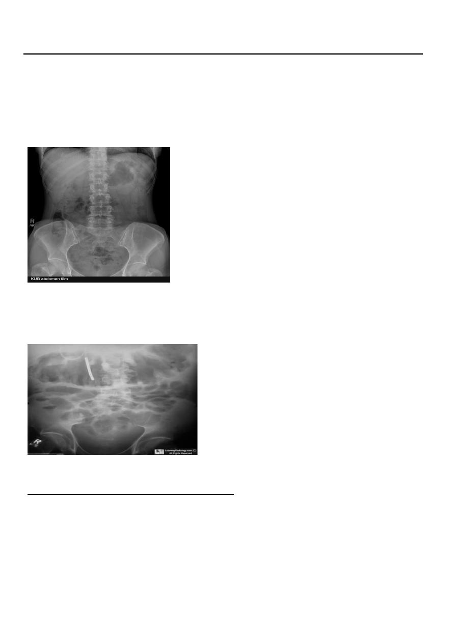
Normal KUB appearance
toxic megacolon (TM) is complication that can be seen in both types of inflammatory bowel
disease more in UC , in infectious colitis, as well as in some other types of colitis.
Radiographic features OF TOXIC MEGACOLON:
The colon (typically transverse colon) becomes dilated to at least 6 cm (usually
greater). There is additional loss of haustral markings
It is serious acute abdominal condition
More in UC > CD
Practical points
barium studies and colonoscopy should be avoided, due to the risk of perforation
2
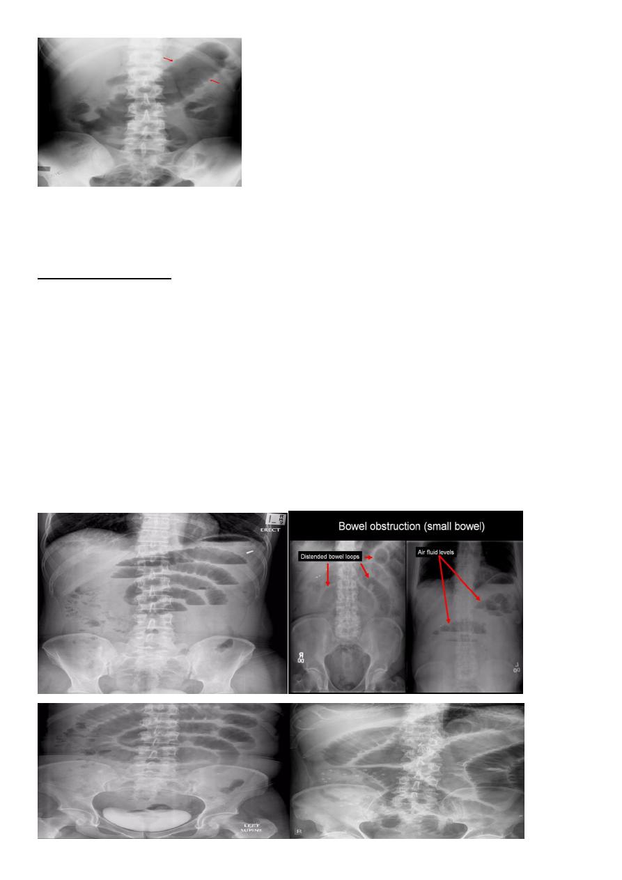
Small bowel obstruction
accounts for 80% all mechanical intestinal obstruction; the remaining 20% result from large
bowel obstruction.
Radiographic features
Abdominal radiograph
Abdominal radiographs are only 50-60% sensitive for small bowel obstruction
.
In most cases, the abdominal radiograph will have the following features:
dilated loops of small bowel proximal to the obstruction
predominantly central dilated loops
three instances of dilatation over 3 cm
valvulae conniventes are visible
fluid levels if the study in erect position
3
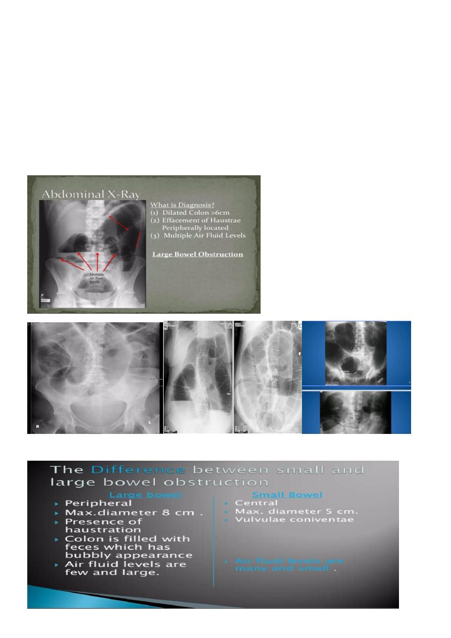
Large bowel obstruction (LBO)
are often impressive on imaging, on account of the ability of the large bowel to massively
distend. This condition requires prompt diagnosis and treatment.
Radiographic features
Colonic distension > small bowel
Peripherally located
Dilated loops Less in no. than SBO
Presence of haustra
4
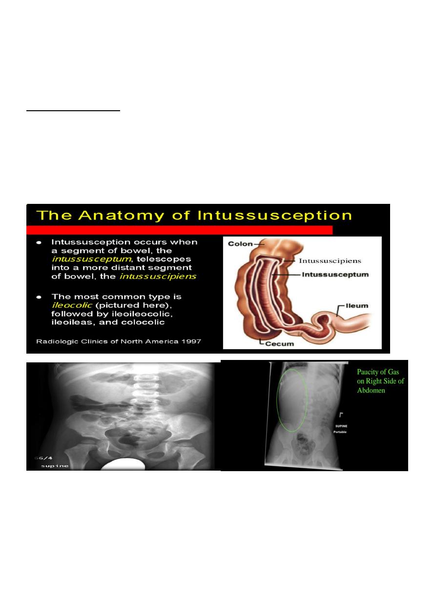
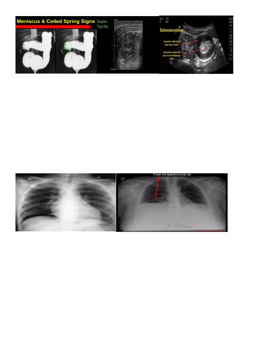
Intussusceptions
occurs when one segment of bowel is pulled into itself (or a neighboring loop of bowel) It is
an important cause of an acute abdomen in children
Intussusceptions may also occur in the adult population where it is usually caused by a focal
lesion acting as a lead point.
Radiographic features
Intussusceptions can occur essentially anywhere, in children there is a
strong predilection for the ileo colic region
Abdominal plain film
Abdominal x-rays may demonstrate an elongated soft tissue mass (typically in the right
upper quadrant in children) with a bowel obstruction proximal to it.
Ultrasound
is a reliable screening tool for children at low risk for intussusceptions
Ultrasound signs include:
target sign (also known as the doughnut sign)
Pseudo kidney shape sign
5
Pneumoperitoneum
describes as gas within the peritoneal cavity, and is often of a critical illness
plain film
Chest radiograph
An erect chest x-ray is probably the most sensitive plain radiograph for the detection of free
intra peritoneal gas as crescent shape of lucency below diaphragm , more in the RT sided
aspect
.
Described as sub diaphragmatic free gas
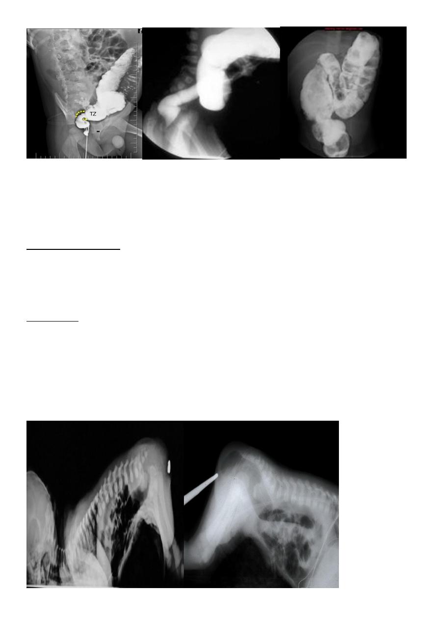
Hirschprung disease
is the most common cause of neonatal colonic obstruction (~15-20%). It is commonly
characterized by a short segment of colonic aganglionosis affecting term neonates,
especially boys .
contrast enema
A carefully performed contrast enema is indispensable in both the diagnosis of Hirschprung
disease but also in assessing the length of involvement. It should be noted however that the
depicted transition zone on the contrast enema is not accurate at determining the
transition between absent and present ganglion cells.The affected segment is of small
caliber with proximal dilatation Fasciculation/saw-tooth irregularity of the aganglion
segment is frequently seen.
6
Anal atresia (or imperforate anus)
refers to a spectrum of ano rectal abnormalities ranging from a membranous separation to
complete absence of the anus.
Abdominal radiograph
can be variable depending on the site of atresia (e.g high or low) , level of impaction with
meconium and physiological effects such as straining
may show multiple dilated bowel loops with with absence of rectal gas
Invertogram
A coin/metal piece is placed over the expected anus and the baby is turned upside down
(for a minimum 3 minutes).
Distance of gas bubble in rectum from the metal piece is noted:
>
3
cm: denotes high type
<3 cm: denotes low type