Fifth stage
RadiologyCT scan1
د. هديل
10/4/2017
Computerized tomography (CT scan)
Introduction
Tomography is one of the imaging investigation, it present from 1960.
Mechanism of Action:
Tomography is the technique of rotating a radiation detector around the patient so that the image obtained gives additional three- dimensional information.
In plain film tomography the source of XR ray & the photographic film move around the patient to produce an image of the structures at a particular depth within the body, bringing them into sharp focus, while deliberately blurring (unclear) structures above & below them.
In computerized tomography, the same technique of usual tomography, but instead of film, there is detectors which attach to computer, this detector will catch the signal & convert it into pulse, then to computer as image of different section.
So CT scan is a form of XR examination , in which the source & detector ( CT scanner ) & the information obtained can be used to produce cross sectional image ( i.e. CT scan can produce an image of slice through the body at a particular level)
N.B. when the no. of the detector increase, the clearance of the image is increase, the usual no. of detectors is used now about 400.
What is the principle of CT scan?
The principle of CT scan is XR penetration & XR attenuation.
The XR can penetrate any organ & when penetrate it, it will face many different density tissues like bone, muscle, fat, fluid or CSF.
Absorption of X-ray by these different tissues depend on:
Thickness
Conc. Of material
Molecular weight of the substance
The higher the thickness, the higher the Molecular weight, the more the absorption of the XR & more attenuation examples:
Bone: is highly attenuated material, because it absorbed much XR so it appear white in color.
Soft tissues have less attenuation, so appear gray in color.
Air has low absorption & low attenuation so it appear black in color.
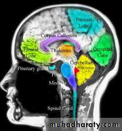
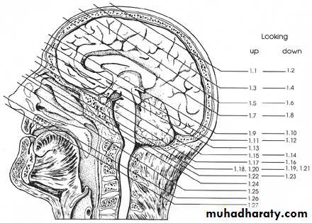
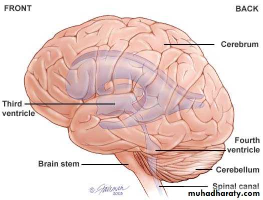
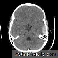
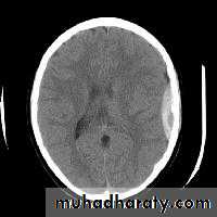
Normal brain (anatomy)
Cerebral hemisphere
Frontal lobe
Parietal lobe
Temporal lobe
Occipital lobe
Posterior fosse
Cerebellum & brain stem
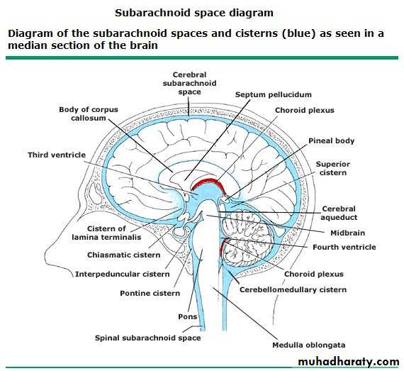
Ventricular system
Lateral ventricle ( frontal horn , body ,temporal horn ,& occipital horn ) .Third ventricle .
Fourth ventricle .
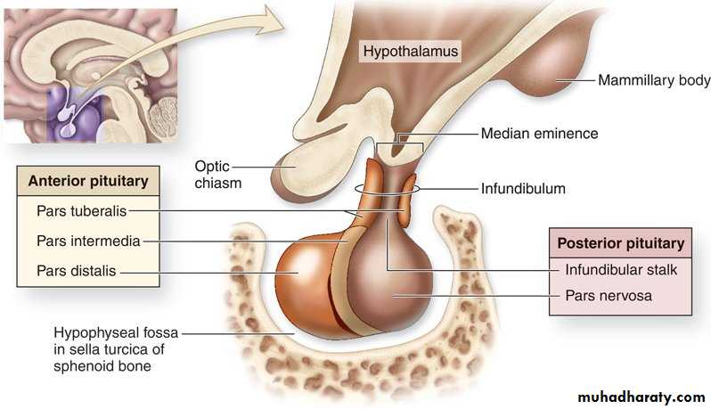
Pituitary fosse
Anteriorcliniod process
The region of the sellatursica
Posteriorcliniod process
Channel like area black in color ( cerebello pontine angle )
Lesser wing of the sphenoid bone & below it immediately the greater wing of spheniod.
CT terms
Isodense, same density to organ examineHyper dense higher than the organ examined
Hypo dense lower than the organ examined
Density levels of different types of tissues
Density levels of almost all soft tissue organs lie within a range 10-90 HU, Water density 0 HU, fluid 10-15 HU depending on their density , air -1000 HU, fat -100 - -50 HU , bone & calcification 100 & above HU , ossification 1000 & even more HU depending on the density
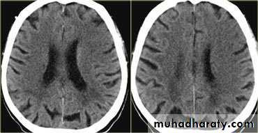
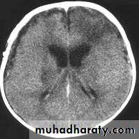
Encephalitis
Age : young & adolescence mostly .
Clinical picture : patient come with headache , vomiting , blurring of vision .
Usually the disease preceded by flue or vaccination , HS virus can also cause encephalitis .
CT finding
Low attenuation of white matter ( due to the edema)
Compressing the ventricular system ( also due to edema ) specially the frontal & the body of the lateral ventricle become slit like .
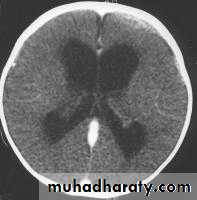
Complication
Brain destruction ( because the disease cause brain necrosis ) .
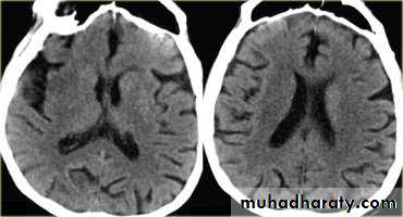
Atrophy of the brain tissues with subsequent loss of volume & dilatation of the ventricular system .
Hydrocephalous .
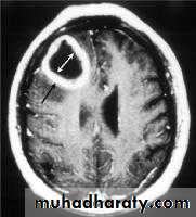
Herpes encephalitis
same as usual encephalitis but there will be low attenuation or hypo density area in the tempo frontal or tempo priato frontal with evidence of patchy whitish area due to the clotted blood ( hemorrhage ) .Brain abscess
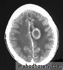
CP: high fever for long duration with focus of infection, patient may be toxic, or present with epilepsy (focal)
CT finding
Hypo dense area surrounded by large area of peri focal edema, which may cause shifting of the midline).
After injection the lesion show ring pattern of enhancement so the peripheral aspect of the lesion become clearer.
DDx
Secondary metastasis, patient have history of primary malignancy, beside absence of the fever.
Primary necrotic tumor, neither Hx of primary, nor fever are present.
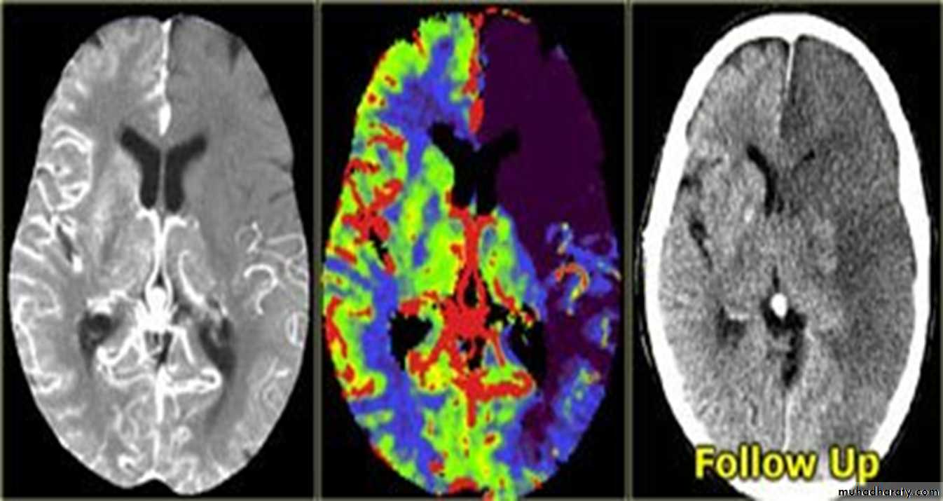
Infarction
AcuteChronic
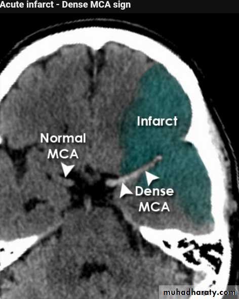
Acute infarction
History: sudden onset of hemiplegia , with slurred speech .
CT features:
Wedge shape area of hypo density involve the tributaries of anterior , middle or posterior cerebral arteries , ( i.e. priato frontal &priato occipital area )
Shift of the midline to the other side .
Compression of the ipsi lateral aspect of the regional ventricle by the edema
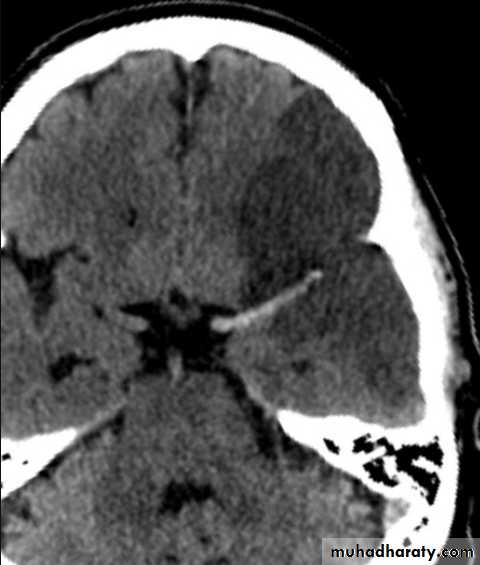
Dense middle artery sign
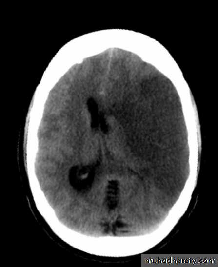
Chronic infarction
The necrotic area will be liquefy & change to the source of fluid (por encephalon ) .
CT finding
Hypo density area as a result of blockage of AC , MC , & PC arteries .
The ventricle will be dilated to compensate for the loosed brain tissue .
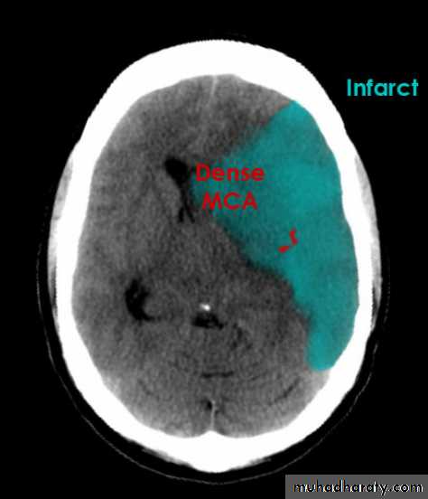
ACUTE INFARCTION
* wedge shape hypo density area.* Shifting of the midline & frontal horn
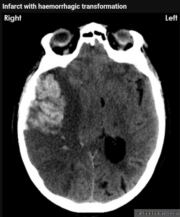
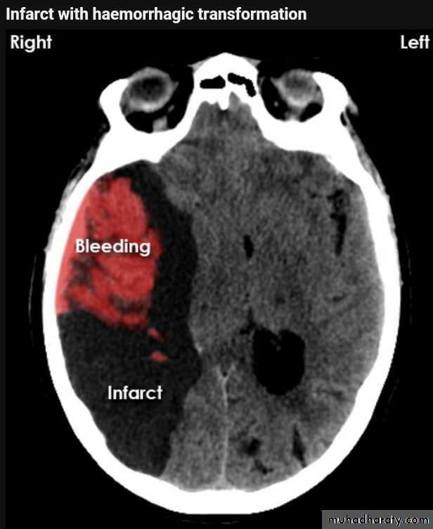
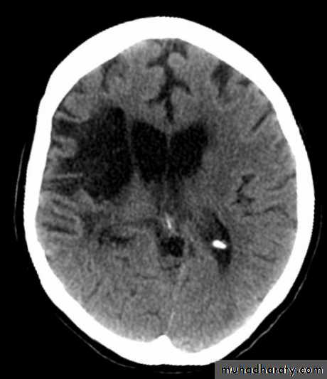
Chronic infarction
Hypo density area(pore encephalic cyst)Dilated ventricle
Hemorrhage
Classify the brain hemorrhage according to the site:Epidural EDH
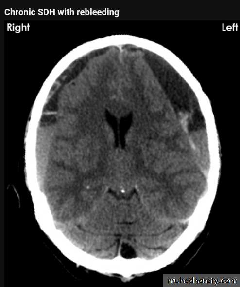
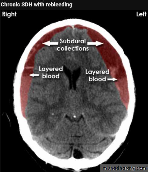
Subdural SDH
Sub arachnoids SAH
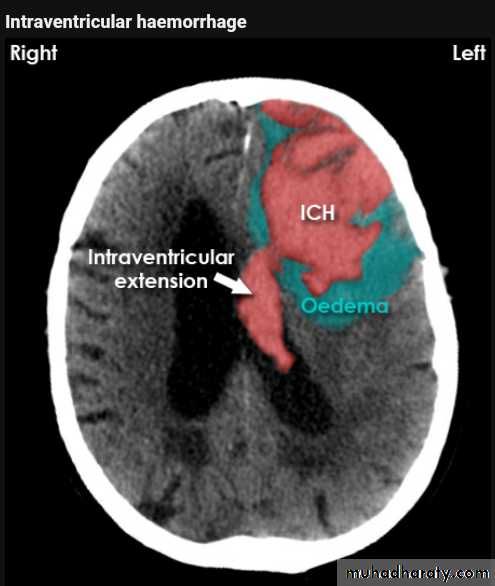
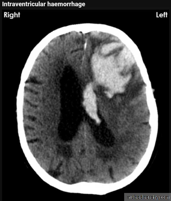
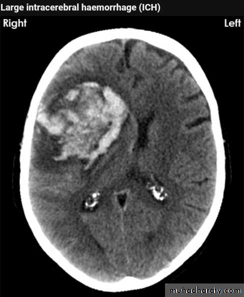
Intra cerebral ICH
Intra ventricular IVH
Other classification according to the onset of bleeding:
Acute
Chronic
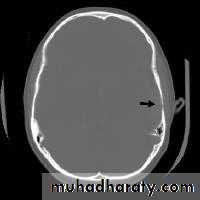
Acute EDH
CT finding
Hyper density area, 50-70 HU, due to the clotted blood, elliptical in shape, no edema around the lesion, but may cause shifting of the midline, or compression of the ipsi lateral ventricle if it is large enough.
The source of bleeding of the haematome is injured middle meningeal artery so the lesion > 90 % associated with skull # at the site of the previous mentioned artery.
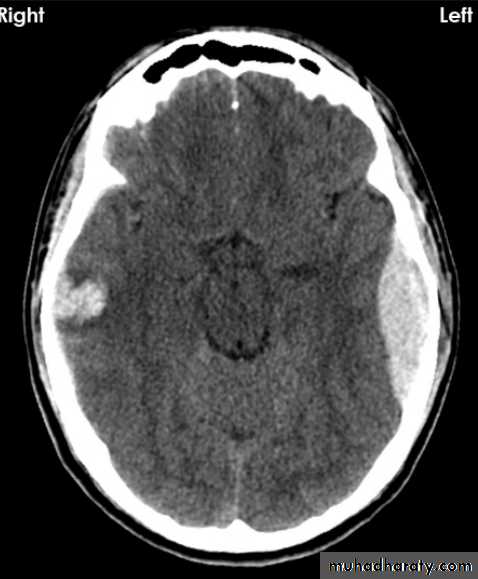
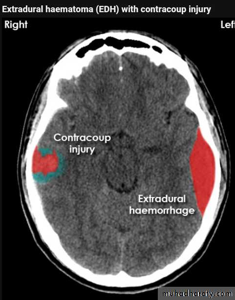
EDH
Biconvex hyper density area
Shifting of the midline
Compression of the ventricles
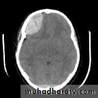
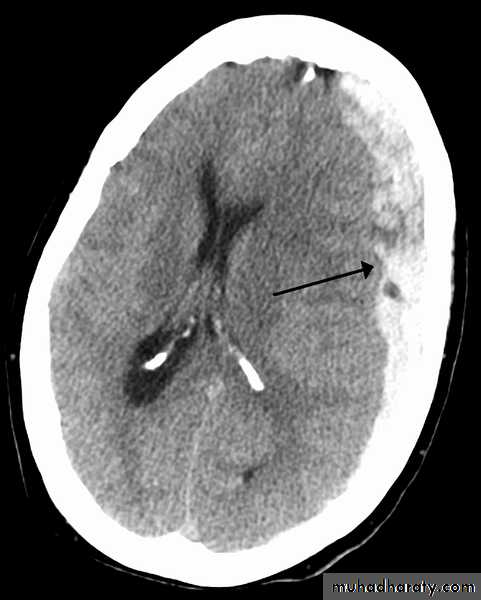
Acute SDH
CT findingCrescent shape or oval shape of hyper density area their inner medial margin is irregular shaggy
Associated with edema which cause shifting of the midline .
The source of bleeding hematoma is venous , not associated with # , but occur as a result of disruption of subdural vein , more commonly to occur in the old age group , due to brain concussion , ( brain atrophied wide SAS ) & in the pediatric age group the SAS &cistern are wide also .
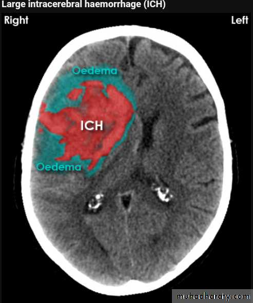
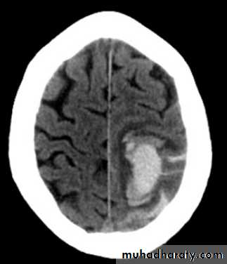
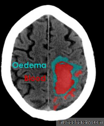
Acute Intra cerebral hematoma
CT finding :Hyper dense area , surrounded by edema , any where within the brain parenchyma.
Shifting of the midline
Compression of the ipsi lateral ventricle .
Associated with infarction , # , trauma concussion , tumor …..
Hypertensive Hemorrhage
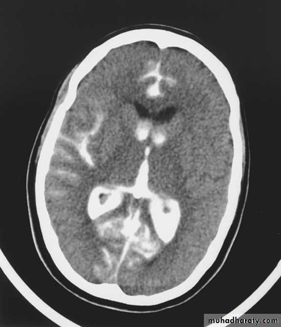
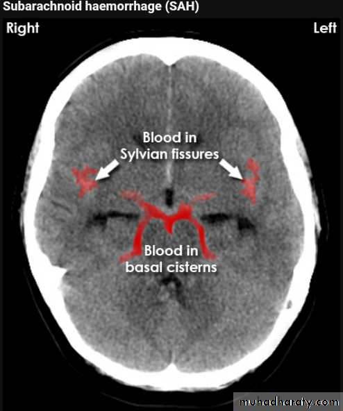
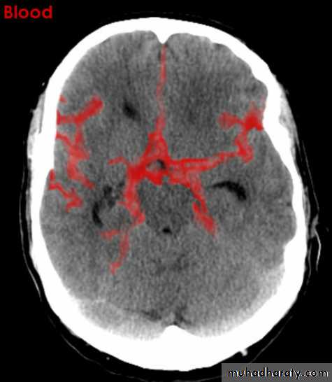
Subarachnoid hemorrhage
causesdue to ruptured aneurysm over 90 % of cases spcially at the circle of Willis .
ruptured AV malformation .
trauma .
CT finding
hyper density is seen within the SAS ( hyperdense sulci , being filled with clotted blood)
opacified inter hemispheric fissure ( become white & more dense )
opacification of the falx cerebri .
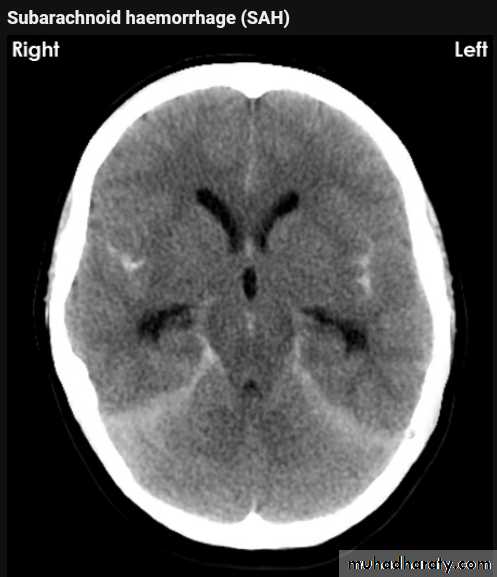
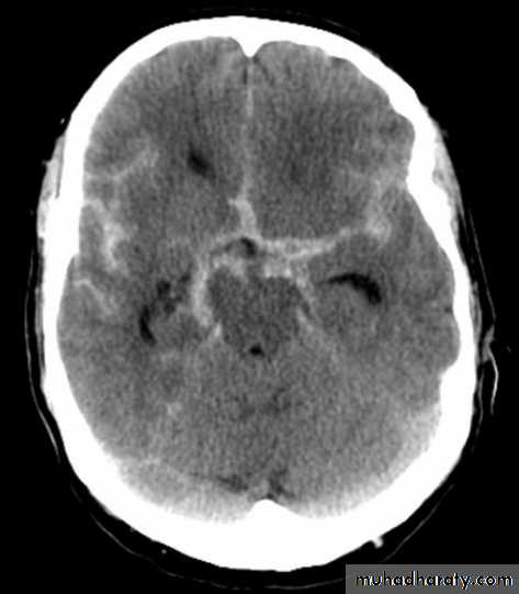
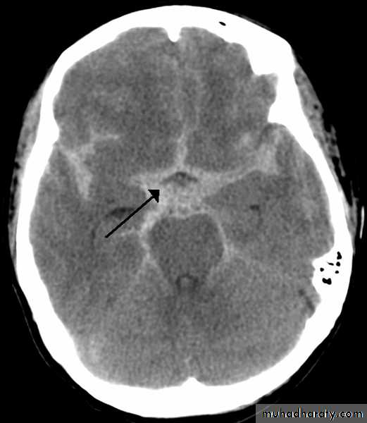
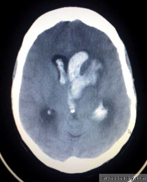
SAH
White sulciOpacified IHF