93
رابع اسنان كركوك \بريو نظري د.حسين
2017/11/16
Lec . 1 PeriodonticsDr.Hussein Al Dabbagh
Periodontics : that branch of dentistry deals with diagnosis and treatment of diseases and condition of the supporting and surrounding tissues of the teeth.
Periodontology : the scientific study of the periodontium in health and disease.
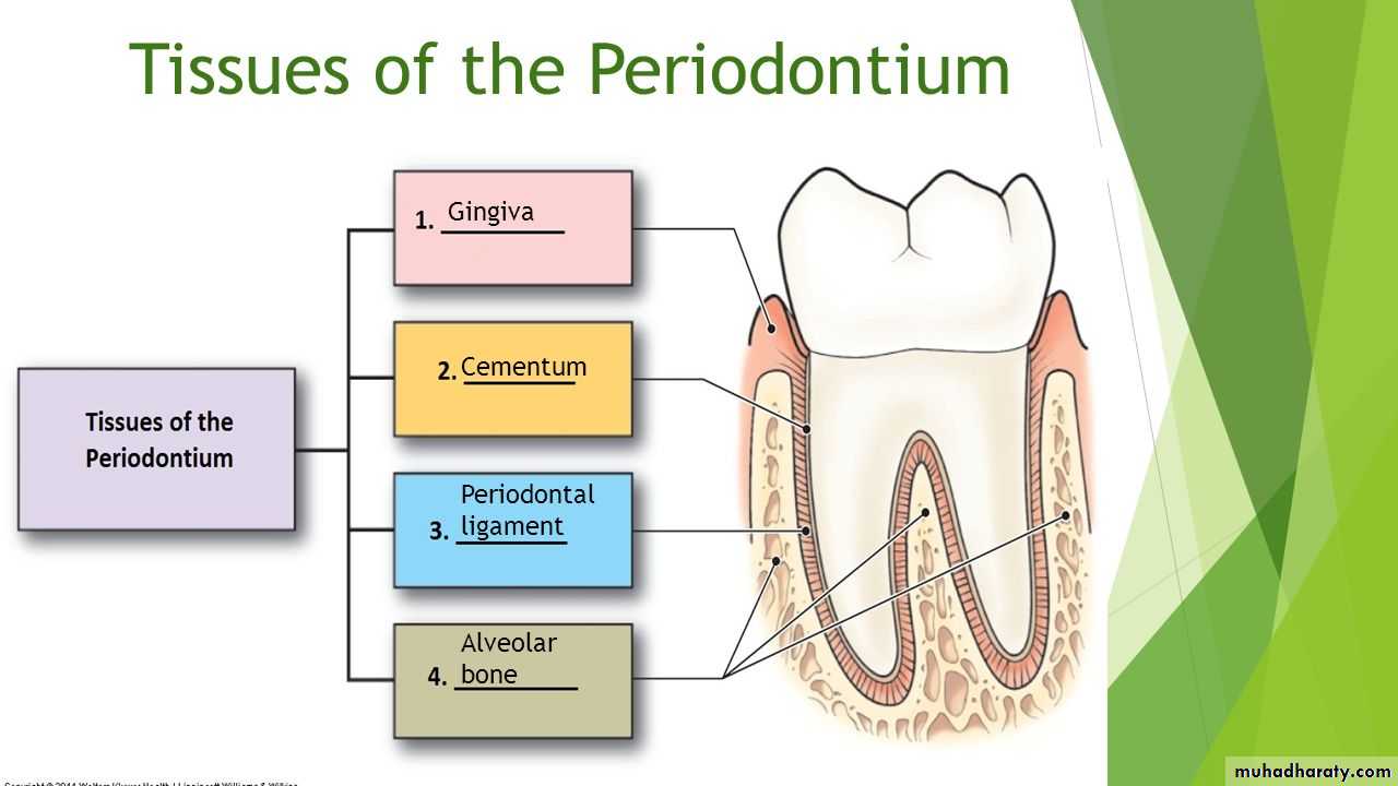
Periodontium : is the tissue that invest and support the teeth.
Periodontium comprises the following tissues:
The gingiva.
The periodontal ligament.
The root cementum.
The alveolar bone.
The main function of the periodontium is to attach the tooth to the bone tissue of the jaws and to maintain the integrity of the surface of masticatory mucosa of the oral cavity.
The periodontium is also called the attachment apparatus , the supporting tissues of the tooth.
The gingiva :
The gingiva is defined as the part of the oral mucosa that covers the alveolar processes of the jaws and surrounds the cervical portion of the teeth.The oral mucosa consists of three zones:
The gingiva and the covering of the hard palate termed (masticatory mucosa).
The dorsum of the tongue covered by (specialized mucosa).
The oral mucous membrane lining the remainder of the oral cavity.
Clinically the gingiva is divided into 3 parts:
Marginal gingiva.
Attached gingiva.
Interdental gingiva.
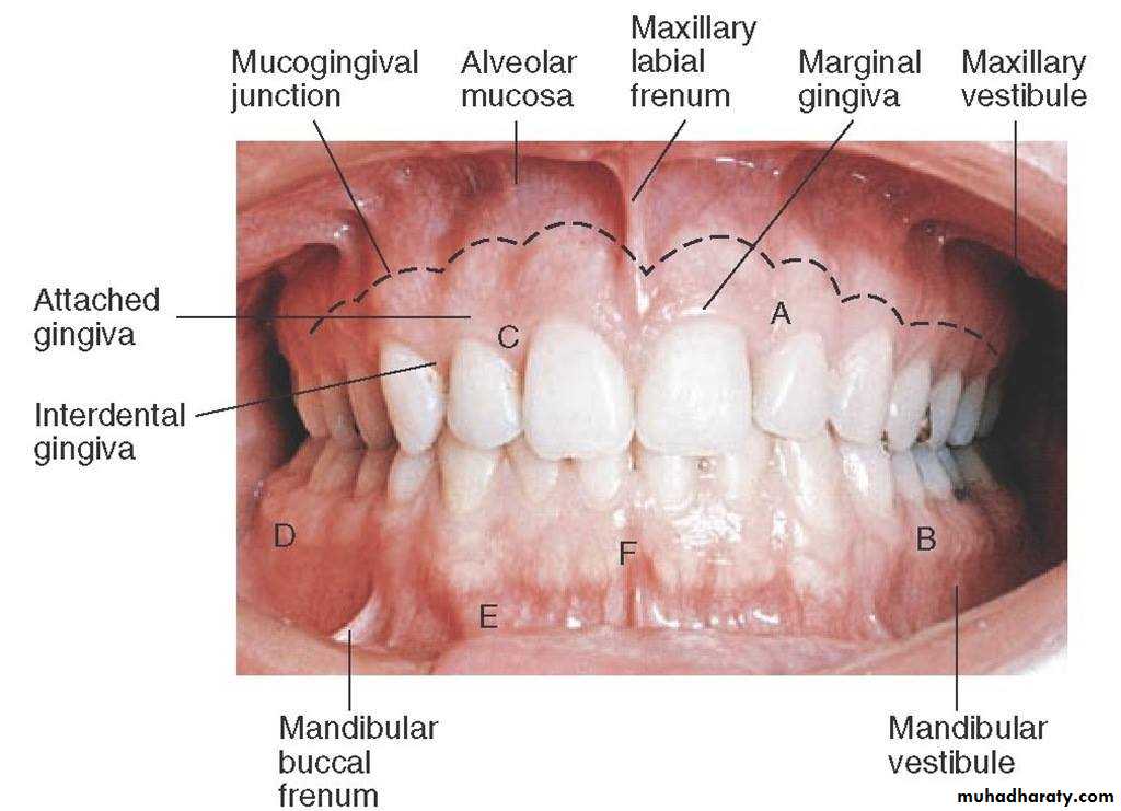
Marginal Gingiva
The marginal, or unattached,or free gingiva is the terminal edge or border of the gingiva surrounding the teeth incollarlike fashion , it is demarcated apically from the adjacent attached gingival by free gingival groove (which is shallow linear depression, usually about 1 mm wide and is positioned at a level corresponding to the level of cement-enamel junction ).
Free gingiva forms the soft tissuewall of the gingival sulcus.
After complete eruption of the tooth , FG margin is located on the enamel surface approximately 0.5-2 mm coronal to the CEJ.
Gingival sulcus :
V-shaped space bounded by the free gingival margin , the tooth , and the most coronal attachment of the junctional epithelium , it is lined by non-keratinized sulcular epithelium. Clinically normal gingival sulcus measures from 1-3 mm deep, but in disease the space become deeper and is referred to as pocket.
Attached gingiva:
It is demarcated coronally from the free gingiva by the free gingival groove and extend apically to the mucogingival junction where it becomes continouseith the alveolar mucosa.It is firm ,collar pink, resilient and tightly bound to the underlying teeth and periosteum of the alveolar bone. The irregular surface texture of the attached gingival (stippling) similar to the surface of orange peel, stippling is only present in 40% of adults and it is considered as a feature of healthy ginginva.
The width of attached gingiva differs in different areas of the mouth, it is greatest on facial aspects of maxillary lateral incisors and narrowest on the facial surfaces of the mandibular canines and first premolars, on the lingual surfaces , it is narrowest in incisors and canines areas, and widest near the first and second molars, the variation range is 1-9 mm.
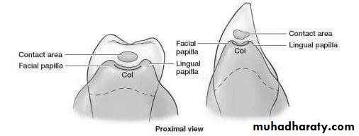
Interdental gingiva (papillae):
It is located in the interproximal space beneath the area of the teeth contact. It is triangular in shape from mesio-distal aspect.The shape of I.D.P is determined by:
The contact relationships between teeth.
The width of the approximal tooth surfaces.
The course of the cementoenamel junction.
In general there are 2 shapes of I.D.P :
Pyramidal shape : present in the anterior region of the dentition where there is approximal contact point between two adjoining teeth.Col shape : the interdental papillae between the posterior teeth are more flattened and there is a concave depression that connects a facial and lingual papilla and confirms to the shape of the interproximal contact surfaces.
In diastema or gingival recession , no Col will be seen, the significance of Colit is covered by thin non-keratinized epithelium , hence represents the most frequent site for initiation of disease process.