Hawler Medical University
College of Health Sciences
Clinical Biochemistry Dept.
Ass. Lec. Amer Ali Khaleel
(M.Sc. Clinical Immunology)
Lab.2 :
Whole Blood, Serum
& Plasma Collections
Practical Immunology and Serology
Copyright © 2016

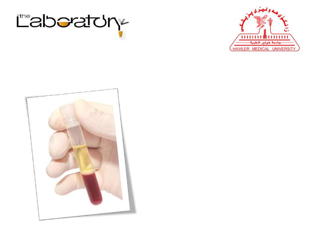
Preparation and Separation of
Plasma:
The blood is transferred from a person’s vein to a test
tube and prevented from clotting (tube containing
anticoagulant e.g. Heparin, EDTA, Sodium Citrate,
Oxalate, Sodium Fluoride, Sodium Iodoacetate ) , this
preparation should be mixed immediately and
thoroughly to avoid clotting, and centrifuged at 2500-
3000 rpm for 5-10 min.

Preparation and Separation of
Plasma:
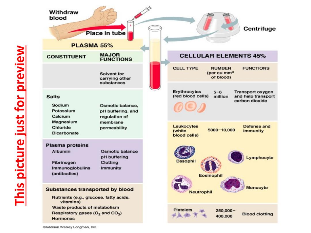
It separates into two layers. The upper liquid layer,
called plasma, represents about 55% of the volume
of whole blood. The lower layer consists of red blood
cells (erythrocytes), white blood cells (leukocytes),
and blood platelets (thrombocytes). Collectively,
these are called the formed (cellular) elements and
represent about 45% of the volume of whole blood.

Preparation and Separation of
Serum:
• If blood is transferred from a person’s vein to a test tube
without containing anticoagulant and then allowed the
blood to clot at room temperature for 15 to 30 minutes,
when the blood has clotted completely and then
centrifuged at 2500-3000 rpm for 5-10 min. it separates into
two layers, the upper liquid layer called serum and the
lower layer consists of formed elements.
• Serum =Plasma- Fibrinogen(coagulant factor)

Laboratory Specimens:
Most Common
•Blood samples
•Urine samples
Swabs
•Vaginal swabs
•Wound swabs
•Nose swabs
•Pernasal swabs
•Ear swabs
•Eye swabs
•Throat swabs
Additional
•Biopsy material
•Cerebrospinal fluid
•Stool samples
•Fungal samples of hair, nail and skin
•Naso-pharyngeal aspirate
•Sputum
•Semen
•Vaginal secretion
•Saliva
•Tears
•Cerumen (earwax)
•Sweat
Body Fluid
•Percardial fluid
•Pleural fluid
•Synovial fluid
•Vitreous humour
•Amniotc fluid
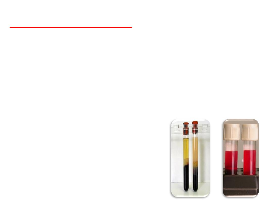
Important note:
• The color of the serum and plasma must be clear yellow
and there is no any turbidity (cloudy) or if any white color
it shows the high proportion of fat in it which affects the
result of the analysis.
• Similarly, if the color is reddish, it indicates the breaking of
red blood cells, which affects a significant impact on some
of the results.

Which is more appropriate Plasma or Serum
or whole blood for doing blood biochemical
estimations?
• It depends on type of analyte to be determined.
• You better use serum rather than plasma.
Anticoagulate in the plasma interferes with many
biochemical parameters particularly trace
elements as lead and mercury are bond
preferably on erythrocytes, therefore for its
determination, the whole blood is preferred.

Method for Serum Preservation
(Storage):
• In normal condition and the right is that the samples are direct do test
without delay but, if necessary keeping the sample must be following
in mind:
• Must separated sample and stored at low temperature in the
refrigerator or freezer.
• Never preserves whole blood samples such as
complete blood count
in the freezer.
• Some samples have special conditions, such as the conservation
Bilirubin
must be kept in the dark.
• There are samples should never delay ,such as analysis of
prothrombin
and
erythrocyte sedimentation analysis
.
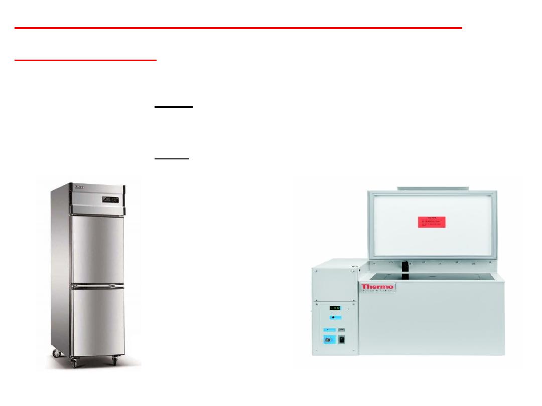
Method for Serum Preservation
(Storage):
1. Preservation for short time,freezing method is followed between
(-4 to -28°C).
2. Preservation for Long time, freezing method is (-86°C).
Benchtop Freezer
Upright Freezer
Notes:
• How long can frozen human serum be stored?
Frozen blood can be stored for 10 years from the date of
blood collection.
• It is important to avoid freeze-thaw cycles because this is
detrimental to many serum components.
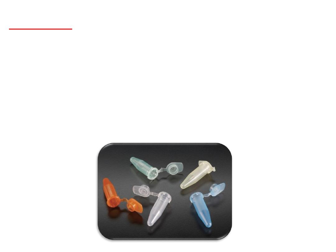
• The serum put in polypropylene microcentrifuge tube.

Common Serum or Plasma Preparation
Errors:
1-Failure to separate serum or plasma from red cells within 30 to 45
minutes of venipuncture.
2-Hemolysis (occurs when the membrane surrounding red blood cells
is disrupted and hemoglobin and other intracellular components
escape into the serum or plasma. Hemolyzed serum or plasma varies in
color from faint pink to bright red, rather than the normal straw color.
Grossly or moderately hemolyzed specimens may be rejected and even
slight hemolysis may alter certain test results).

How To Prevent Hemolysis:
1. Do not leave tourniquet for longer than one minute.
2. Allow alcohol to dry completely before puncturing the
skin.
3. Use a properly sized needle for routine collections.
4. Do not remove the needle from the vein with the vacuum
tube still engaged.
5. Make sure sample is not exposed to extreme heat or cold.
6. Allow blood to clot completely prior to centrifugation.
7. Avoid vigorous mixing or shaking of tubes.
8. Do not centrifuge specimens at higher speed or for longer
than necessary.

Differences between in vitro, in vivo, and in
silico studies
There are three broad categories of experiments:
In vitro
(Latin for within the glass) refers to the technique of performing
a given procedure in a controlled environment outside of a living
organism.
In vivo
(Latin for “within the living”) refers to experimentation using a
whole, living organism as opposed to a partial or dead organism.
In silico
is an expression used to mean “performed on computer or via
computer simulation.

Serological
Reactions:
• Antigen-antibody reaction (binding) in vitro are known as
serological reactions, its useful for detection and
measurement either antibody (Ab) or antigen (Ag) in the
serum.
Uses:
1- Diagnosis of infections.
2- Evaluation of the immunological status.

• Antigens (Ag) : Are molecules (substances) recognised by
the immune system, which induce an immune response
examples
,
,
&
fungi
.
• Antibodies (Immunoglobulin): Are proteins produced by
plasma cells in response to stimulation of B cell by foreign
antigen, basic structure of immunoglobulin is Y shaped
structure, there are five immunoglobulin classes of antibody
molecules found in serum:
IgG, IgM, IgA, IgE
and
IgD
.
Some Concept:

Serological Techniques and Immune
Assays:
•Numerous types of serologic test differ in their speed and
sensitivity, so the most effective test have high specificity and
sensitivity, some are strictly qualitative (positive or negative)
and others are quantitative (amount measured).

Serological Techniques and Immune
Assays
:
There are several different methods used in serological tests:
1.Agglutination test.
2.Precipitation test.
3.Labeled Immune assay (immunoassay).
A.Radioimmunoassay (RIA).
B.Fluorescent immunoassay (FIA).
C.ElectroChemiluminescent immunoassays (ECL).
D.Enzyme immunoassay (EIA) & Enzyme linked immunosorbent assay
(ELISA).

Principle of Serological Tests:
• A reaction between antigen (Ag) and antibody
(Ab) to produce a DETECTABLE reaction.
• Antigen-antibody reaction are visible by
clumps,
precipitates, color changes,
emitting photons &
release of radioactivity.

Agglutination Reaction
(test):
• The interaction (immune reaction) between antibody and a
particulate antigen resulting a visible clumping called
agglutination. Antibodies that produce such reactions are
called agglutinins while antigens are called agglutinogen.
• Agglutination: This reaction done between Ab & insoluble
Ag is part of the surface of some particulate material (such
as Erythrocytes, Bacterial cells, Inert carriers such as
polystyrene latex particles).
• Purpose: Agglutination reactions used to detect either the
presence of antigen or antibody in a sample.
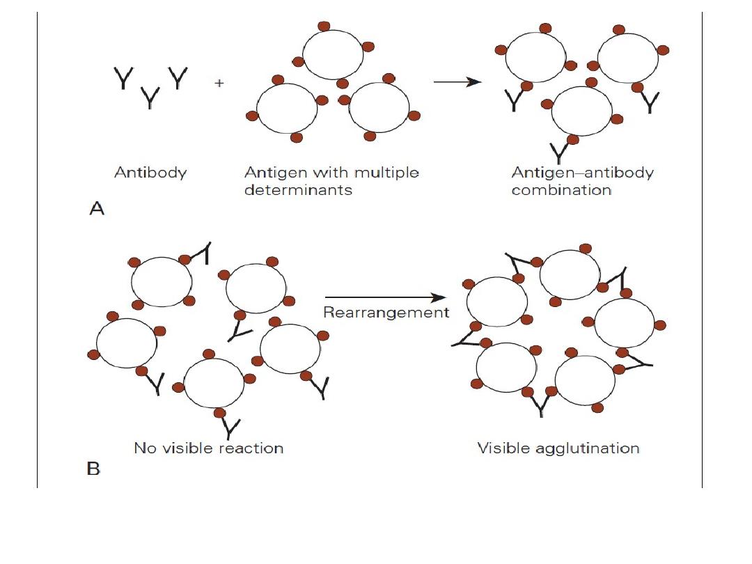
Figure:
Phases of agglutination. (Stevens, 2010) A) Sensitization, physical
attachment of Ag & Ab. B) Network formation.

Clinical Applications of agglutination test:
(Reagents)
• ANA (Antinuclear antibody)
test for diagnosis nuclear autoantibodies.
• ASO (Antistreptolysin O)
test for diagnosis of Streptococcus bacteria.
• Brucella agglutination
test for diagnosis of brucellosis.
• CRP (C-Reactive Protein)
to check for inflammation in the body.
• IM (Infectious Mononucleosis) or Monospot
screening test for infections
mononucleosis.
• Latex test
for rheumatoid factor.
• PT (Pregnancy testing)
for detection Beta-HCG.
• RF (Rheumatoid factor test)
detects autoantibodies present in Rheumatoid
arthritis.
• TPHP (Treponema pallidum particle agglutination assay)
screening test for syphilis.
• VDRL (Venereal Disease Research Laboratory test)
screening test for syphilis.
• Widal test
for diagnosis of salmonellosis.
1-Prepartion of serum.
2-Prepartion of plasma.
3-Preservation methods.
Practical Part