Urinary system
IVU & to a lesser extent CT provide both functional & anatomical informations.
US & MRI provide anatomical informations.Radionuclide scanning provides functional informations only.
Intravenous Urography (IVU):
Also known as excretory urography.It means the visualization of kidney parenchyma, calyces and pelvis after intravenous injection of iodinated contrast medium to the patient, followed by a series of x-ray films.
Its use has been decreased lately as it is replaced by US and Computerized Tomography (CT).
Remains the primary modality for visualization of the pelvicalyceal system and ureters, providing both functional and anatomical information.
Indications
When detailed demonstration of the pelvicalyceal system and ureters is required.
-The assessment of suspected acute ureteric colic.
-Investigation of renal calculi (stones).
-The investigation of hematuria.
Contraindications:
Renal failure.Uncontrolled diebetes.
Multiple myeloma.
In patients receiving drugs for chonic bronchitis, emphysema, asthma and metformin for DM.
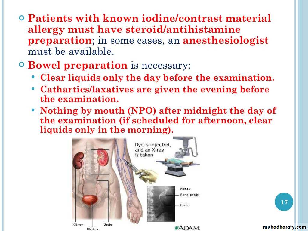
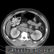
Contrast medium and its excretion:
Urographic contrast media are highly concentrated solutions containing iodine, also known as iodinatedLarge volume of CM (50-100mi) is injected intravenously passes to glomerular filtrate concentrated in renal tubules passes to-pelvicaticeal systems ---ureters---bladder.

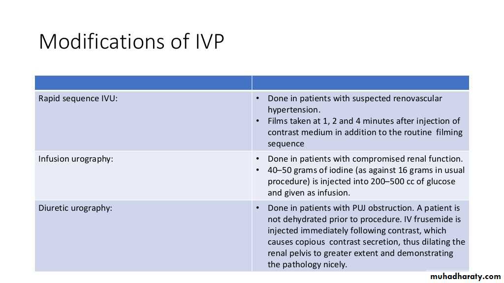
The procedure of performing IVU:
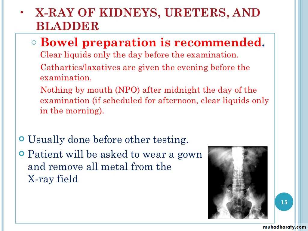
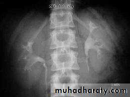
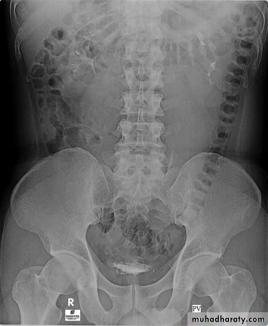
A. First a plain x-ray of the abdomen is taken before the injection of the contrast media, also known as A KUB (kidney, Ureter and Bladder). Calcification & stones may be obscured & missed by contrast media if plain film not takenfirst .B. Films taken after injection of contrast’ medium:
A series of x-ray films are taken after injection of the contrast.
Each film is taken at a time interval determined by the radiologist who is supervising the procedure.
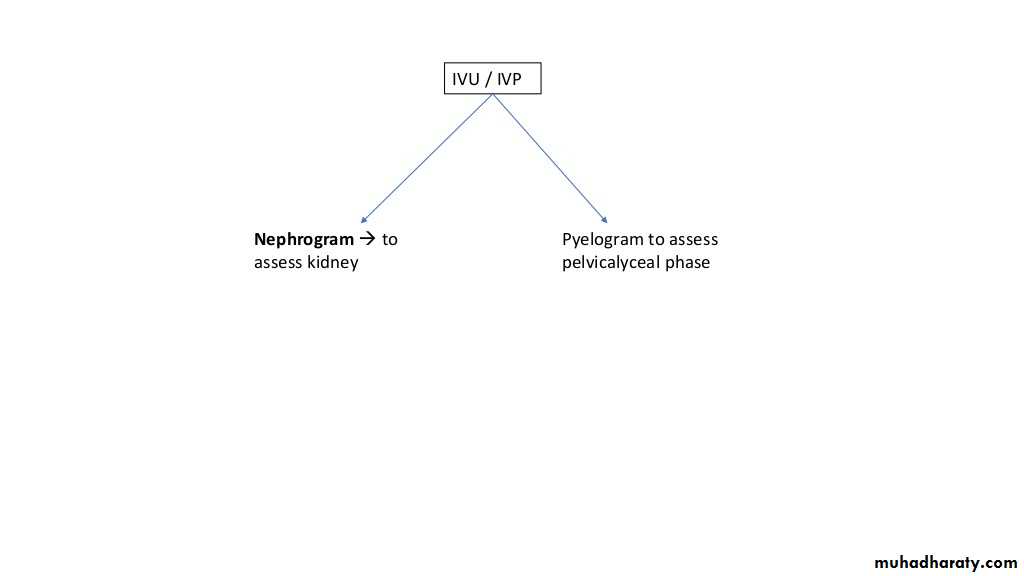
1.Nephrogram phase (Immediately after injection of contrast) .
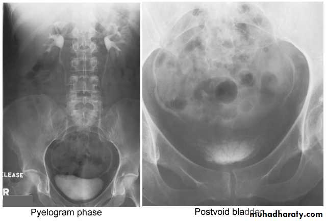
2. Pyelogram Phase(l-5 minutes after injection of contrast) .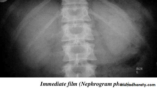
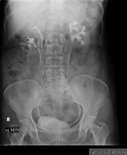
3. After 10 minutes with compression, to get better distention of the pelvis and calyces.
4. Full length film after release of compression .
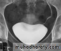
5. A full bladder film (with the urinary bladder fully distended with contrast)
6. Post voiding full length film
Interpretation of IVU films (what to look for?):
Interpretation of IVU films (what to look for?):
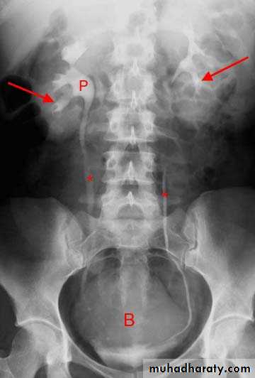
The kidneys:Check their position (left kidney is usually higher).
Identify the whole of both renal outlines, look for any indentations or bulges:
Renal parenchymal width should be uniform(2-2.5cm)
measure renal lengths:
Normal length of adult kidney at IVU is 10-16cm. This is higher than in ultrasound due to image magnification.
2.Calyces:
-should be evenly distributed -symmetrical.
-Cup shaped (normal shape) -Club shaped (when dilated)
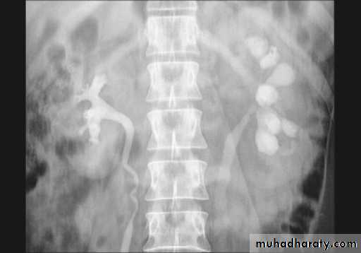
3. Renal pelvis and ureter:
-The normal renal pelvis and pelvi-ureteric junction are funnel shaped.-Ureters are seen only in part of their length on any one film due to obliteration by peristalsis.
Dilatation of pelvis and ureter may be due to:
-Obstruction (by stone , tumors or external compression).-secondary to vesico-ureteric reflux
Look for filling defects which could be due to three common causes
stonestumors.
Blood clots.
4. Bladder
The bladder is a centrally located structure.
Smooth in outline.
Smooth indentation from above by uterus on the right or sigmoid colon on the left and from below by muscles of the pelvic floor is normal.
Other than that, any filling defect, wall irregularity or diverticula must be carefully looked for.
Dense nephrogram:
Faint nephrogram becoming increasingly dense overhours to days.
1. Acute obstruction.
2. Acute hypotension.
3. Acute tubular necrosis.
4. Acute pyelonephritis.
5. Multiple myeloma.
6. Renal vein thrombosis.
7. Acute glomerulonephritis.
8. Amyloid.
9. Acute papillary necrosis.
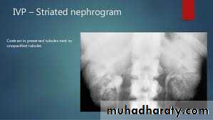
Striated nephrogram:
Linear bands of contrast extending from renal medulla to cortex.1. Acute ureteric obstruction.
2. Infantile polycystic disease.
3. Medullary sponge kidney.
4. Acute pyelonephritis.
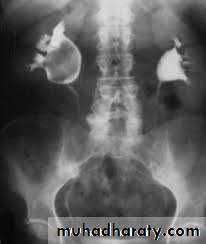
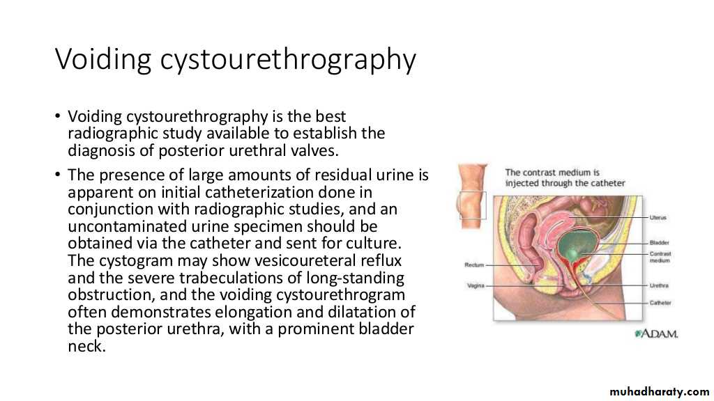
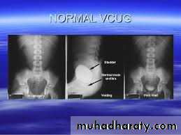
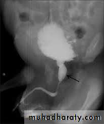
Posterior urethral valve
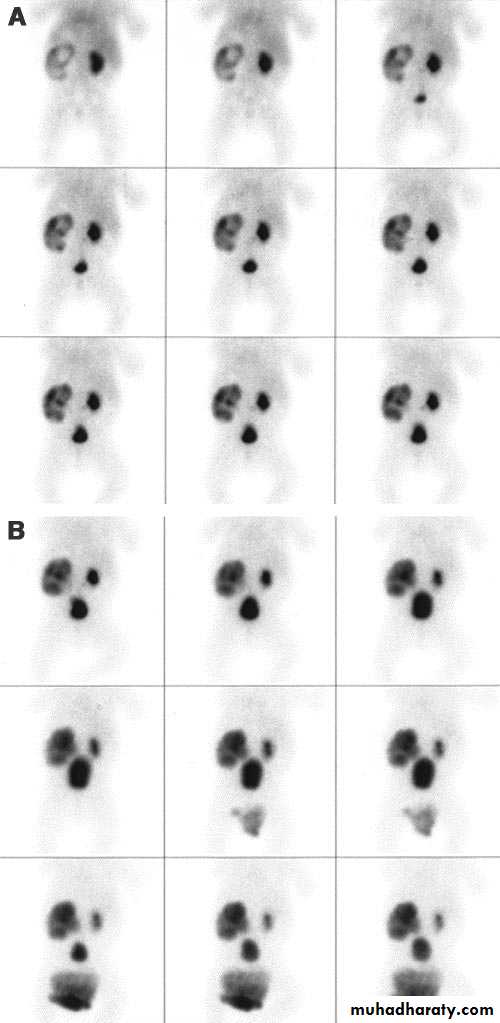
Renal scintigraphy
Indications1. renal perfusion and function
2. Obstruction3. Renovacsular hypertension
4. Infection
5. Presurgical assessment
6. Renal transplant
7. Congenitalanomalies and masses
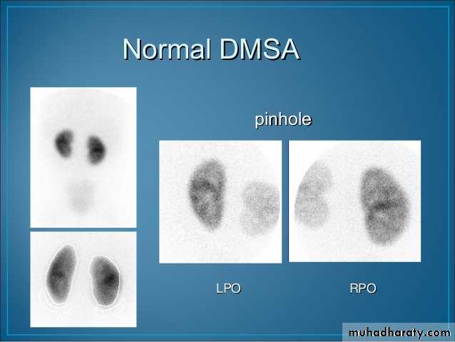
DMSA (dimercaptosuccinic acid)
MAG 3( mercaptoacetylglycylglyine)DTPA ( diethylentriamine penta acetic acid)
DMSA scan (renal morphology scan)
Applications
Renal ectopic and anomalies
Renal masses
Renal infection and scarring
Normal DMSA scan
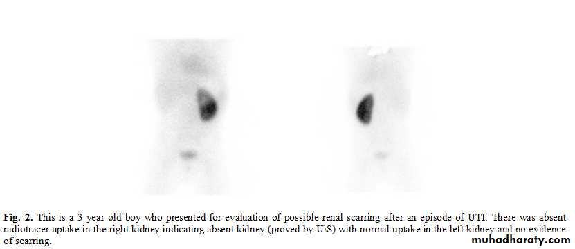
Abscent right kidney
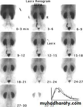
Dynamic renal studies
(DTPA) and (MAG3)Filtered through glomeruli so uses for evaluating
1. Renal perfusion ( vascular supply)
2. Filtration (measuring renal function) ( GFR)3. Drainage ( detect obstruction)
4. Diminished renal function (MAG3)
5. Renal transplant (MAG3)
Normal DTPA scan
Normal MAG3 scan
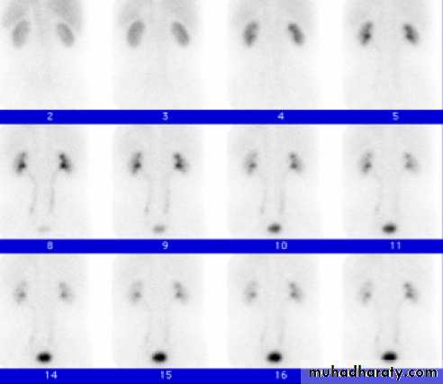
HN of right kidney