CHEST RADIOLOGY
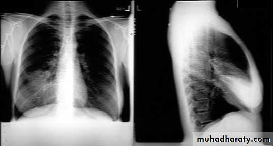
X-ray views1- PA view.
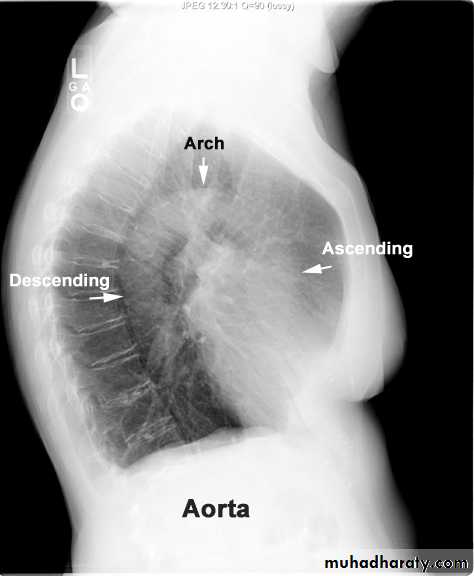
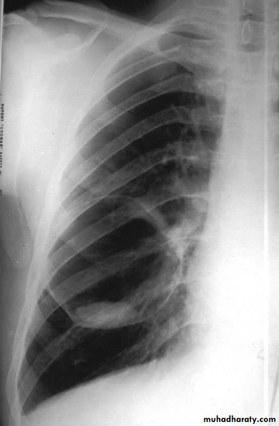
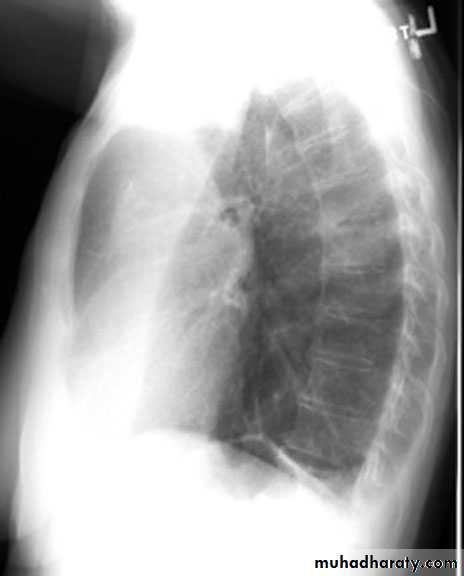
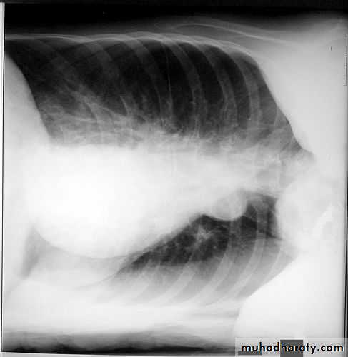
2- lateral.
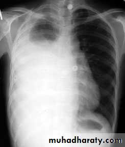
AP ) ) 3-Lordotic
4-P A penetrated view.
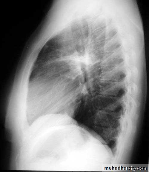
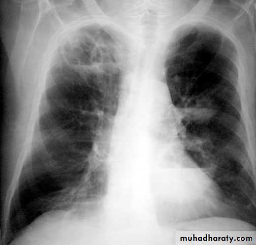
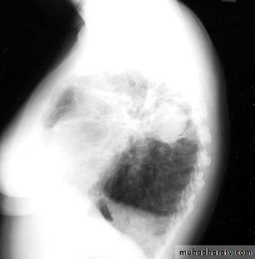
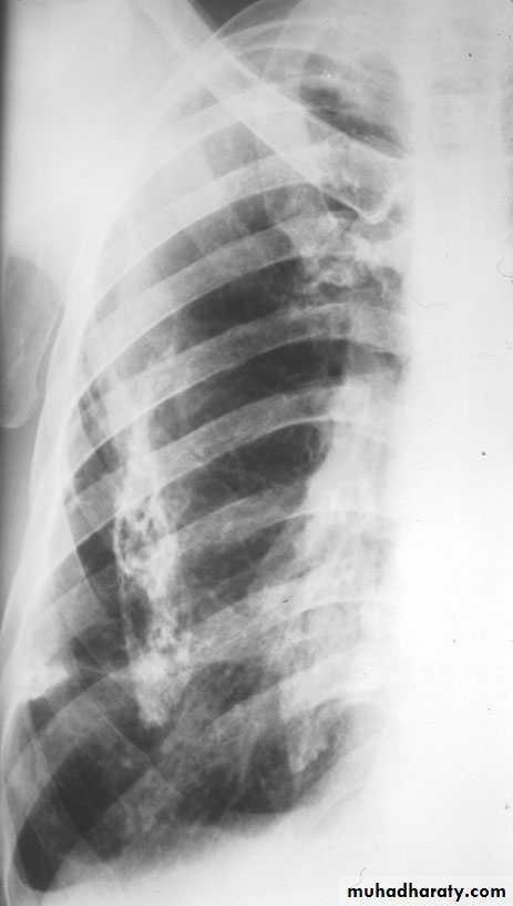
lateral PA view


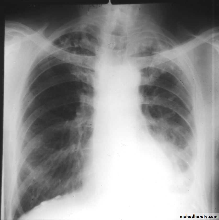
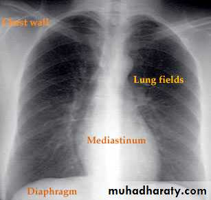
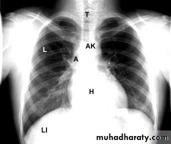
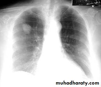
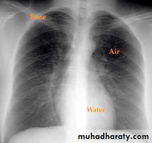
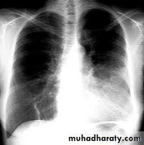
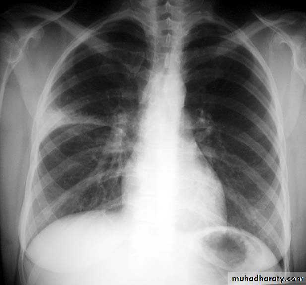
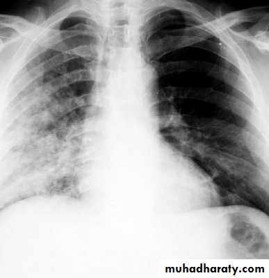
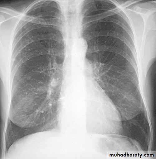
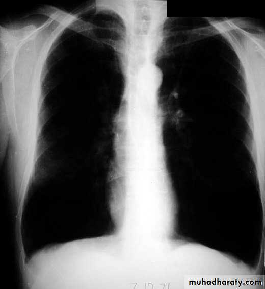
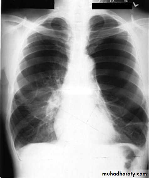
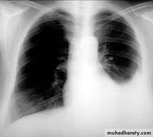
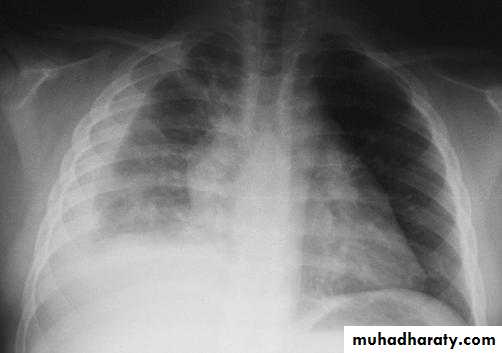
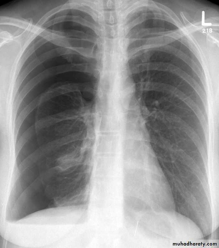
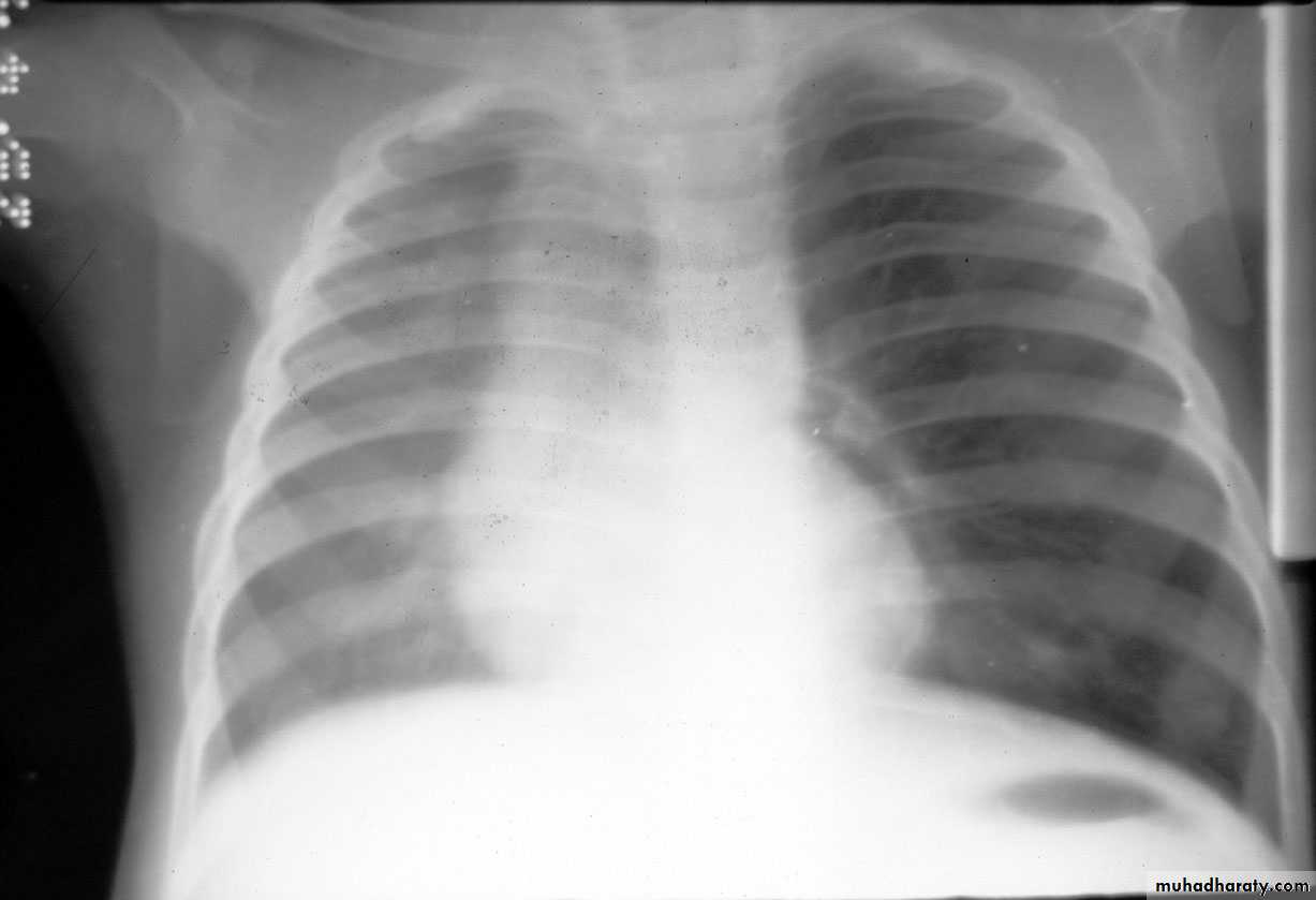
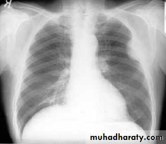
NORMAL PA view
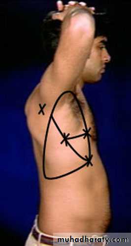
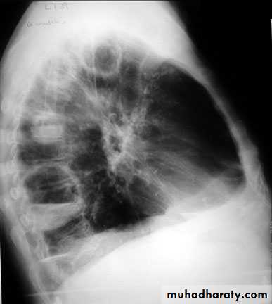
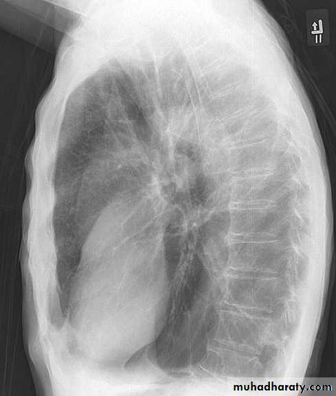
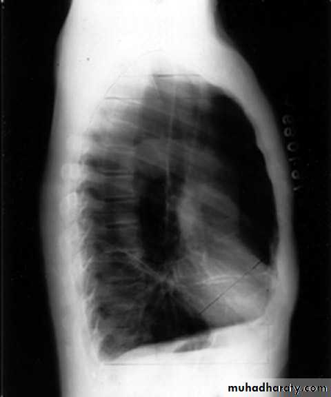
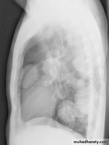
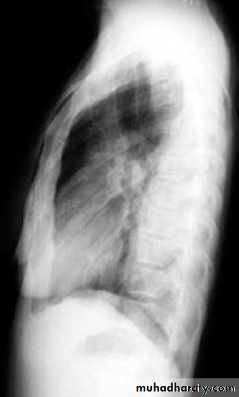
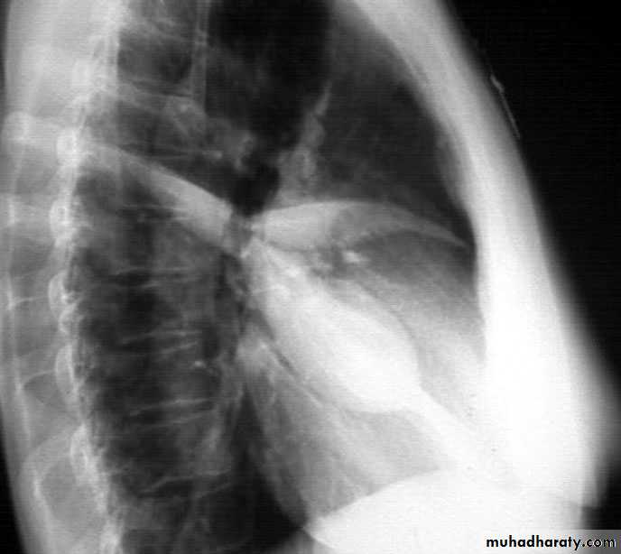
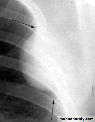
Lateral view position
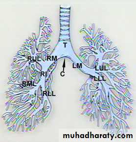
Normal bronchial tree
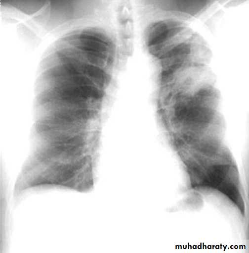
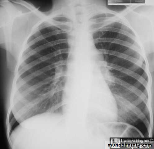
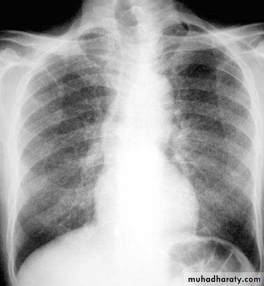
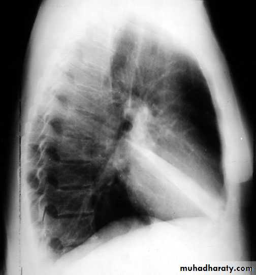
Lordotic view
How to interpret CXR
Pulmonary infection
1- Pneumonia.(a) Lobar ( consolidation) pn.
(b) Lobular or broncho pn.
2- Lung Abscess.
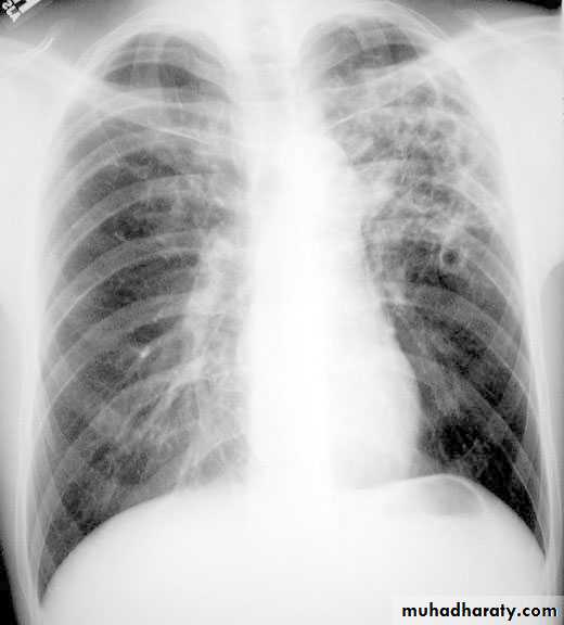
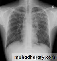
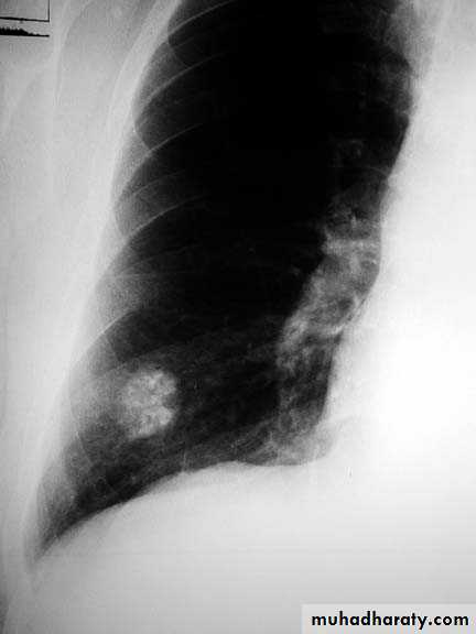
3- Tuberculosis.
Lobar pneumonia with bronchogram.
Middle lobe pneumonia
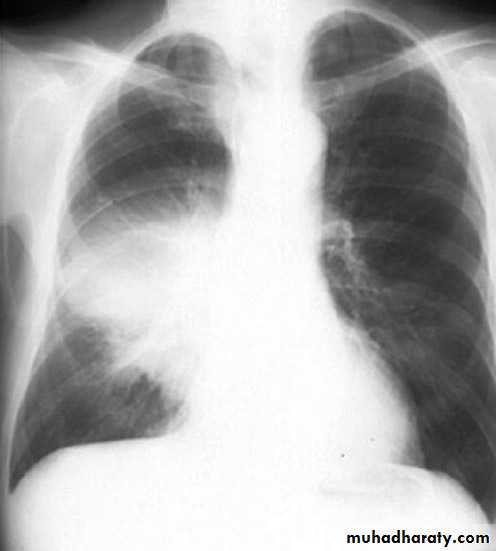
rt.upper lobe pn. Left lower lobe pn.
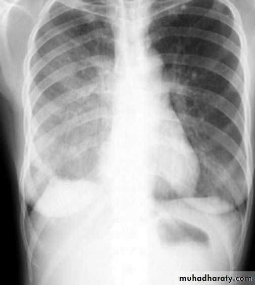
Klebsiella pn. Massive pn.
Resolving consolidation
Cavitations in consolidation
Bronchopneumonia
Lung abscess
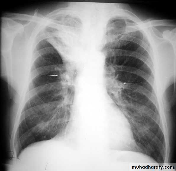
Tuberculosis primary TB
Post primary tb
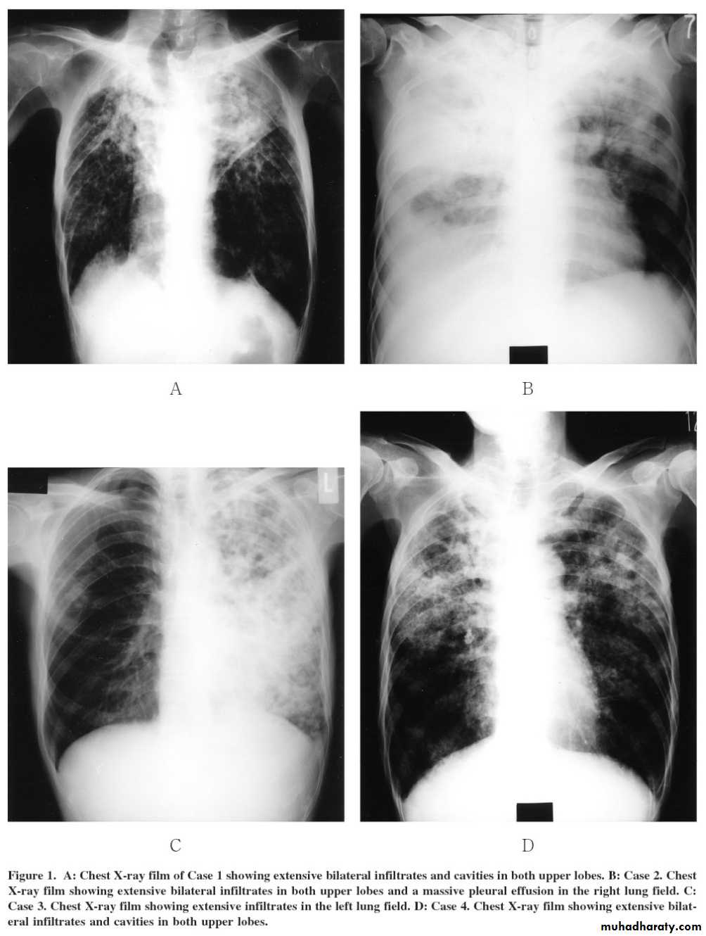
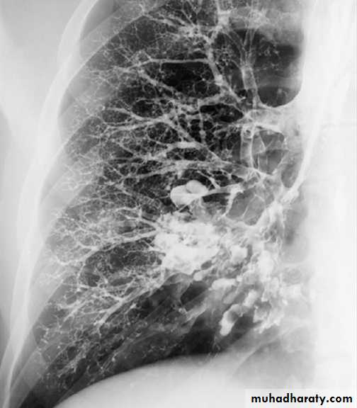
TB lung
Tuberculoma mycetoma
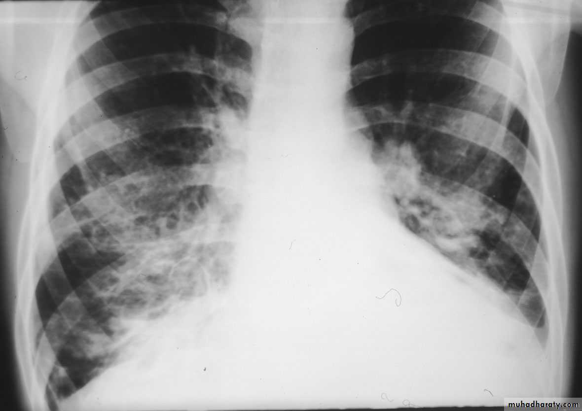
Miliary tb

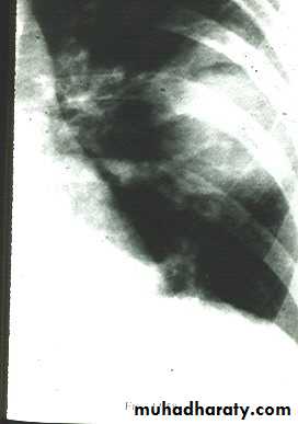
bronchiectasis
bronchiectasis
emphysema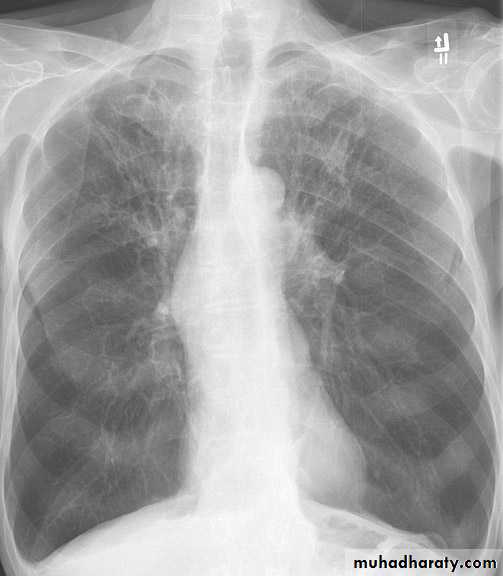
emphysema
Bullous emphysema
Upper lobe c. Middle lobe c.
rt lower lobe c. Left lower lobe c.

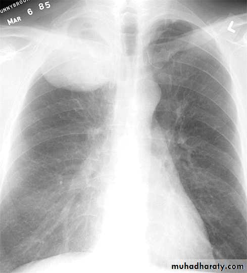
Left upper lobe collapse
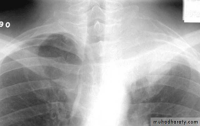
Complete rt. & left Lung collapse
Hydatid cyst early rupture
Water lilly sign (ruptured hydatid cyst)
Lung tumour
Pancoast tumour
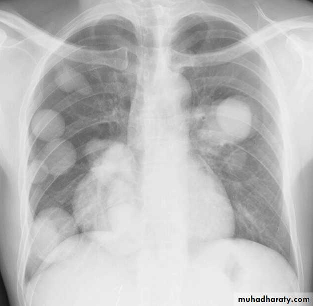
Cannon ball secondaries
Pleural diseasespleural effusionpneumothorax hydro pneumothoraxpleural calcifications & thickeningpleural tumour
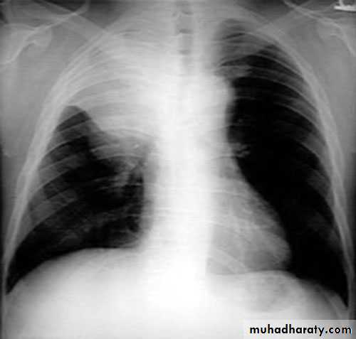
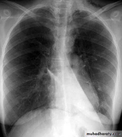
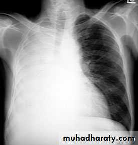
Pleural effusion ( free)
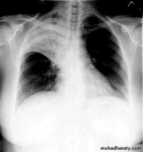
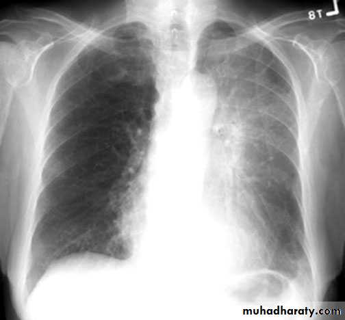
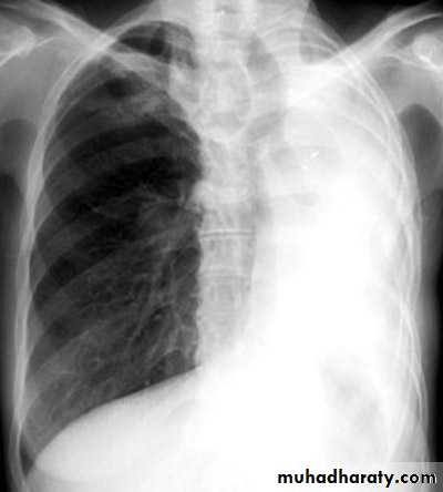
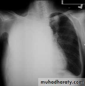
Massive pleural effusion
Lamellar pleural effusion
Encysted peural effusion
Sub costalFissural
Sub pulmonary
Mediastinal
Subcostal encysted effusion
Encysted fissural effusion
Sub pulmonary encysted effusion
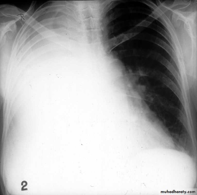
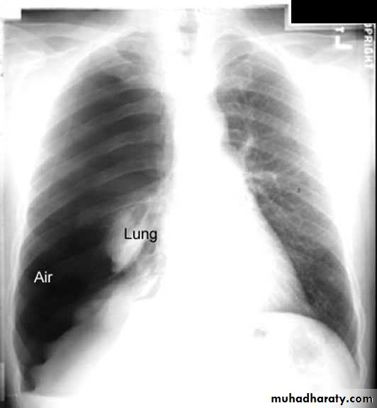
pneumothorax
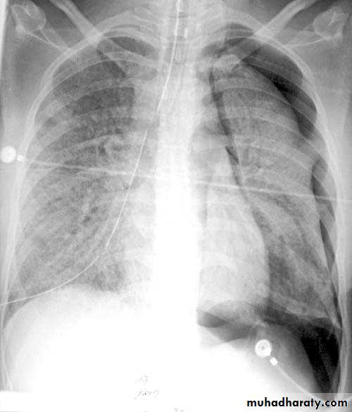
Pneumothorax hydropneumothorax
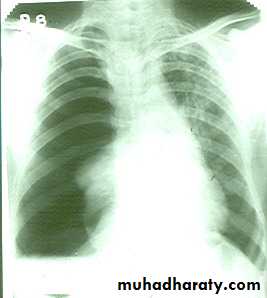
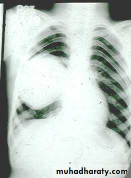
Tension pneumothoarax
Pleural tumour
Pleural calcification
Pleural thickenning