Gestational trophoblastic disease
GTDASS. PROF
Dr.AHMED JASIMMBChB.DOG.FICOG
Gestational trophoblastic disease(GTD)
Is term applied for a group of pregnancy-related disorders arising from abnormal placental trophoblast cells.The main categories of GTD are:
A.The pre-malignant conditions:Hydatidiform mole (benign):
Complete hydatidiform mole
Partial hydatidiform mole.
B.Malignant gestational trophoblastic neoplasias (Gestational trophoblastic tumour (GTT))(malignant disorders):
invasive mole
choriocarcinoma
placental site trophoblastic tumour. very rare

Invasive mole

all forms of GTD produce β -human chorionic gonadotrophin (β-hCG ).The amount of β-hCG correlates with disease volume and so monitoring this hormone is an accurate biomarker for screening, diagnosis, therapeutic response and follow up of women with GTD.
Hydatidiform mole
Are abnormal conception events, which produce rapidly growing, highly vascular, placenta-like structures. It is a Benign tumour of trophoblast and the commonest form of GTD.Rarely, a molar pregnancy may develop as a twin to a normal embryo.
16% of complete hydatidiform mole and 0.5% of partial hydatidiform mole undergo malignant transformation
Incidence:
Complete mole 1 / 1000 pregnancy.Partial mole 3 / 1000 pregnancy.
Risk factors:
1. maternal age (≤15 and ≥40 years old).
2.previous history of molar pregnancies (0.5-2% risk of recurrence)3. ethinic variation and increased rates in Asian women.
4.familial syndrome of recurrent complete hydatidiform mole (inherited in autosomal recessive pattern) but it is extremely rare.
Pathophysiology (cytogenitics)
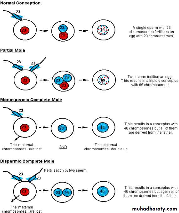
In normal fertilization single sperm with 23 chromosome fertilize an ovum with 23 chromosome.Hydatidiform mole occurs because of an abnormal fertilization process.
Complete hydatidiform mole
Are androgenic in origin and have diploid chromosomal constitution totally derived from paternal (male) genome results from the fertilization of an ‘empty’ oocyte it is usually resulting from:the single haploid sperm duplicating it's own chromosome after meiosis leading to homozygous 46XX karyotype. In majority of cases.
Complete hydatidiform mole
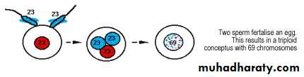
• less frequently , two haploid sperm fertilize an empty ovum leading to heterozygous 46XX OR 46XY.Partial mole
Are genetically biparental ,Usually with a triploid karyotype and arise due to two sperm fertilize an ovum or reduplication of paternal sperm haploid set and having 2 set of chromosome from paternal origin and one from maternal origin 69XXX, 69XXY, 69XYY.Partial mole:
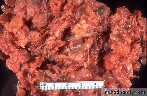
Pathology:
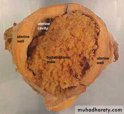
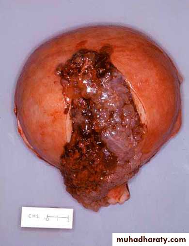
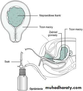
Hydatidiutiform mole appears as multiple vesicles as bunch of grapes in complete mole


In partial mole less clear cut clear picture, vesicles and fetus and placenta.
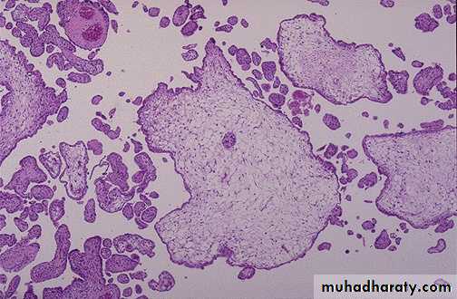
Microscopical:complete mole
Generalized swelling of villous tissueDiffuse trophoblastic hyperplasia
No embryonic or fetal tissue
Absence of fetal blood vessel
The avascular villi of molar pregnancy are quite large. However, these must be distinguished from simple "hydropic degeneration" seen in placentas of fetuses undergoing intrauterine demise.
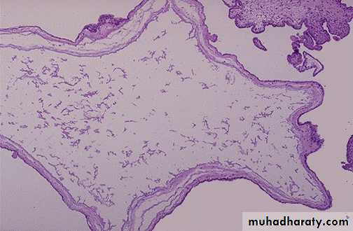
Partial mole
Focal swelling of villous tissuefocal trophoblastic hyperplasia
there is embryonic or fetal tissue
Fetal vessels seen
In partial moles, some villi (as seen here at the lower left) appear normal, whereas others are swollen. There is minimal trophoblastic proliferation.
Clinical features
1.Typical clinical feature in complete mole:amenorrhoea.
Vaginal bleeding is most common sign of variable amount mostly in early pregnancy around 12-14 weeks.
Usually painless but sometimes associated with pain due to uterine contractions.
Symptome of pregnancy in exaggerated form.
2. symptoms of complications of hydatidiform mole:
a. pre-eclampsia early onset before 20th weeksb. hyperemesis gravidarum.
c. Anaemia.
d. hyperthyroidism.
e. complication of theca lutein cyst of ovary (rupture, torsion).molar pregnancy produces excessive hCG , which stimulates excessive growth of ovaries.
h. pelvic infection.
g. perforated uterus.
j. disseminated intravascular coagulopathy (DIC).
k. embolization and respiratory symptoms.
3. spontaneous expulsion of vesicles from vagina around 16 weeks.(if undiagnosed before).
4. discovered accidentally by ultrasound at booking which make the gestational age at evacuation of hydatidiform mole is about 9-10 weeks.
Examinations:
General condition and vital sign can be :Normal or reflect preeclampsia or hyperthyroidism or bleeding or anaemia and pallor
Chest examination: finding if respiratory embolization.
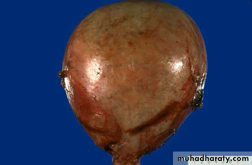
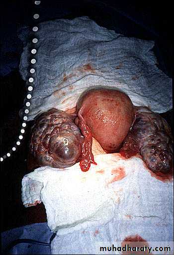
Abdominal examination:
Uterus is often large for date and feel very soft and doughy in consistency.No fetal part or fetal heart can be detected.
Ovarian enlargement occurs in 1/3 of cases and difficult to palpated until uterus evacuated
This is only a second trimester pregnancy, but note how large for dates the uterus is because of a molar pregnancy. An ultrasound in this case revealed no fetus, only a "snowstorm" effect.
Pelvic examination
Sometimes grape like vesicles of mole may be detected which is confirmation to diagnosis if spontaneous expulsion occurs only.Investigations:
Clinical finding of amenorrhoea and vaginal bleeding with larger than expected size uterus arouse suspicion for molar pregnancy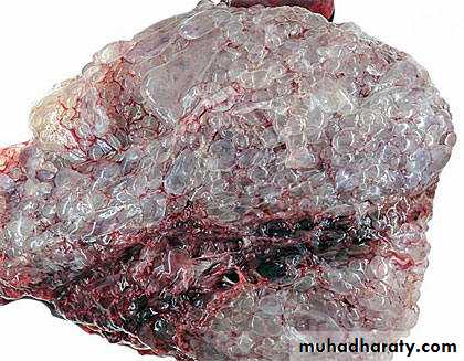

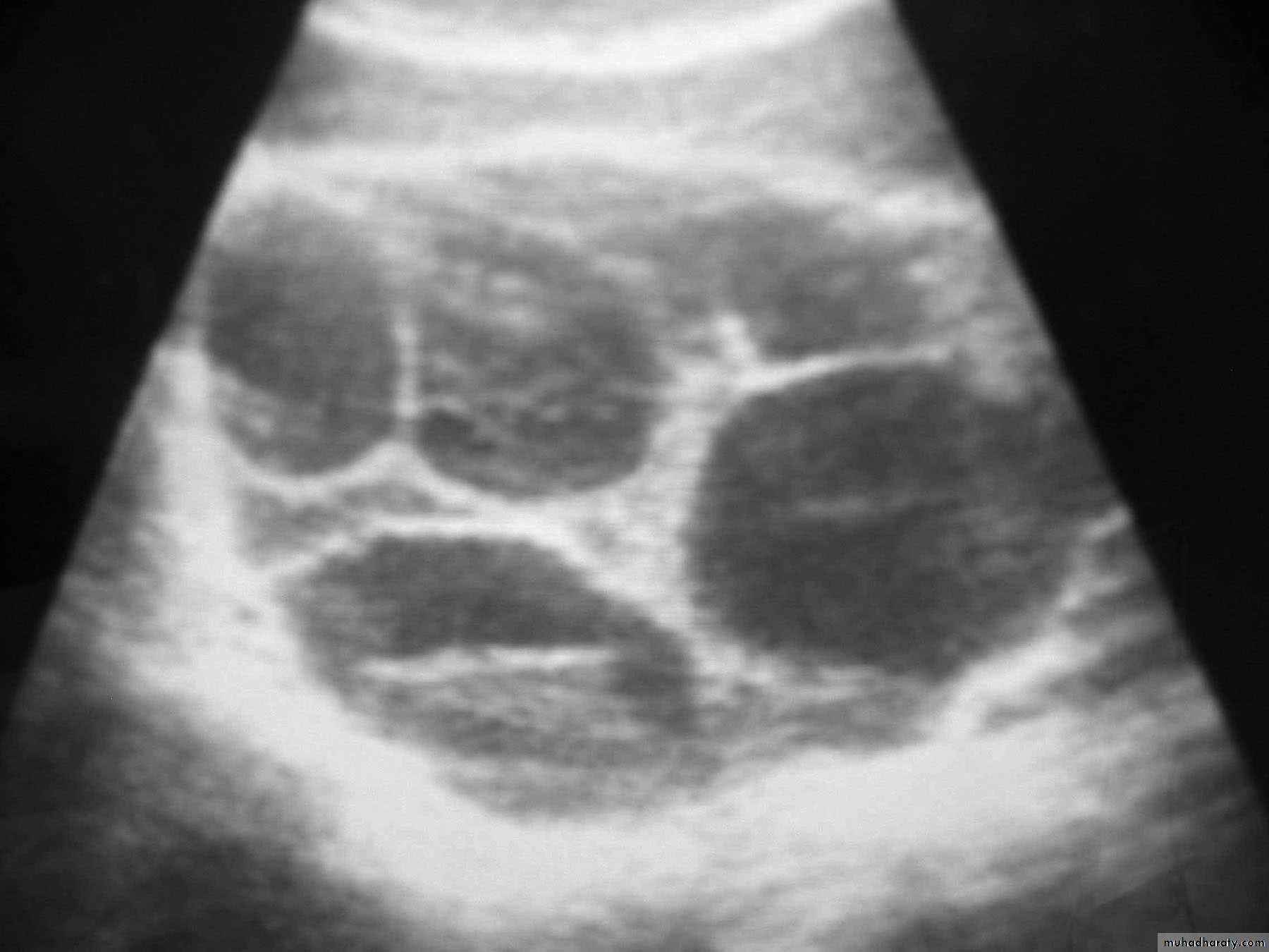
Investigations
ultrasound:a. which confirm diagnosis of mole as show snowstorm appearance (uterine cavity filled with multiple sonolucent area of varying size and shape).
b. absence of fetus.
Theca Lutein cyst of ovary.
β-human chorionic gonadotrophin (β-hCG ) :
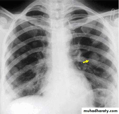
It is high but does not give useful distinction between mole and normal pregnancy. it is pivotal in diagnosis and follow up of GTD.Chest x- ray to exclude metastasis
others:
Full blood count, coagulation screen, liver function test, renal function test, blood type and cross matchpartial mole
In partial mole many of the feature which seen in complete hydatidiform mole may be absent and clinical feature may be less obvious and patients with partial mole present with signs and symptoms of an incomplete or missed abortion, and have bleeding, a small uterus, and low hCG levels.Treatment:
All molar pregnancy has the potential to develop into potentially fatal carcinoma.Treatment consist of two phases:
A. Immediate evacuation of mole
B. Subsequent follow up for detection of persistent trophoblastic proliferation or malignant changes.
termination of molar pregnancy
Termination of mole more common than spontaneous abortion of mole so adequate evaluation of women should be done and combatable blood should be available*If preeclampsia , thyrotoxicosis is present ,may require treatment.
*Evacuation by suction curettage is performed regardless uterine size under general anesthesia (other form of surgical intervention or use of oxytocic drug to induce uterine contraction increase the likelihood of needing chemotherapy and the risk of trophoblastic embolism).
*There is no clinical indication for the routine use of a second uterine evacuation in the management of molar pregnancies. Uterine evacuation may be recommended, in selected cases.
*Hysterectomy is indicated for:
Older women or those of high parity
Severe uncontrolled haemorhhage.
*Theca lutein cyst regress in size as the hCG level falls to normal and need no special treatment unless complicated (rupture , torsion).
*If patient present with Spontaneous abortion of mole:
May required gentle curettage and oxytocics drug .
Send for histopathological examination.(must be)
*Anti D should be given to non sensitized Rh negative mother.
*Prophylactic chemotherapy:
It is not advised for all patient as 80% has spontaneous remission and risk of drug toxicity.Management of Gestational Trophoblastic Neoplasia
Best done by registering all molar pregnancies with a Specialist CentreMethotrexate is the 1st line drug but treatment requires individualization
And multi-agent chemotherapy may be requiredSecond curette rarely necessary
A few patients require surgery as part of their care
FIGO 2000 Score for GTN
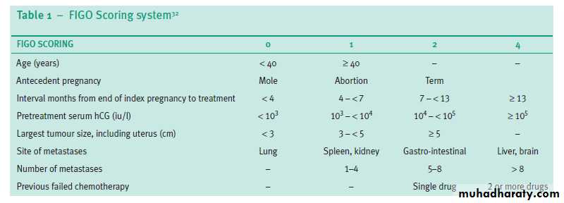
Chemotherapy for GTN is based on FIGO Score..
For score ≤ 6 Methotrexate only:
Alternate daily for a week
With Folinic acid rescue on the alternate days
Then rest for 6 days and measure HCG
Repeat as necessary until HCG is normal
Then weekly HCG for 6w and monthly for 12m
For score ≥ 7 Multi-agent chemotherapy:
DactinomycinCyclophosphamide
Vincristine
Etoposide
Prognosis after chemotherapy for GTN
Cure rates in excess of 97% should be possibleRisk of another molar pregnancy is 1:80
No increased obstetric risk
Unless the pregnancy is conceived within 12m of chemotherapy
Increased risk of pregnancy loss (some by TOP)
But no increased risk of fetal malformation
Menopause occurs slightly earlier
By a mean of 12m or 3 yrs after multi-agent chemo
And some women at risk of developing secondary cancers if chemo continued >6m
Leukemia (RR 16.6)
Ca colon (RR 4.6), melanoma (RR 3.4), Ca breast (RR 5.6)
Women should be advised to avoid pregnancy until hCG levels have been normal for six months following evacuation of a molar pregnancy and for one year
following chemotherapy for gestational trophoblastic tumour.
Advice patient to use barrier method as contraceptive method at beginning and then combined oral contraceptive pill (if no other contraindications) which can be used safely after the hCG levels have returned to normal (if taken while hCG levels are raised, may increase the need for treatment)
Postmolar Surveillance (Follow up):
Aim is to prompt detection of any change suggestive of malignancy.16% of complete hydatidiform mole and 0.5% of partial hydatidiform mole undergo malignant transformation.
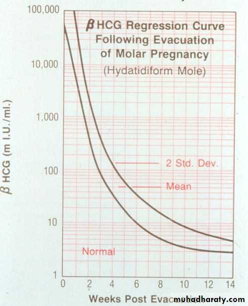
1.β-human chorionic gonadotrophin (β-hCG )
postmolar surveillance with serial quantitative serum -hCG levels should be the standard. Titers should be monitored following uterine evacuation at least every 1 to2 weeks until they become undetectable.
6 months of surveillance after achieving an undetectable -hCG levels (hCG become negative) was recommended for all patients with molar gestation.
hCG should be progressively fall to undetectable levels ,if not decrease main trophoblast persist and trophoblast proliferation which main either:
malignant changes
or b. pregnancy.
Physical examination:
Including pelvic examination at regular interval for first year until involuation of pelvic organ.
Chest x ray
3. Chest x ray: If hCG titer plateau or rises.Chemotherapy is indicated if
-hCG titer rises or plataue during follow up.Evidence of choriocarcinoma.
Persistant uterine bleeding and positive hCG .
hCG levels >20000IU/Lit serum at 4-6 weeks post evacuation.
5. In subsequent pregnancy
An early ultrasound should be performed in all subsequent pregnanciesbecause of the 1–2% risk of a second molar pregnancy.
b. In subsequent pregnancy in any patient had prior trophoblastic tumour of any type should have hCG assay at 6 and 10 weeks post-delivery.
Risk of hydatidiform mole:Before evacuation:
• pre-eclampsia early onset before 20th weeks• hyperemesis gravidarum.
• Anaemia.
• hyperthyroidism.
• complication of theca lutein cyst of ovary (rupture, torsion).molar pregnancy produces excessive hCG , which stimulates excessive growth of ovaries.
• pelvic infection.
• perforated uterus.
• disseminated intravascular coagulopathy (DIC).
• embolization and respiratory symptoms.
During evacuation
• Bleeding can be profuse.
Sepsis.
Perforation of uterus.
Air embolism.
Incomplete evacuation of uterus.
After evacuation:
Choriocarcinoma.Increase risk of recurrence of mole.