Obstruction of renal tract
Causes:-Within the lumen
Calculi
Blood clot
Sloughed papilla (papillary necrosis)
-Within the wall of the collecting system Tumor (transitional cell carcinoma) Infective stricture (TB or Schistosomiasis) Intrinsic PUJ obstruction
-Extrinsic pathology:
Tumors (CA cervix or recto-sigmoid junction).
Retroperitoneal fibrosis.,Aberrant renal artery or retrocaval ureter.
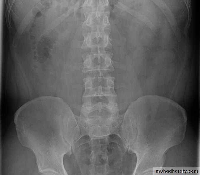
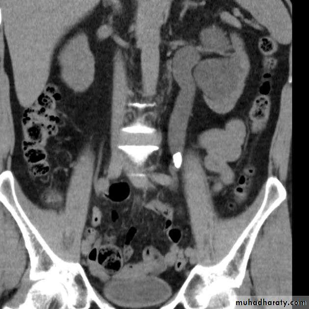
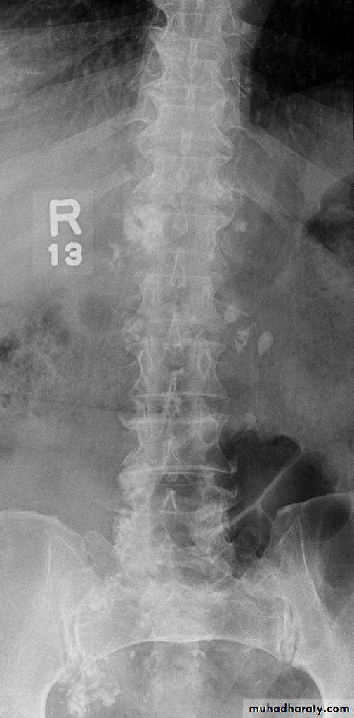
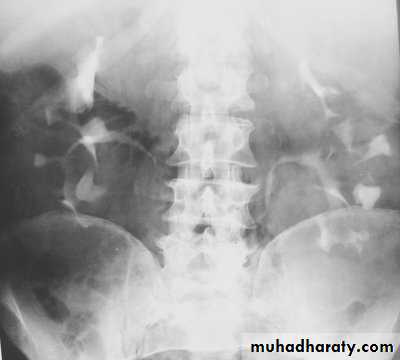
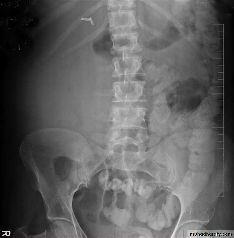
Ureteric calculi (stones):
-Over 90% of calculi are radiopaque on plain films and virtually all on CT as very sensitive for detection of calculi, even those that appear radiolucent on plain film.-Most of these stones are a mixture of calcium oxalate and phosphate.
-Only pure uric acid and xanthine stones are radiolucent on plain x-ray but CAN be identified by CT or US, uric acid stones are associated with increased uric acid excretion in urine as in gout.
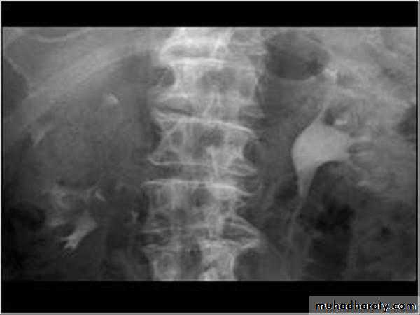
-Principal feature is dilatation of the pelvicalyceal system and ureter.
• The degree of dilatation depends on chronicity (long standing obstruction=more
dilatation).
• The dilatation is down to the level of pathology
The prime objective of imaging is to determine the level and the cause of obstruction.
l.V.U. findings:
-Plain film may be useful in demonstrating calculi.-After contrast injection:
Acutely obstructed kidney shows a dense nephrogram (dense opacification of the renal parenchyma).excretion of contrast (opacification of the collecting system which may take many hours), then the level and degree of obstruction can be determined as dilated pelvi-caliceal system and ureter are followed down to the point of obstruction (point of hold up).
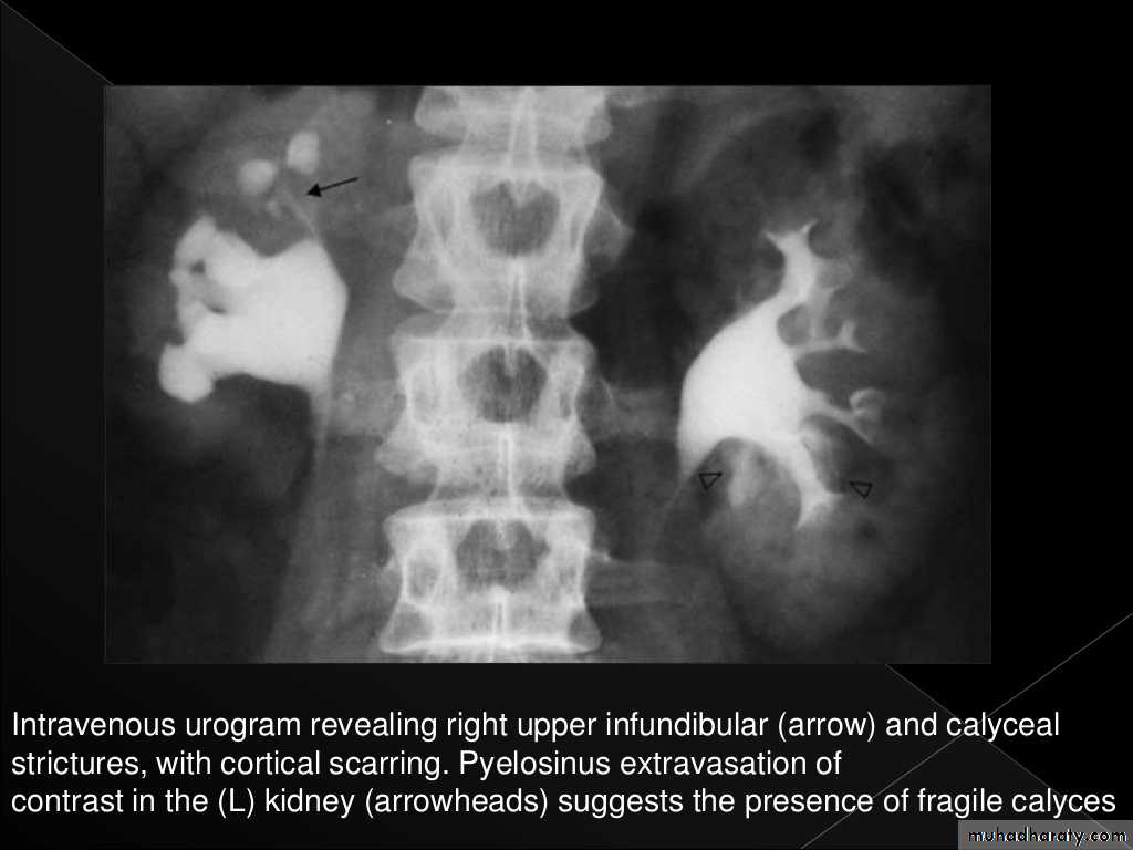
Pyeloxinus reflux may result from rupture of a fornix precipitated by contrast-induced diuresis superimposed on the increased hydrostatic pressure of an obstructed pelvicaliceal system. Urine and contrast extravasate into the renal sinus and perirenal space .
Radiolucent stones appear as filling defect on IVU.
Now CT scans used during acute renal stone-Non contrast CT sensitively identify calculi and non opacified collecting system down to the level of obstruction. it has a sensitivity of 97% and specificity of 96% for detection of ureteral calculi
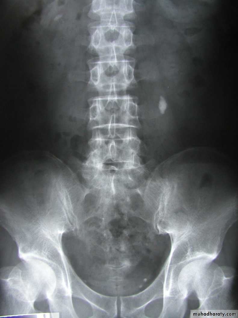
• Ddgx of stone on KUB :
• 1. Gall stone• 2. calfied LN , cartilage ,fibroid,
• 3. Phlebolith: round, lucent centre.
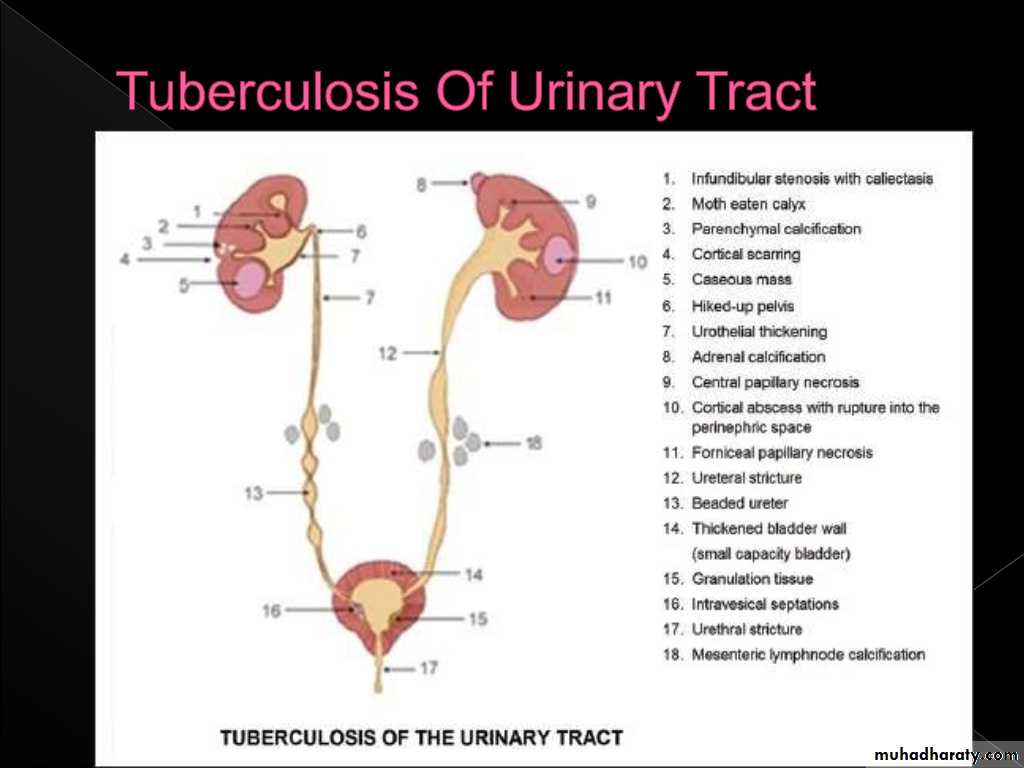
Infection:
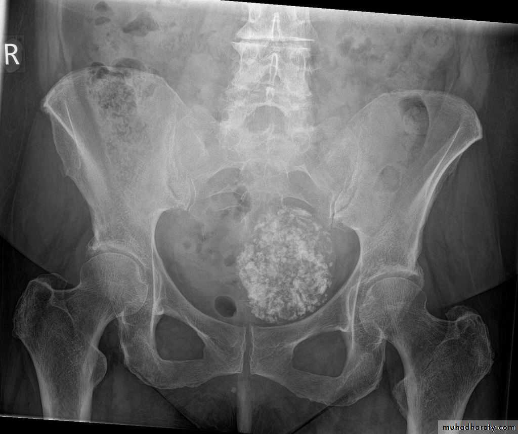
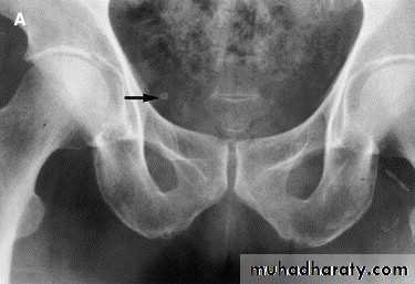
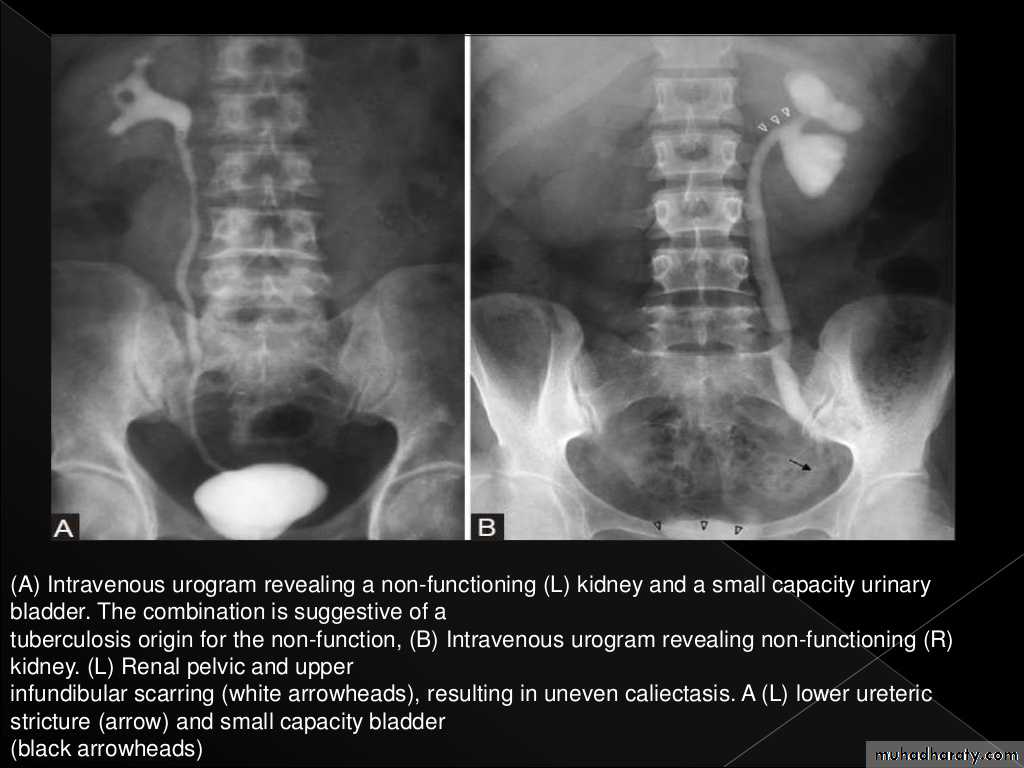
Renal TB:GU tract second most common site of tuberculous infection after lungs, 2ndry to TB infection everywhere.
Spread is hematogenous
Features :
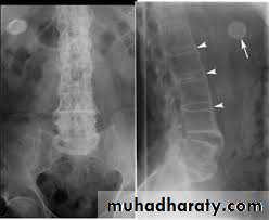
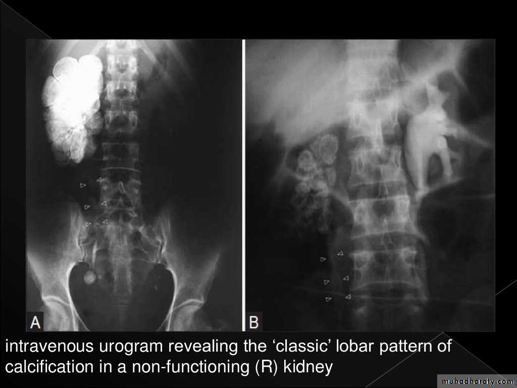
Plain films may show large globular, amorphous calcificationsIVU :
Cortical scarring
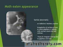
"Smudged" papillae (moth-eaten) –irregular due to inflammation and necrosis
Infundibular strictures
Hydrocalyces without dilatation of renal pelvis, or Hydronephrosis
Autonephrectomy – small, shrunken kidney with dystrophic calcification
When ureters are involved, usually the upper or lower third (more common)
Bladder involvement rarely leads to calcification of wall (think schistosomiasis)
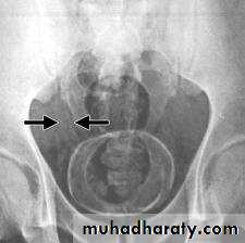
Schistosomiasis
Infestation by s.hematobium
Calcification is most important feature, mainly in bladder & lower ureters , but may involve whole ureters .
In early stage inflammation may cause cobble stone appearance.
Bladder capacity not affected.Ddgx of bladder calcification :
1. schistosomiasis .2.tumor , TB, …
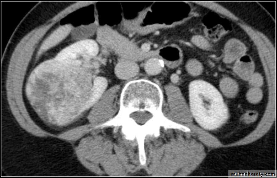
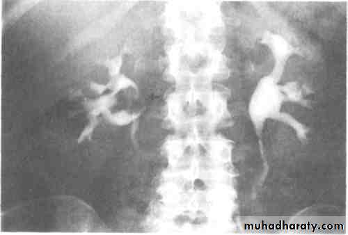
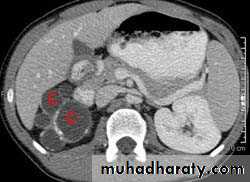
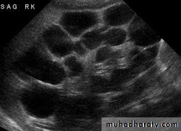
Poly cystic disease
Adult typePresent after the third decade of life , Familial.
Renal parenchyma is replaced by numerous cysts containing fluid , The cysts are of variable size ,
Clinically renal colic, loin mass , heamaturia and hypertension, Renal tissue interposed between the cysts after time dssimcted ended with renal failure
Almost bilateral.
IVU
Large kidney .
Lobulated out-line.
Distortion of pelvi- calyceal system depend on cyst size, number and position.
In advanced cases there is elongation and stretching of minor and major calyces ( spider leg).
In advanced cases IVU shows non-functioning kidney .
Infantile type :
Usually affect liver, spleen and pancreas , Incompatible with life .I.V.U.
- Bilateral Large kidney due to numerous small cysts (1-2 mm size ).- The out-line is not lobulated as in adult.
- Nephrogram shows minute filling defects.
• Tumors
• Renal cell carcinoma:• Comprise 85% of renal malignancy
• Features
• 1. Soft tissue mass on KUB.
• 2. Irregular filling defect with destruction of calyces on IVU.
• Heterogeneous necrotic mass on CT scan may extend to the perinephric area, renal vessels with regional LN and distant metastasis to the lungs or bone.
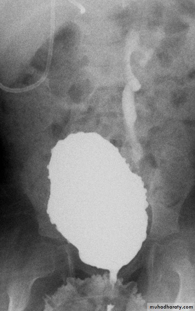
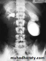
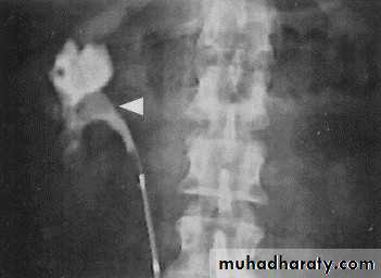
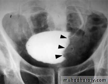
Urothelial tumors
. 85.90% of tumors arising within the collecting systems of the kidneys are transitionalcellcarcinomas(TCC) . May occur at multiple sites, so pelvicalyceal system, ureter and bladder must be well examined.. IVU plays an important role in displaying upper tract tumors. . They appear as a filling defects projecting into the lumen, within the collecting system or urinary bladder.. Must be differentiated from blood clots or radiolucent calculi .. if confused with overlying gas shadow, then tomography may be required during an IVU to solve this problem and now replaced by CT scan.
TCC of kidney
TCC of UB
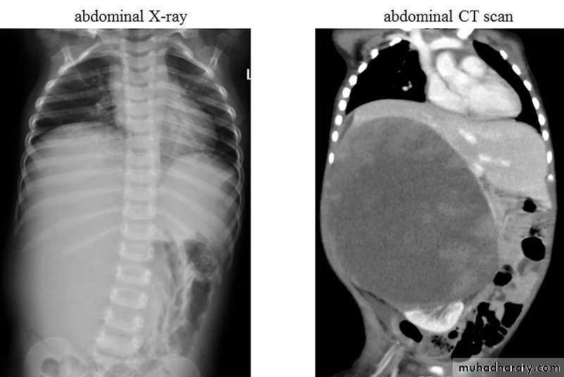
Wilms Tumor ;Nephroblastomma
Most common malignant abdominal neoplasm in children.Peak age: 2-3 years.
It arise from kidney, 90 % unilateral, 10 % bilateral.
Clinical presentation: Palpable abdominal mass, hypertension or hematuria.
Radiological features:
Appear as soft tissue opacity on plain x ray displacing the bowel loops.
On CT scan appear as a large heterogeneous well defined solid mass.
Sometimes there is a calcification and cystic formation.It cause displacement of adjacent structures and may cross the midline.
Treatment: surgery and radiotherapy.
Prognosis: cure rate about 90 %
Multicystic Dysplastic Kidney
It is a type of non heritable pediatric renal cystic disease, it results in multiple cysts being formed in utero.Usually it is unilateral in 80-90 %, if bilateral in compatible with life.
Diagnosis either during antenatal obstetric US or during neonatal period as loin mass.
Sometimes associated with contra lateral renal abnormalities like VU reflux or PUJ obstruction.
imaging appearance of MCDK varies by age:
√ large kidney with lobulated contour in infancy√ often incidental finding of small kidney in adult.
√ calcification: curvilinear / ringlike in wall of cysts in 30% of adults, rarely in children
√ Absence of renal sinus and minimal or absent renal parenchyma.
√ peripheral non communicating cysts ± calcifications
√ absence / severe atrophy of ipsilateral ureter + renal collecting system + renal vasculature
√ contralateral renal hypertrophy
√ contralateral vesicoureteral reflux (VUR) in 5–43%
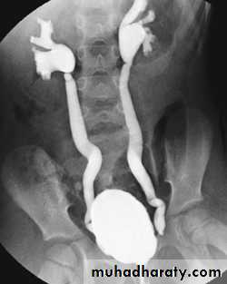
Vesicoureteric Reflux
Abnormal valve mechanism at UVJ resulting in reflux, ureteral dilatation, clubbed calices, eventual renal scaring.Clinical presentation: recurrent UTI, if long standing lead to renal scarring.
Two Types:
A. CONGENITAL REFLUX = PRIMARY REFLUX
Occurs in children, due to short submucosal ureteral tunnel, unilateral or bilateral.
Resolve spontaneiusly.
B. ACQUIRED REFLUX = SECONDARY REFLUX causes:
1. Duplication with ureterocele.2. Cystits.
3. Urethral obstruction.
4. VU reflux.
Diagnosis:
Mainly by voiding cystourethrography( VCUG) by urinary bladder catheterization and distension to confirm reflux.Also by US for assessment of renal parenchyma and scarring.
Primary VUR
Secondary VUR