Lec. : 2 Physiology
1
Cell membranes
The cell membrane is an extremely pliable structure composed primarily of
back-to-back phospholipids (a ―bilayer‖). Cholesterol is also present, which
contributes to the fluidity of the membrane, and there are various proteins
embedded within the membrane that have a variety of functions.
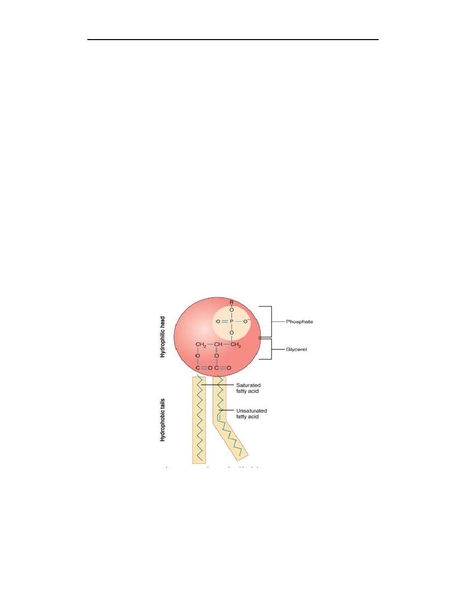
A single phospholipid molecule has a phosphate group on one end, called the
―head,‖ and two side-by-side chains of fatty acids that make up the lipid tails. The
phosphate group is negatively charged, making the head polar and hydrophilic or
―water loving.‖ A hydrophilic molecule (or region of a molecule) is one that is
attracted to water. The phosphate heads are thus attracted to the water molecules of
both the extracellular and intracellular environments. The lipid tails, on the other
hand, are uncharged, or nonpolar, and are hydrophobic—or ―water fearing.‖
A hydrophobic molecule (or region of a molecule) repels and is repelled by water.
Some lipid tails consist of saturated fatty acids and some contain unsaturated fatty
acids. This combination adds to the fluidity of the tails that are constantly in
motion. Phospholipids are thus amphipathic molecules. An amphipathic molecule
is one that contains both a hydrophilic and a hydrophobic region.
1. Intracellular fluid (ICF) is the fluid interior of the cell.
2. Extracellular fluid (ECF) is the fluid environment outside the enclosure of the
cell membrane.
Lec. : 2 Physiology
3. Interstitial fluid (IF) is the term given to extracellular fluid not contained
within blood vessels.
Membrane proteins
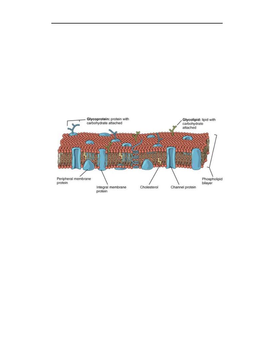
The lipid bilayer forms the basis of the cell membrane, but it is peppered
throughout with various proteins. Two different types of proteins that are
commonly associated with the cell membrane are the integral proteins and
peripheral protein. As its name suggests, an integral protein is a protein that is
embedded in the membrane. A channel protein is an example of an integral
protein that selectively allows particular materials, such as certain ions, to pass into
or out of the cell.
Peripheral proteins are typically found on the inner or outer surface of the
lipid bilayer but can also be attached to the internal or external surface of an
integral protein. These proteins typically perform a specific function for the cell.
Some peripheral proteins on the surface of intestinal cells, for example, act as
digestive enzymes to break down nutrients to sizes that can pass through the cells
and into the bloodstream.
Transport across the cell membrane
One of the great wonders of the cell membrane is its ability to regulate the
concentration of substances inside the cell. These substances include ions such as
Ca
++
, Na
+
, K
+
, and Cl
–
; nutrients including sugars, fatty acids, and amino acids; and
waste products, particularly carbon dioxide (CO
2
), which must leave the cell.
The membrane’s lipid bilayer structure provides the first level of control. The
phospholipids are tightly packed together, and the membrane has a hydrophobic
interior. This structure causes the membrane to be selectively permeable. A
Lec. : 2 Physiology
3
membrane that has selective permeability allows only substances meeting certain
criteria to pass through it unaided.
1. Passive transport: is the movement of substances across the membrane
without the expenditure of cellular energy. In contrast, active transport is the
movement of substances across the membrane using energy from adenosine
triphosphate (ATP).
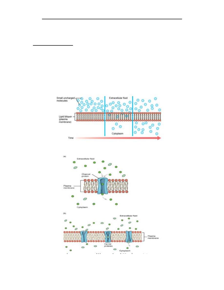
A. Simple diffusion across the cell (plasma) membrane: The structure of the
lipid bilayer allows small, uncharged substances such as oxygen and carbon
dioxide, and hydrophobic molecules such as lipids, to pass through the cell
membrane, down their concentration gradient, by simple diffusion.
B. Facilitated diffusion of substances crossing the cell (plasma) membrane takes
place with the help of proteins such as channel proteins and carrier proteins.
+
Lec. : 2 Physiology
Water also can move freely across the cell membrane of all cells, either through
protein channels or by slipping between the lipid tails of the membrane itself.
Osmosis is the diffusion of water through a semipermeable membrane.
The movement of water molecules is not itself regulated by cells, so it is
important that cells are exposed to an environment in which the concentration of
solutes outside of the cells (in the extracellular fluid) is equal to the concentration
of solutes inside the cells (in the cytoplasm). Two solutions that have the same
concentration of solutes are said to be isotonic (equal tension). When cells and
their extracellular environments are isotonic, the concentration of water molecules
is the same outside and inside the cells, and the cells maintain their normal shape
(and function).
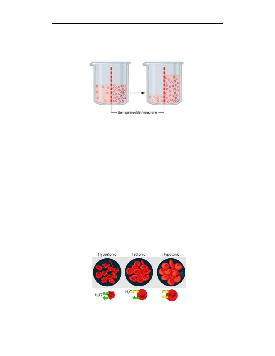
Osmosis occurs when there is an imbalance of solutes outside of a cell versus
inside the cell. A solution that has a higher concentration of solutes than another
solution is said to be hypertonic, and water molecules tend to diffuse into a
hypertonic solution. Cells in a hypertonic solution will shrivel as water leaves the
cell via osmosis. In contrast, a solution that has a lower concentration of solutes
than another solution is said to be hypotonic, and water molecules tend to diffuse
out of a hypotonic solution. Cells in a hypotonic solution will take on too much
water and swell, with the risk of eventually bursting.
Lec. : 2 Physiology
5
2. Active transport: During active transport, ATP is required to move a substance
across a membrane, often with the help of protein carriers, and usually against its
concentration gradient.
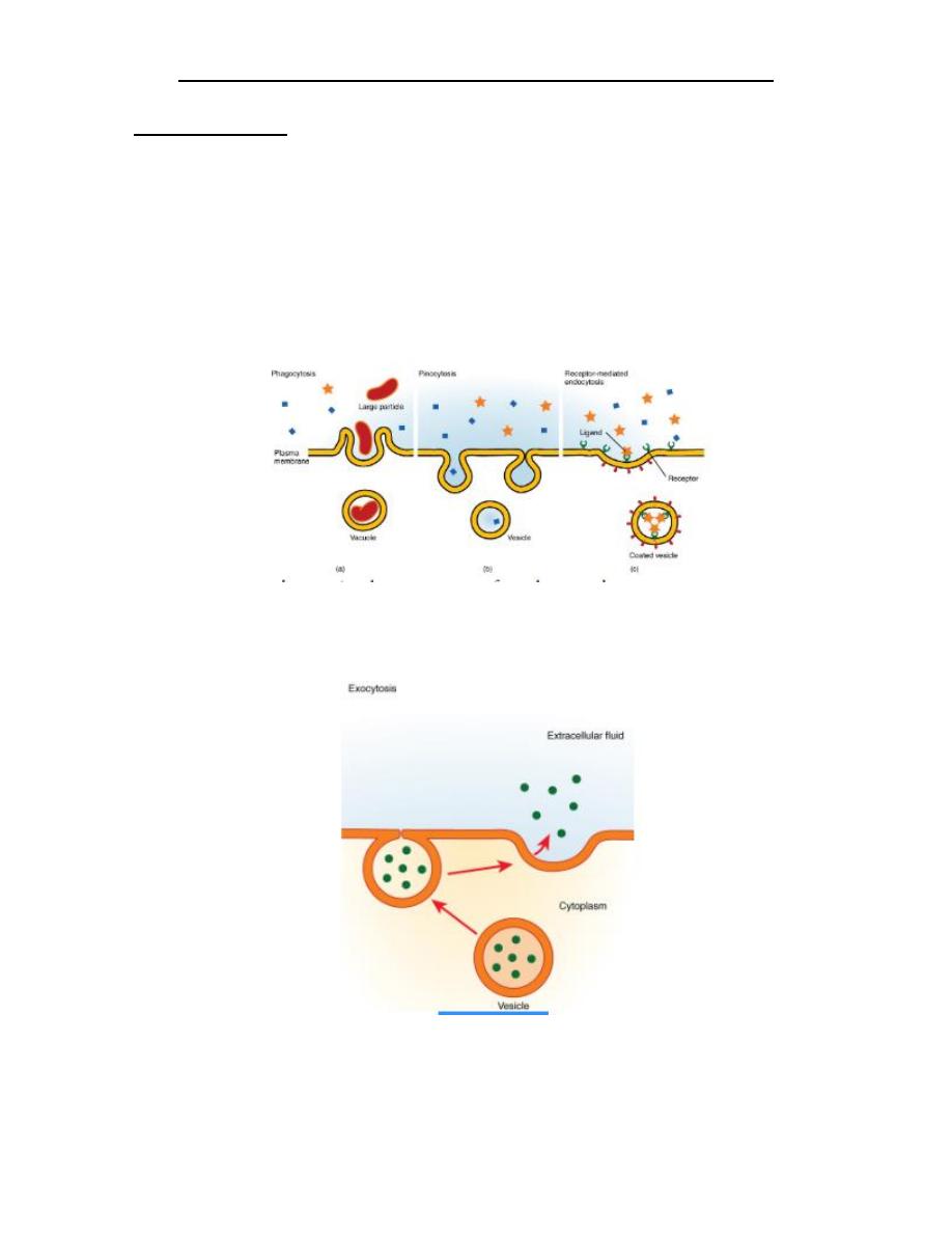
A. Endocytosis is a form of active transport in which a cell envelopes extracellular
materials using its cell membrane. (a) In phagocytosis, which is relatively
nonselective, the cell takes in a large particle. (b) In pinocytosis, the cell takes
in small particles in fluid. (c) In contrast, receptor-mediated endocytosis is
quite selective. When external receptors bind a specific ligand, the cell responds
by endocytosing the ligand.
B. Exocytosis: is much like endocytosis in reverse. Material destined for export is
packaged into a vesicle inside the cell. The membrane of the vesicle fuses with
the cell membrane, and the contents are released into the extracellular space.