Lec. : 3 Physiology
1
Contraction of Skeletal Muscle
Skeletal Muscle Fiber
Figure below
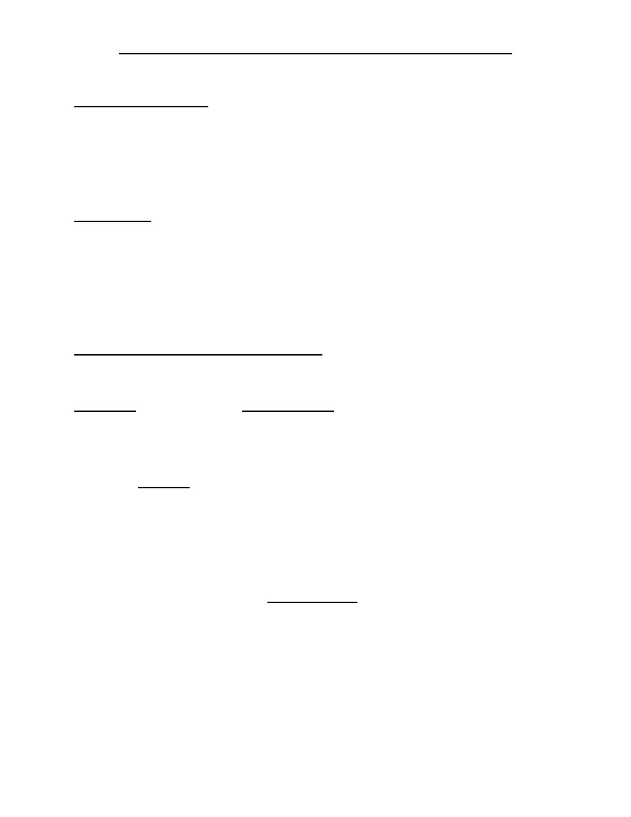
shows the organization of skeletal muscle, demonstrating that all
skeletal muscles are composed of numerous fibers ranging from 10 to 80
micrometers in diameter. In most skeletal muscles, each fiber extends the entire
length of the muscle. Except for about 2 per cent of the fibers, each fiber is usually
innervated by only one nerve ending, located near the middle of the fiber.
Sarcolemma
The sarcolemma is the cell membrane of the muscle fiber. The sarcolemma
consists of a true cell membrane, called the plasma membrane, and an outer coat
made up of a thin layer of polysaccharide material that contains numerous thin
collagen fibrils. At each end of the muscle fiber, this surface layer of the
sarcolemma fuses with a tendon fiber, and the tendon fibers in turn collect into
bundles to form the muscle tendons that then insert into the bones.
Myofibrils; Actin and Myosin Filaments
Each muscle fiber contains several hundred to several thousand myofibrils,
which are demonstrated by the many small open dots in the cross-sectional view of
Figure (C). Each myofibril (Figure D and E) is composed of about 1500 adjacent
myosin filaments (thick) and 3000 actin filaments
(
thin), which are large
polymerized protein molecules that are responsible for the actual muscle
contraction.
Note in Figure E that the myosin and actin filaments partially interdigitate and
thus cause the myofibrils to have alternate light and dark bands. The light bands
contain only actin filaments and are called I bands because they are isotropic to
polarized light. The dark bands contain myosin filaments, as well as the ends of the
actin filaments where they overlap the myosin, and are called A bands because
they are anisotropic to polarized light. Note also the small projections from the
sides of the myosin filaments in Figure E and L. These are cross-bridges. It is the
interaction between these cross-bridges and the actin filaments that causes
contraction.
The ends of the actin filaments are attached to a so-called Z disc. From this
disc, these filaments extend in both directions to
interdigitate with the myosin
filaments. The Z disc, which itself is composed of filamentous proteins different
from the actin and myosin filaments, passes crosswise across the myofibril and
also crosswise from myofibril to myofibril, attaching the myofibrils to one another
Lec. : 2 Physiology
all the way across the muscle fiber. Therefore, the entire muscle fiber has light and
dark bands, as do the individual myofibrils. These bands give skeletal and cardiac
muscle their striated appearance.
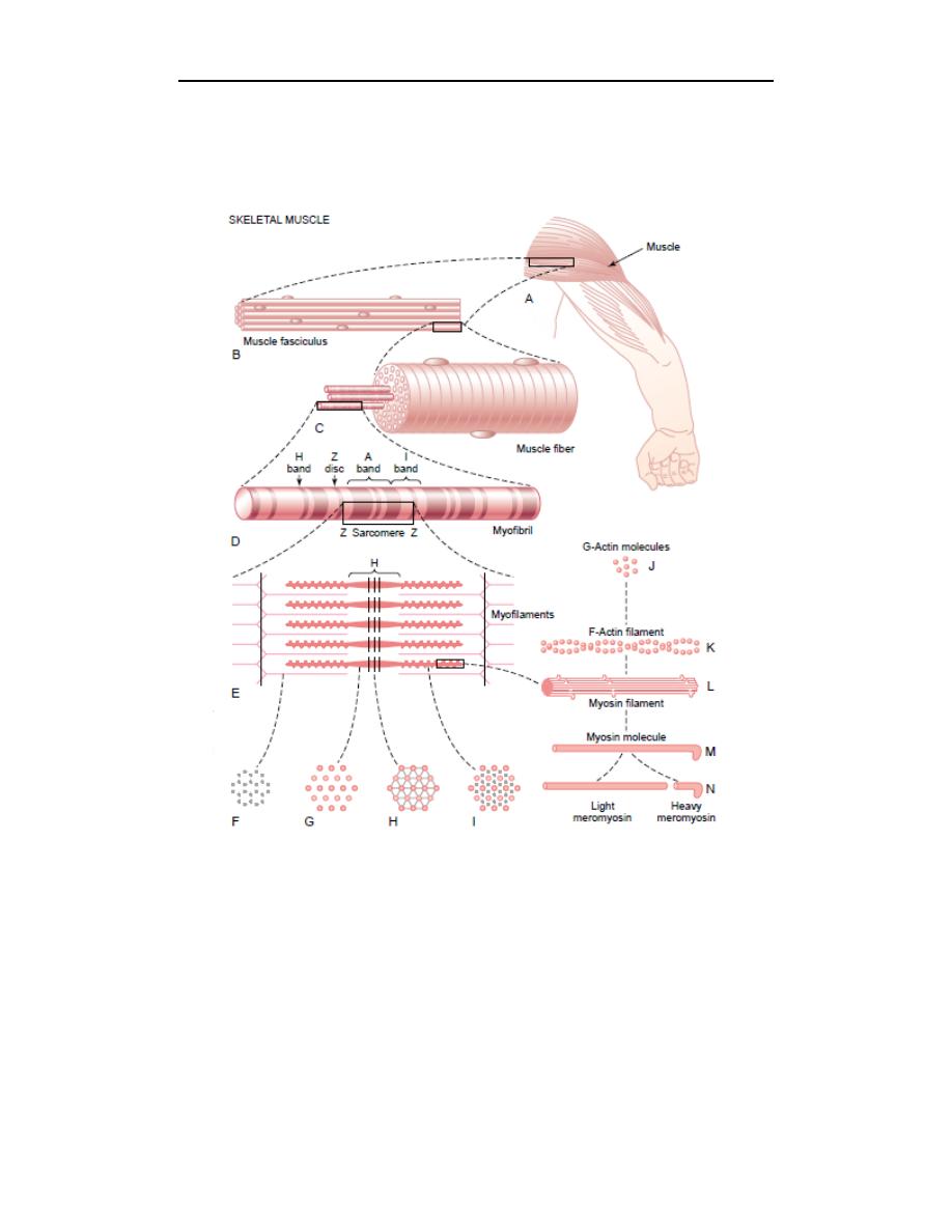
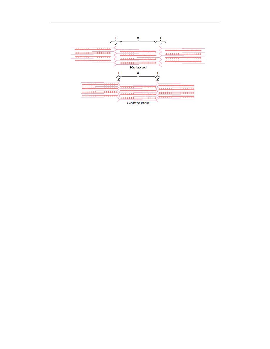
The portion of the myofibril (or of the whole muscle fiber) that lies between two
successive Z discs is called a sarcomere. When the muscle fiber is contracted, as
shown at the bottom of Figure, the length of the sarcomere is about 2 micrometers.
At this length, the actin filaments completely overlap the myosin filaments, and the
tips of the actin filaments are just beginning to overlap one another. We will see
later that, at this length, the muscle is capable of generating its greatest force of
contraction.
Lec. : 3 Physiology
3
General Mechanism of Muscle Contraction
The initiation and execution of muscle contraction occur in the following
sequential steps.
1. An action potential travels along a motor nerve to its endings on muscle fibers.
2. At each ending, the nerve secretes a small amount of the neurotransmitter
substance acetylcholine.
3. The acetylcholine acts on a local area of the muscle fiber membrane to open
multiple “acetylcholinegated” channels through protein molecules floating in
the membrane.
4. Opening of the acetylcholine-gated channels allows large quantities of sodium
ions to diffuse to the interior of the muscle fiber membrane. This initiates an
action potential at the membrane.
5. The action potential travels along the muscle fiber membrane in the same way
that action potentials travel along nerve fiber membranes.
6. The action potential depolarizes the muscle membrane, and much of the action
potential electricity flows through the center of the muscle fiber. Here it causes
the sarcoplasmic reticulum to release large quantities of calcium ions that have
been stored within this reticulum.
7. The calcium ions initiate attractive forces between the actin and myosin
filaments, causing them to slide alongside each other, which is the contractile
process.
8. After a fraction of a second, the calcium ions are pumped back into the
sarcoplasmic reticulum by a Ca
++
membrane pump, and they remain stored in
the reticulum until a new muscle action potential comes along; this removal of
calcium ions from the myofibrils causes the muscle contraction to cease.